Neurofibromatosis type 1 is a autosomal dominant disease, with an estimate frequency of 1 per 2500–3000 births.Reference Trevelyan, Been and Patel1 Neurofibromatosis type 1 may have different vascular manifestations, such as aneurysms and stenosis that can affect different arterial territories including the great vessels, cerebral, visceral, and renal arteries.Reference Syed and Lesch2 Coronary involvement is extremely rare and only anecdotical cases have been reported so far. Age, clinical presentation, and underlying pathophysiology are also variable, encompassing aneurysms formation complicated by acute thrombosis, vasospasm, and stenosis. The most radical therapeutic option contemplated in the literature is aneurysm ligation and bypass graft.Reference Pontailler, Vilarem, Paul and Deleuze3
The presentation in paediatric population is even more exceptional and acute, and follow-up management is particularly challenging given the paucity of evidences and data about the natural history.
We report a case of a 13-year-old boy with neurofibromatosis type 1 presenting with myocardial infarction and multi-vessel aneurysmal coronary dilatation.
Case report
A 13-year-old boy with a previous diagnosis neurofibromatosis type 1 (17q11.2 exon 22 c3790G →aa1264), with no previous cardiac symptoms, presented with acute myocardial infarction with ST elevation in the inferior and lateral leads.
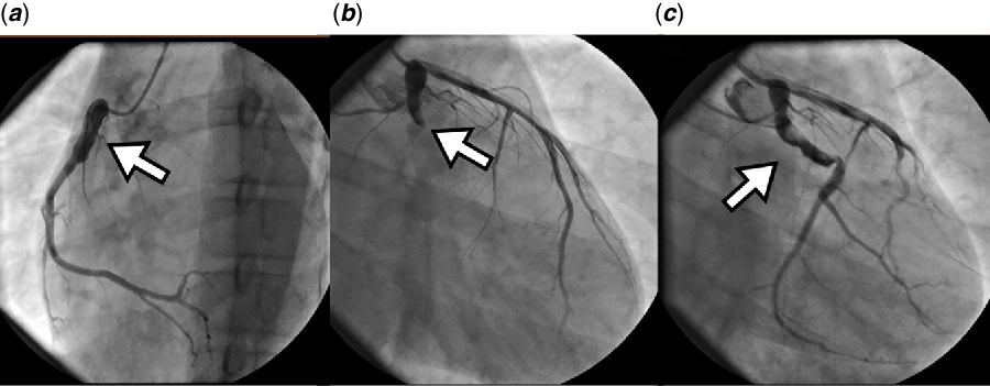
Coronary angiography revealed an aneurysm of the circumflex artery complicated by acute thrombosis. Right coronary artery also had a proximal aneurysm with normal flow (Fig 1a and b).
( a ) Right oblique view angiogram showing aneurysm of the first tract of the right coronary artery. ( b ) Left coronary angiogram in the same view showing acute occlusion of the circumflex. ( c ) Left coronary angiogram showing reperfusion after thromboaspiration and ballon dilatation. White arrow indicates residual thrombotic material in the aneurysm.

Thromboaspiration and plain old balloon angioplasty) were performed with immediate achievement of a good flow that was maintained at a second angiography performed 10 days later because of recurrent atypical chest pain (Fig 1c).
He was discharged asymptomatic on aspirin and warfarin (INR range 2–2,5), with no evidence of ischaemia/Q waves at the ECG and with normal ventricular function.
At 2 months follow-up, ECG and ECHO persistently showed completely normalised findings.
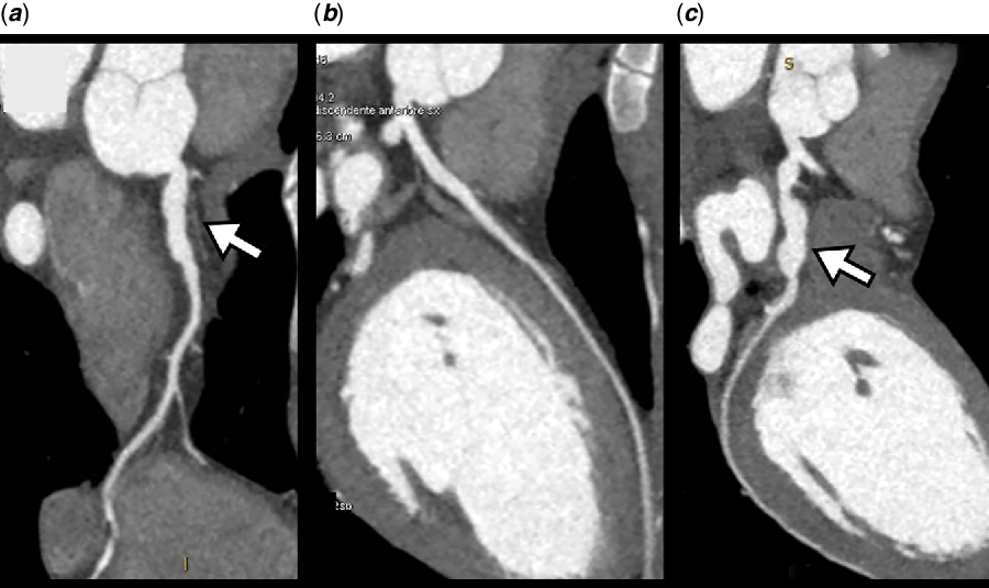
A coronary CT, performed 5 months later, confirmed segmental aneurysmal dilatation of the proximal tract of the right coronary and circumflex arteries with a completely normal filling profile. The wall of the aneurysm was smooth without evidence of associated parietal lesions. The rest of the coronary anatomy was completely normal (Fig 2). As compared with the angiographic images, no evolution of the aneurysm sizes was noted. One year follow-up is completely uneventful.
Coronary CT cross-sectional images. ( a ) Right coronary. ( b ) Interventricular artery. ( c ) Circumflex.

Discussion
Coronary involvement is a rare but recognised complication of neurofibromatosis type 1. This is the first report of ST elevation acute coronary syndrome, in a patient of this age. This case has some clinical and pathophysiological hints.
The specific pathophysiologic mechanism involved in this case was purely acute thrombosis in an aneurysmatic vessel with apparently smooth contour. The pathogenesis of vascular involvement and, in particular, aneurysm development is still incompletely understood. Cases of post-mortem examination have reported some common findings characterised by intimal and medial proliferation and/or dysplasia.Reference Kanter, Graham, Fairbrother and Smith4 Our patient was elected to undergo thromboaspiration avoiding the implant of any stent. There is no consensus about the most appropriate acute management of patients with coronary aneurysms and acute coronary syndromes, and several interventional strategies (including thromboaspiration and stent implantation) and antithrombotic regimens have been proposed.Reference Ruggieri, D’Arrigo, Abbate, Distefano and Upadhyaya5,Reference Cereda, Tiberti, Pera, Cantù, Ferri, Savonitto and Piatti7 A 68-year-old woman with neurofibromatosis type 1 and recurrent acute coronary syndrome associated with coronary aneurysms was successfully chronically treated with anti-platelets and anticoagulation, with no further events after 14 months follow-up.Reference Molina-Martin de Nicolas, Jurado, Rubio and Garcia6
The rationale of long-term treatment may also depend on the presence of associated stenosis, the size and extension of the aneurysm. Since the most obvious mechanism of coronary occlusion was intra-aneurysm thrombotic occlusion, our patient was initially treated with aspirin and warfarin after mechanical coronary recanalisation.
The coronary CT performed at follow-up confirmed aneurysms involving the circumflex and the right coronary arteries without evidence of residual thrombus and/or any wall irregularities; therefore, we decided to maintain full antithrombotic regimen, including aspirin and warfarin. It is worth underscoring that, differently from other reports of older patients, in our case coronary disease was very segmental and characterised purely by aneurysms without evidence of pathology involving the vessel wall, neither inside the dilated segment nor at the entry and exit edges where the parietal stress is allegedly higher.Reference Ruggieri, D’Arrigo, Abbate, Distefano and Upadhyaya5
This case report highlights that cardiac ischaemic complication, although rare, should not be overlooked even in very young patients with neurofibromatosis type 1. In this regard, new generation coronary CT scans may play a role in active surveillance of selected cases. A possible alternative explanation of coronary aneurysm formation is Kawasaki disease sequele. However, the particular angiographic presentation of this case, characterised by multiple “cobblestone like” dilatations, further supports the etiopathogenetic link with neurofibromatosis type 1.Reference Kawakubo, Uehara, Minatsuki, Amiya and Komuro8 Furthermore, as expected, acute thrombosis in absence of arterial wall pathology is the most relevant mechanism; therefore, long-term single anti-platelet associated with low intensity anticoagulation therapy appears to be a reasonable option to minimise the risk of ischaemic recurrences.
Supplementary material
To view supplementary material for this article, please visit https://doi.org/10.1017/S1047951120002413
Acknowledgements
None.
Financial support
This research received no specific grant from any funding agency, commercial or not-for-profit sectors.
Conflicts of interest
None.
Ethical standards
Not applicable.