Case
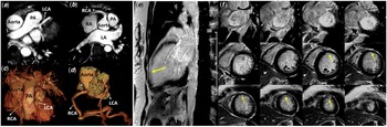
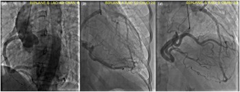
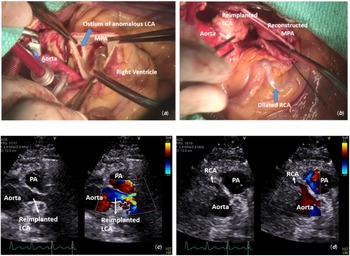
A 29-year-old female with presumed peri-partum cardiomyopathy presented in cardiac arrest secondary to ventricular fibrillation and was successfully defibrillated to sinus rhythm. Hypothermia protocol was initiated with complete neurologic recovery. Transthoracic echocardiogram post-arrest revealed severely depressed left ventricular systolic function (ejection fraction 15%) and trivial mitral regurgitation. Cardiac MRI revealed anomalous origin of left coronary artery from pulmonary artery (Fig 1a–d), with mildly dilated left ventricle (indexed end diastolic volume 119 cc/m2) and severe global systolic dysfunction (ejection fraction 27%). Delayed enhancement was noted in the mid to apical anterior and anterolateral segments with thinning and akinesis (Fig 1f), identifying the substrate for arrhythmia. Cardiac catheterisation with aortic root angiography revealed normal origin of the right coronary artery, without distal coronary artery disease; a coronary artery was not seen arising from the left cusp (Fig 2a–c). Mean pulmonary artery pressure was 28 mmHg. Patient underwent successful two coronary artery repairs with coronary artery reimplantation into the ascending aorta and pulmonary artery reconstruction (Fig 3a and b), with autologous pericardium. Intraoperative pulmonary artery pressures were normal (mean pressure ∼ 20 mmHg); no major macro collaterals were identified. There was good antegrade flow in the reimplanted coronaries (Fig 3c and d) and improved left ventricular function at discharge (ejection fraction 44%). Patient was discharged on a life vest and had implantable cardiac defibrillator placement 6 weeks later.
( a ) Whole heart sequence axial view demonstrating left coronary artery arising anomalously from main pulmonary artery. ( b ) WHS axial view demonstrating dilated right coronary artery arising normally from the aorta. WHS sequence ( c ) and coronaries only ( d ) volume-rendered images showing LCA from PA and dilated RCA arising normally from the aorta. ( e and f ) Areas of thinning and near transmural late enhancement in the mid to apical left ventricular anterior and anterolateral segments on short-axis stack ( f ) and two-chamber view ( e ) on delayed gadolinium enhancement imaging. RA = right atrium; LA = left atrium.

( a ) Aortic root angiography (AP view) demonstrating dilated right coronary artery from right cusp and no coronary artery arising from left cusp. ( b and c ) Selective right coronary angiography demonstrated late filling of the left coronary artery, seen anomalously from the pulmonary artery.

( a ) Intraoperative pictures (before repair): Ostium of anomalous left coronary artery from the main pulmonary artery. ( b ) Intraoperative pictures (after repair): Reimplanted left coronary artery anastomosed to the aorta, with dilated right coronary artery. ( c ) Parasternal short-axis view (2D and colour compare) with robust antegrade flow in the reimplanted left coronary artery. ( d ) Parasternal short-axis view (2D and colour compare) with antegrade flow in the dilated right coronary artery. MPA = main pulmonary artery.

Discussion
Anomalous origin of the left coronary artery from pulmonary artery is a rare entity affecting 1/300,000 live births; Reference Boutsikou, Shore and Li1,Reference Chattranukulchai, Namchaisiri and Tumkosit4 90% die in infancy if undiagnosed but a subset may survive into adulthood. Reference Chattranukulchai, Namchaisiri and Tumkosit4 Two primary mechanisms of survival into adulthood have been described; well-formed inter-coronary collaterals perfusing the left ventricular myocardium or presence of high pulmonary artery pressures allowing antegrade flow into the left coronary artery. Reference Peña, Nguyen, Merchant and Dennie5,Reference Qiu, Yang and Wang6 Clinical presentation in adulthood typically entails angina, myocardial infarction, exertional dyspnoea, or sudden cardiac death. Reference Qiu, Yang and Wang6,Reference Dilaveris, Koutagiar, Alexopoulos, Tsiachris and Gatzoulis7 The latter could be the first presentation accounting for 90% of cases presenting beyond infancy. Reference Yau, Singh, Halpern and Fischman2,Reference Alexi-Meskishvili, Berger, Weng, Lange and Hetzer3
Our patient presented 2 months peri-partum with witnessed aborted cardiac arrest, leading to timely diagnosis. The presence of an ischaemic aetiology and a surgically correctable lesion excluded the differential diagnosis of peri-partum cardiomyopathy. Successful prompt resuscitation enabled appropriate surgical correction. Intraoperative pulmonary artery pressures were normal without any epicardial inter-coronary macro collaterals. This enigmatic presentation and survival into adulthood highlight the potential role of microvascular collaterals as another mechanism of survival.
The chronology of clinical presentation was also significant. Haemodynamic changes during pregnancy and delivery could have precipitated myocardial dysfunction in the setting of chronic subendocardial ischaemia. During pregnancy, an increase in blood volume, cardiac output, and heart rate occur with a decrease in both systemic and pulmonary vascular resistance. Pregnant women are susceptible to pulmonary oedema given reduction in the colloid osmotic pressure/pulmonary capillary wedge pressure gradient. Reference Soma-Pillay, Nelson-Piercy, Tolppanen and Mebazaa8 Patients with anomalous origin of the left coronary artery from pulmonary artery appear to be even more susceptible due to shifting of blood away from higher pressure coronary circulation to lower pressure pulmonary circulation causing coronary steal. This, along with increased cardiac workload can contribute to ischaemia and cardiomyopathy. Although the differential diagnosis with this presentation can include peripartum cardiomyopathy, a thorough investigation must be done to elicit the underlying pathology. Echocardiogram revealed left ventricular dysfunction, but cardiac MRI confirmed the diagnosis, emphasising the role of multimodality imaging in anomalous origin of the left coronary artery from pulmonary artery.
While echocardiography is instrumental to diagnosis in infancy, cardiac MRI is more useful in adults providing information on myocardial perfusion, fibrosis, and viability. Reference Kim, Cho, Hwang and Nah9 Restoration of a dual coronary system will prevent further ischaemia and arrhythmias, but not alter the underlying substrate for malignant ventricular arrhythmias. Delayed enhancement on cardiac MRI and history of aborted cardiac arrest established class I candidacy for Implantable Cardioverter Defibrillator for secondary prevention. Surgical myocardial revascularization is the standard of care recommended by the American Heart Association for Adult Congenital Heart Disease treatment guidelines. Reference Stout, Daniels and Aboulhosn10
A high index of suspicion for anomalous origin of the left coronary artery from pulmonary artery should be maintained in patients presenting with ischaemic cardiomyopathy in the pregnant/post-partum patient. Our case reiterates knowledge gaps in mechanisms leading to survival into adulthood and highlights the role of multimodality imaging to assess both functional and anatomical cardiac reserve.
Conflicts of interest
None.