INTRODUCTION
Marine biofouling refers to growth (usually undesirable) that occurs on any structure introduced into seawater. New surfaces introduced into the marine environment in Singapore, especially the many artificial structures such as jetty pilings, buoys and pontoons present in Singapore Port, provide novel substrata for colonization by sessile animals such as ascidians. As highlighted by Lim et al. (Reference Lim, de Voogd and Tan2009), marine fouling communities in Singapore's coastal waters are diverse and substantial. Ascidians form a significant component of the fouling present on artificial structures, making up at least 20% cover of the fouling community on the surface of buoys (S.S.-C. Lee, personal observation). However, little is known about the ascidian fauna in Singapore though the island state is found in the Indo-Pacific, a region which hosts approximately 75% of the world's coral reefs (Bruno & Selig, Reference Bruno and Selig2007) and where biodiversity of several major taxa such as corals, fish and crustaceans is heralded as the highest in the world (Roberts et al., Reference Roberts, McClean, Veron, Hawkins, Allen, McAllister, Mittermeier, Schueler, Spalding, Wells, Vynne and Werner2002).
Given the poor records of ascidians in the South China Sea, little is known of the status of potential invasive ascidians. This information has become important in recent years as a result of the rapid spread of pest species such as Didemnum vexillum Kott, Reference Kott2002 (Coutts & Forrest, Reference Coutts and Forrest2007; Lambert, Reference Lambert2009; Lengyel et al., Reference Lengyel, Collie and Valentine2009). Singapore Port is recognized as one of the major shipping intersections in the world (Kaluza et al., Reference Kaluza, Kölzsch, Gastner and Blasius2010), and the risk of transfer of species between vessels through ballast water uptake and from hull and sea chest fouling is high (Floerl et al., Reference Floerl, Inglis, Dey and Smith2009). However, the difficulty of managing invasive species in tropical South-East Asian waters is compounded by a lack of useful historical biodiversity baselines, poorly resolved systematics and the unknown biogeographical status of many marine invertebrate phyla in the South China Sea (Yeo et al., Reference Yeo, Carlton, Teo, Ng, Galil, Clark and Carlton2011).
A search through the Ascidiacea World Database (http://www.marinespecies.org/ascidiacea) (Shenkar et al., Reference Shenkar, Gittenberger, Lambert, Rius, Rocha, Swalla and Turon2012) reports close to 300 species of ascidians documented in the seas of the Central Indo-Pacific. Shenkar & Swalla (Reference Shenkar and Swalla2011) reported inventories of 187 species from the Western Pacific, 117 and 102 species from Guam and the South China Sea area, respectively. The number is actually much higher: detailed monographs on ascidians have been published for some locations such as New Caledonia (Monniot C., Reference Monniot1987a, Reference Monniotb, Reference Monniot1988, Reference Monniot1989, Reference Monniot1991a, Reference Monniotb, Reference Monniot1992; Monniot F., Reference Monniot1987, Reference Monniot1988, Reference Monniot1989, Reference Monniot1990, Reference Monniot1992, Reference Monniot1993, Reference Monniot1994, Reference Monniot1995) and Australia (Kott, Reference Kott1985, Reference Kott1990, Reference Kott1992, Reference Kott2001, Reference Kott2003, Reference Kott2002a, Reference Kottb, Reference Kott2004a, Reference Kottb, Reference Kott2005a, Reference Kott2006, Reference Kott2007, 2008, Reference Monniot2009), while there are also some records for the tropical Western Pacific covering Micronesia, Papua New Guinea, Philippines, Malaysia and Indonesia (Sluiter, Reference Sluiter1904, Reference Sluiter1909; Van Name, Reference Van Name1918; Tokioka, Reference Tokioka1942, Reference Tokioka1950, Reference Tokioka1952, Reference Tokioka1967, Reference Tokioka1970; Millar, Reference Millar1975; Kott, Reference Kott1980, Reference Kott1981, Reference Kott1982, Reference Kott1995; Vorontsova & Cole, Reference Vorontsova and Cole1995; Monniot & Monniot, Reference Monniot and Monniot1996, Reference Monniot and Monniot2001, Reference Monniot and Monniot2006, Reference Monniot and Monniot2008; Cole & Vorontsova, Reference Cole and Vorontsova1998; Lambert, Reference Lambert2003; Monniot F., Reference Monniot2009a,Reference Monniotb, Reference Monniot2010). Checklists of ascidians for neighbouring regions include the southern coasts of India (Jaffar Ali & Sivakumar, Reference Jaffar Ali and Sivakumar2007; Jaffar Ali et al., Reference Jaffar Ali, Sivakumar and Tamilselvi2009; Tamilselvi et al., Reference Tamilselvi, Sivakumar, Jaffar Ali and Thilaga2011) and China (Zheng, Reference Zheng1995).
There are only 14 species of solitary ascidians listed in published records for Singapore, most of which were derived from Millar's (Reference Millar1975) description of ascidian specimens collected from South-East Asia, mostly from low tide collections from the early 1900s. While the exact locations were not stated, the collections could possibly have been from intertidal reef areas, as the south-west coast of Singapore was largely fringed by intertidal coral reefs while the south-east coast was dominated by intertidal sand and mud flats. Apart from Millar's (Reference Millar1975) work, there are no details of the habitats for the specimens reported in the other literature.
Large-scale coastal development in the last five decades has changed the marine biodiversity in the coastal waters around Singapore, including a major reduction of coastal reefs (Hilton & Manning, Reference Hilton and Manning1995). While waters in the southern Singapore Strait experience good flushing, the Johor Strait in the north is estuarine (Gin et al., Reference Gin, Holmes, Zhang, Lin and Wolanski2006). Nevertheless, the coastal waters around Singapore retain pockets of biologically diverse habitats ranging from mangroves to coral reefs, nestled within a highly built-up urban shipping port. Such ecosystems are typical in many urban coastal cities in South-East Asia.
Unlike earlier studies, the present study focused on ascidians found on artificial structures, which are known to contain diversity significantly different from that found in natural habitats. Connell (Reference Connell2001) showed that floating pontoons provide very different habitats compared to rocky reef and pilings. Glasby et al. (Reference Glasby, Connell, Holloway and Hewitt2007) found that non-indigenous species (NIS) were greater on pontoons and pilings compared to rocky reefs while on reefs, numbers of native species were as much as three times greater than NIS. Lambert (Reference Lambert2002, Reference Lambert2003) reported that most non-indigenous ascidians in Guam were confined to harbour structures. Tyrell & Byers (Reference Tyrrell and Byers2007) also showed that exotic ascidians tend to thrive and expand better on artificial surfaces over time. In the present work, solitary ascidians on artificial structures in Singapore's coastal waters are described to provide preliminary baseline information of the fouling species present on artificial hard substrata in tropical South-East Asian ports. A similar paper is planned for the colonial species.
MATERIALS AND METHODS
Field surveys were carried out from June 2009 to October 2011. Of the 38 sites visited, solitary ascidians were encountered at 33 (Figure 1). Ascidians were collected from navigational buoys, floating fish farms, jetty pilings and submerged pontoons of marinas. All navigational buoys surveyed in this study carry fouling communities that were no more than 3 years old, as buoys are routinely brought back for cleaning and re-deployment no later than a 3-year period. Most of the samples were collected from structures at less than 5 m depth, on ropes, chains, fish net cages, plastic panels and pipes. Specimens on jetty pilings were accessed by SCUBA diving. The ascidians were carefully removed from the substratum with forceps, or removed with bits of chipped off substratum. Specimens were relaxed in menthol–saturated seawater for at least 2 hours before fixing in 10% buffered seawater–formalin for storage. Ascidians were dissected, examined and sorted to family/genus based on description and keys by Kott (Reference Kott1985) and Monniot C. et al. (Reference Monniot, Monniot and Laboute1991).
Map showing all 33 survey sites: (1) Changi FC 12E Fish Farm; (2) CAAS Buoy 2; (3) Northeast Tekong Oyster Farm; (4) Paku Buoy; (5) Angler Buoy; (6) Jambi Jaya Buoy; (7) Eastern Buoy; (8) Airway Buoy; (9) Lower Shoal Buoy; (10) Main Fairway Buoy; (11) Kukor Buoy; (12) West Buran Buoy; (13) Pulau Tekukor; (14) Kusu Island; (15) St John Island; (16) Sister's Island (Laut side); (17) Sisters Buoy; (18) Pulau Hantu Besar; (19) Berkas Buoy; (20) Raffles Light House; (21) Balok Buoy; (22) Raffles Buoy; (23) Kuda Buoy; (24) Salu Buoy; (25) CC2 Buoy; (26) North Cyrene Buoy; (27) Pusing Buoy; (28) Republic of Singapore Yacht Club (RSYC); (29) Retan D Buoy; (30) Tembuan Buoy; (31) Tengeh Buoy; (32) Raffles Marina; (33) Laba buoy. Map adapted from Charts for small craft published by the Hydrographic Department, Maritime and Port Authority of Singapore, 2009.

SYSTEMATICS
Order PHLEBOBRANCHIA
Family ASCIDIIDAE Herdman, 1882
Genus Ascidia Linnaeus, 1767
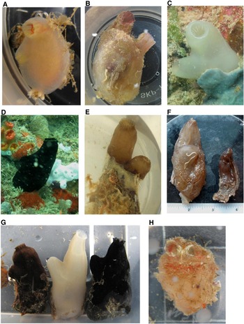
Ascidia gemmata Sluiter, Reference Sluiter and Semon1895
(Figure 2A)
Sites (Figure 1): 7, 9, 10, 11, 12, 15, 25, 27, 29, 30, 31, 33.
(A) Ascidia gemmata; (B) Ascidia sydneiensis; (C) Phallusia arabica; (D) Phallusia nigra; (E) Phallusia philippinensis; (F) two colour morph individuals of P. philippinensis; (G) three species of Phallusia. Left to right: P. philippinensis, P. arabica and P. nigra; (H) Rhodosoma turcicum.

DIAGNOSIS
Animals are laterally flattened and usually attached entirely on the left but occasionally at the posterior end. When alive, individuals are of translucent lime-yellow or rarely slightly pinkish with distinct red stripes lining the inside of the oral siphon and sometimes along the endostyle. The tunic is smooth, cartilaginous and often free from fouling. The oral siphon is terminal, the short atrial siphon opens at about halfway to 2/3 down the body and sometimes directed posteriorly. Musculature on the right body wall is made up of longitudinal and transverse muscle bands criss-crossing, forming an irregular mesh. However, these muscles join to form regular parallel transverse bands at the ventral margin. Only longitudinal muscles are observed on the left and anterior to gut loop. Thirty-five crowded, simple tentacles with two orders of lengths are counted from a specimen measuring 2 cm tall and 1 cm wide, and 72 in a specimen 5 cm in length. Prepharyngeal area has tiny sparse scattered papillae. Dorsal tubercle is large, occupying almost the entire V-shaped prepharyngeal area, and has a U-shaped slit. Neural ganglion is positioned dorsally halfway between the two siphons. Dorsal lamina is a prominently ribbed membrane; ribs project beyond the free margin of membranes. No papillae were observed on the lamina at the level of the oesophagus in one specimen, but tiny sparse papillae were present in another. There are about 8–9 straight stigmata per mesh in the pleated branchial sac. Blunt-ended antero-posteriorly flattened papillae, longest near the dorsal lamina, are supported on internal longitudinal vessels; there are no intermediate papillae. The gut loop is large with a greatly expanded stomach and takes up most of the body on the left side; it forms a closed deeply curved double loop most of which is isodiametric, ending near the atrial opening with a modestly dilated rectum. The anus may be smooth-rimmed or with small irregular lobulations. The ovary is situated in the primary pole of the gut loop, while male follicles are scattered across most of the surface of the entire digestive tract.
REFERENCES
Tokioka (Reference Tokioka1950, Reference Tokioka1952), Kott (Reference Kott1985), Monniot C. (Reference Monniot1991), Nishikawa (Reference Nishikawa1991), Monniot & Monniot (Reference Monniot and Monniot2001).
Ascidia sydneiensis Stimpson, Reference Stimpson1855
(Figure 2B)
Sites 1, 3, 4, 7, 11, 15, 24, 25, 26, 27, 28, 32.
DIAGNOSIS
This species is usually covered with epibionts, leaving only the slightly reddish striped siphons visible. When cleaned of fouling, the tunic is thin, cartilaginous, translucent and somewhat flaccid; both apertures have a tentacular fringe. Specimens are laterally flattened and attached entirely on the left side. The body is loosely attached to the tunic and when removed, a border of parallel short but strong muscle bands are seen that encircle the right side, including between the siphons, finer along the posterior margin. The ventral horizontal muscle bands are longer than the dorsal ones, extending almost to the middle of the right side. Circular muscle bands are found only in the siphons along with a few longitudinal muscles along the ridges of the lobes. Numerous long fine branchial tentacles are densely packed; 123 were counted in a 6 cm long specimen, with additional tiny ones interspersed. The dorsal tubercle is large and very variable, with many long or short convoluted slits, and sits in the widened V-shaped region of the prepharyngeal cavity which elsewhere is narrow and does not appear to be papillated. The neural ganglion is situated just posterior to the dorsal tubercle. The dorsal lamina is a single ribbed membrane, not papillated at the level of the oesophagus. Blunt-ended or rounded roughly conical papillae are supported on internal longitudinal vessels on the deeply pleated branchial sac and there are 8–10 stigmata per mesh; there are no intermediate papillae. The gut loop is large and forms a deep recurved closed loop; the posterior intestine is greatly dilated. There is a sharp U turn where the posterior intestine ends and the straight ascending rectum begins. The anus rim has many shallow lobes. The ovary is ramified over the mesial side of the posterior stomach region and gut loop while the testis follicles are widely scattered over both sides of the gut loop and anterior rectum in very ripe individuals.
REMARKS
Individuals presenting dark-grey siphons were occasionally collected, but on closer examination of the internal morphology, it was concluded that the red and grey specimens are colour morphs of the same species. Many colour morphs are known worldwide.
REFERENCES
Kott (Reference Kott1985), Monniot C. (1983), Monniot & Monniot (Reference Monniot and Monniot1987).
Genus Phallusia Savigny, Reference Savigny1816
Phallusia arabica Savigny, Reference Savigny1816
(Figure 2C, G)
Sites 6, 7, 9, 10, 11, 13, 15, 16, 19, 21, 22, 23, 24, 25, 26, 27, 30.
DIAGNOSIS
Animals have a translucent white cartilaginous tunic which is smooth and free from fouling when alive. The dense ramified tunic vascular system, mentioned by Monniot & Monniot (Reference Monniot and Monniot1996) is easily seen through the tunic surface. This species is laterally flattened and either attached entirely on its left or on its posterior end. Siphons are long, often directed upwards; the edges of both siphons are irregularly undulating with the lobes obscure but with one yellow ocellus (when freshly preserved) corresponding to each lobe. Atrial siphon is positioned at the mid-point of body. Musculature on the right of body is well developed, longitudinal and transverse muscle bands criss-crossing giving rise to an irregular mesh. Muscles on the left are only found anterior to the gut loop. Fifty-eight robust yellow oral tentacles of three orders of lengths were counted in one specimen (8 cm tall, 3 cm wide), 59 in a 4 cm tall individual and 92 in a 7 cm tall animal. Prephargyneal area is narrow and papillated; there is no obvious V-shaped prepharyngeal region. The tiny dorsal tubercle contains a small, short slit; some specimens have multiple tubercles while others only one. In one examined specimen no tubercle was visible. There are however multiple accessory openings along the neural duct in all examined animals. Neural ganglion is situated at the base of atrial siphon. Dorsal lamina is a double membrane anteriorly and ribbed, the ribs extending beyond the free margin. Curved but flattened branchial papillae are supported at the junctions of the longitudinal and horizontal vessels. There are two slight protuberances one on either side of each papilla along the longitudinal axis. There are approximately 10–12 stigmata in each mesh of the deeply plicated branchial sac. Gut loop takes up all the posterior half of the body wall on the left side. The primary loop is a deeply curved closed U-shape; the rectum is greatly distended with mud and ends in an anal rim with more than 20 lobes. The ovary is tucked into the primary gut loop while male follicles are more widely scattered on the gut loop.
REMARKS
Kott (Reference Kott1985) and Monniot & Monniot (Reference Monniot and Monniot1996) counted about ten or more stigmata per mesh in their specimens of P. arabica, whereas Monniot C. (Reference Monniot1997) recorded 5–7.
REFERENCES
Kott (Reference Kott1985), Monniot & Monniot (Reference Monniot and Monniot1996), Monniot C. (Reference Monniot1997), Monniot F. (Reference Monniot2010).
Phallusia nigra Savigny, Reference Savigny1816
(Figure 2D, G)
Sites 5, 8, 9, 10, 19, 21, 24, 25, 26, 27, 29, 31, 32.
DIAGNOSIS
Animals are glossy black when alive and the cartilaginous tunic (1–2 mm thick in most regions) is smooth, without fouling except sometimes at the posterior end. This species is laterally flattened but usually attached to the substrate on its posterior end. Edges of both siphons are lightly lobed with about one red ocellus (when freshly preserved but turned black over time) corresponding to each lobe; there may be 8–15 lobes on each siphon. The atrial siphon, which appears short externally, is positioned at the anterior 1/3 of the body and can either project side-ways, or upwards alongside the oral siphon. The body when removed from tunic is usually greenish-black after preservation. Right side musculature is well developed with longitudinal and transverse muscle bands criss-crossing giving rise to an irregular mesh. Muscles on the left are only found anterior to gut loop; circular muscles are concentrated on the siphons. Oral tentacles, quite variable in number (47 in a 9 cm tall animal, 65 in a 6.5 cm animal), are of three orders of length with the shortest sometimes presented as short stumps and are not joined at the base. Prepharyngeal area is very narrow and minutely papillated, only widening slightly near the endostyle region. Dorsal tubercle in the V-shaped prepharyngeal area varied in samples examined; five specimens had a single, small U-shaped slit, sometimes with one horn turning outwards, while three specimens had multiple small slits (some U-shaped, some straight); in one specimen it was shaped like an inverted S. In specimens where the dorsal tubercle has multiple slits, the V-shaped prepharyngeal area is especially wide. Multiple accessory openings along the neural duct are present in all examined animals. Neural ganglion is situated at the base of atrial siphon. Dorsal lamina is a double membrane and ribbed anteriorly. At about one-third distance down the body, only the left ribbed membrane remains while the right membrane regresses into ribs corresponding to transverse vessels that only reach the base of the lamina. The left membrane continues down to the oesophagus where transverse ribs extend beyond the free margin. At the region of the oesophagus, ribs from the right side of the branchial sac fuse to form shovel-like projections (about 2 ribs to 1 projection). Posterior to the oesophagus, the same projections are observed on both sides of the body. Curved papillae without side protuberances are found at the junction of the longitudinal and transverse vessels. There are about 8–9 stigmata per mesh in the deeply plicated branchial sac. The stomach is a large rounded bag with internal folds; the intestine and rectum are isodiametric and not inflated. The primary gut loop is deeply curved into a closed U; the anal rim is rolled with a few shallow elongated lobes and is slightly posterior to the apex of the primary gut loop. The entire digestive tract is covered with numerous renal vesicles. The ovary is mostly confined to within the pole of the primary gut loop, while the testis follicles when well developed are scattered over and around much of the digestive tract. Ovary turned black in 10% formalin and sometimes some of the testis follicles also.
REMARKS
The pigments of this species leach and cause the 10% formalin preservative fluids to turn black or dark brown after soaking overnight. After several changes of solution, some specimens appeared grey.
REFERENCES
Savigny (Reference Savigny1816), Van Name (Reference Van Name1945) (as Ascidia nigra).
Phallusia philippinensis Millar, Reference Millar1975
(Figure 2E–G)
Sites 6, 7, 9, 10, 11, 21, 23, 24, 25, 26, 27, 29.
DIAGNOSIS
The living colour of these animals can be variable, ranging from dark-brown to light-grey. The cartilaginous tunic is smooth. Siphons tend to be cylindrical and slightly bulbous at the openings; both siphons are marked by a constriction before the lobes flare open in living specimens. If fouling is present, it is mostly found on the posterior end of the body where it attaches to the substratum. The atrial siphon arises from mid-point of body and is often directed upwards alongside the oral siphon. Edges of both siphons are lightly lobed with red ocelli (when freshly preserved but disappeared over time) and there are 8–14 lobes on each siphon. Body when removed from tunic is usually light-brown after preservation. Right side musculature is well developed with longitudinal and transverse muscle bands criss-crossing giving rise to an irregular mesh. Muscles on the left are only found anterior to gut loop. Circular muscles are concentrated on siphons; tentacles are of three orders of length with the shortest sometimes presented as short stumps and joined at the base. In a 7 cm animal there were 48 oral tentacles; 44 were counted in an 8.5 cm specimen. Prepharyngeal area is densely papillated. Rounded papillae are also found along the upper edge of the prepharyngneal band. Dorsal tubercular area is very shallow and opening is usually a single slit or U-shaped, or a slightly inrolled C opening anteriorly. Numerous accessory openings line both sides of the neural duct to the neural ganglion which is situated at the base of atrial siphon. Dorsal lamina is a single ribbed membrane from the left, with ribs extending slightly beyond the rim of the lamina, to form a fringe. Transverse vessels from right side of body only reach the base of the lamina. At oesophagus, transverse vessels on right extend into tentacular projections (~2 vessels to 1 projection). Dorsal lamina on left side regresses after oesophagus to be replaced by similar projections. Branchial papillae slightly curved at the tip and without lateral protuberances, are at the junctions of the internal longitudinal and horizontal vessels. Minute button-like intermediate papillae are sometimes present between the main papillae. There are about 12–14 stigmata in each elongated mesh; the sac is deeply pleated. The stomach is expanded, and the gut loop is isodiametric and deeply curved into an elongated U-shape, occupying about two-thirds of the body on the left. Anus rim is rolled, divided into a few shallow lobes, and is posterior to the pole of the gut loop. Ovary (pinkish in formalin) is tucked in the pole of the gut loop while male follicles are scattered.
COMPARISON WITH PHALLUSIA NIGRA
There do not seem to be any clear distinctions in number of siphonal lobes between P. nigra and P. philippensis. Also the form of the dorsal lamina is rather similar in most aspects. However, there are several morphological features that differentiate the two species:
-
(1) both species have a papillated prepharyngeal area (although much narrower in P. nigra than P. philippinensis based on observations though no measurements were taken). Only P. philippinensis has rounded projections on the anterior blade of the prepharyngeal band (see Monniot & Monniot, Reference Monniot and Monniot2001);
-
(2) Phallusia nigra's tentacles are not joined at the base unlike P. philippinensis;
-
(3) oral tentacles: 44–54 in four large P. philippinensis (54 in 5 cm animal, 48 in 7 cm animal, 44 and 52 in two 8.5 cm animals). In P. nigra 47–65 in four large animals (60 in 6 cm animal, 61 in 8 cm animal, 47 in 9 cm animal and 65 in 11.5 cm animal) (plus a few tiny ones);
-
(4) branchial sac longitudinal vessels: 98–117 in the same four P. philippinensis (58 right, 52 left in 5 cm animal; 54 right, 44 left in 7 cm animal; 61 right, 56 left and 62 right, 55 left in two 8.5 cm animals). In P. nigra 143–185 in same four animals (66 right, 77 left in 6 cm animal; 94 right 76 left in 8 cm animal, 98 right 80 left in 9 cm animal and 90 right 95 left in 11.5 cm animal);
-
(5) Phallusia nigra's ovary stained black in preservation in 10% seawater formalin, while in P. philippinensis it is pinkish;
-
(6) the body of preserved P. nigra is dark black or dark greenish-black while in P. philippinensis it is light-brown. Individuals of P. philippinensis collected from a cave of buoy are often of a lighter shade compared to those attached to the outsides of a buoy.
Tokioka's (Reference Tokioka1970) description of Phallusia depressiuscula from Mindoro Island in the Philippines is rather similar to our described species especially in terms of the external form, tunic colour and morphology of the papillated prepharyngeal area, including presence of rounded papillae along the anterior edge of the prepharyngeal band. The exception is the absence of minute buttons between papillae, which are actually easily overlooked. Van Name (Reference Van Name1918) remarked that P. depressiuscula was the commonest and most widely distributed ascidian in the Philippines region. Although his description did not mention presence of accessory openings, which is the defining character for the Phallusia genus, or features of the prepharyngeal area, he did note that his collections of P. depressiuscula had mixtures of almost colourless and fairly clear or smoky-brown individuals. This colour variation coincides with our collections. However, Van Name also found no intermediate papillae in his Philippines specimens as with Tokioka (Reference Tokioka1970). Millar (Reference Millar1975) proposed a new species Phallusia philippinensis to replace P. depressiuscula, as he felt that there were insufficient details in Heller's (Reference Heller1878) original account of Ascidia depressiuscula to support it as a Phallusia. Our specimens closely resemble P. philippinensis described by Millar (Reference Millar1975) and Monniot & Monniot (Reference Monniot and Monniot2001).
REFERENCES
Van Name (Reference Van Name1918), Tokioka (Reference Tokioka1970), Millar (Reference Millar1975), Monniot & Monniot (Reference Monniot and Monniot2001).
Family CORELLIDAE Lahille, 1888
Genus Rhodosoma Ehrenberg, 1828
Rhodosoma turcicum (Savigny, Reference Savigny1816)
(Figure 2H)
Site 28: one specimen, on a rope, ~3 m below water surface.
DIAGNOSIS
Animal is upright, attached by its posterior end and is slightly laterally flattened. When alive, the translucent cartilaginous tunic was light-pink underneath epibionts. Both siphons are close together and directed upwards. Extension of the tunic from right side of animal results in a lid that closes over the siphons when animal is startled. Orange pigments lined the edge of the lid in the living animal. Pointed papillae are concentrated around the lid; some papillae were found more posteriorly on the tunic but less concentrated. When body is removed from tunic, thick longitudinal muscle bands are found crowded around the anterior part of body close to siphons. These muscles originated from approximately one-third down dorsal and ventral region and are subdivided into thinner bands before crossing circular muscles. Some longitudinal muscles originating from ventral side are found to cross over to atrial siphon. There are no muscles further down the rest of the body. Branchial tentacles of about equal size are simple and well-spaced; 17 tentacles were counted in this sole specimen measuring 2.5 cm tall. Prepharyngeal area is wide, with very minute papillae that can only be seen after staining, and dorsal tubercle has a U-shaped slit. Dorsal lamina consists of a series of languets; internal longitudinal vessels are supported on curved papillae originating from transverse vessels. There are about four straight stigmata in each mesh. Stomach and gut loop occupy about three-quarters of body wall on the right. Stomach has internal folds and is positioned vertically. Gut loop is narrow and makes a U-shaped curve along the left side of stomach hence enclosing it. The anus has shallow lobes that are almost indiscernible and opens at the atrial siphon anterior to the level of the gut loop pole. Gonads are enclosed within pole of gut loop with extensive branching of male follicles over the gut loop.
REFERENCES
Van Name (Reference Van Name1945), Tokioka (Reference Tokioka1952), Kott (Reference Kott1985), Nishikawa (Reference Nishikawa1991).
Family DIAZONIDAE Seeliger, 1906
Genus Rhopalaea Philippi, 1843
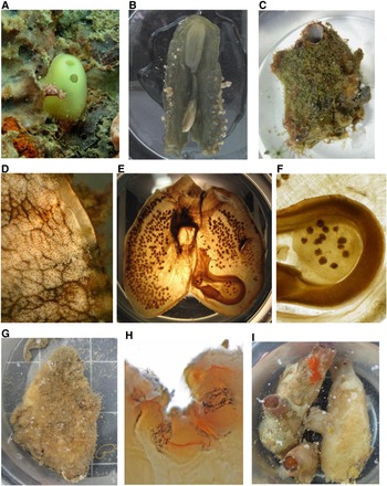
Rhopalaea crassa (Herdman, Reference Herdman1880)
(Figure 3A, B)
Sites 10, 12, 15, 19, 20, 21, 23, 24.
(A) Rhopalaea crassa; (B) dissected Rhopalaea crassa showing thickened tunic around abdominal region; (C) Polycarpa argentata; (D) close-up of P. argentata siphon; (E, F) dissected P. argentata with branchial sac removed to show distribution of gonads. Gonads are normally attached to body wall but this sole specimen was found with gonads on flat top endocarp within gut loop; (G) Polycarpa aurita; (H) close-up of P. aurita siphon; (I) Polycarpa captiosa.

DIAGNOSIS
The living colour of this species is lime-yellow. Its thick cartilaginous smooth tunic is free from fouling and especially thick around the abdominal area. Animal can be attached either at its posterior end or along the length of the abdomen. The two 6-lobed siphons, placed next to each other and directed anteriorly, are very short and often appear as sessile openings. A pigment spot is present between each lobe. When removed from the tunic, the pharynx is very delicate while the posteriorly placed gut loop is made up of a very compact mass that can be as long as 1.5 times that of pharynx. Musculature on the pharynx is largely longitudinal. Circular muscles are confined to siphons. Longitudinal muscles are only ramified on siphons and along the ventral line. Branchial tentacles (up to about 25) are simple, widely spaced with tiny ones interspersed between the larger ones. Dorsal tubercle contains a single longitudinal slit in a wide V-shaped prepharyngeal groove. Prepharyngeal area wide, not papillated. Dorsal lamina is made up of long languets. The branchial sac, widest at the anterior end, is flat (not pleated) and has up to about 100 rows of stigmata with 3–4 stigmata per mesh. Approximately 90–100 longitudinal vessels are supported on rounded conical papillae; in the specimens examined many of the longitudinal vessels were incomplete. The neck connecting pharynx to abdomen is very narrow, is embedded in the substrate, and tends to break when animal is removed from tunic or from its substrate. Due to the dense nature of the gut loop, it was not possible to discern the stomach and the gonads from the few specimens that were dissected. Anus opens at the base of atrial siphon and has short finger-like projections. In one specimen, a single test vessel was found to extend from the end of the abdomen.
REFERENCES
Millar (Reference Millar1975), Kott (Reference Kott1990), Monniot & Monniot (Reference Monniot and Monniot2001).
Family STYELIDAE Sluiter, Reference Sluiter and Semon1895
Genus Polycarpa Heller, 1877
Polycarpa argentata (Sluiter, Reference Sluiter1890)
(Figure 3C–F)
Site 15.
DIAGNOSIS
Animals are often found covered entirely with epibionts, leaving only the siphons visible. The siphonal lining is mottled with orange, brown and white pigments that disappear in formalin. The tunic is leathery and thin, and translucent on the inside. Both siphons are short; the branchial aperture is directed anteriorly while the atrial aperture which is positioned one-third down the body is directed at a right angle relative to the former. Body turns dark-brown to black in preservation. Musculature forms a dense mesh with finer fibres extending over the gut loop region on the left side. Branchial tentacles (36 in 7 cm tall animal), are simple, widely spaced with three orders of length and are set very close to the prepharyngeal band. The dorsal tubercle bears a slightly raised slit, usually elongated and sinuous, occupying a V-shaped prepharyngeal area. Neural ganglion is found immediately after dorsal tubercle. The smooth dorsal lamina increases in height near the oesophagus. Branchial sac has four folds on each side that do not overlap. Folds closest to the dorsal lamina are low and tend to disappear posteriorly. Branchial formula of 7 cm tall and 5 cm wide specimen is as follows: RE 3 (7) 4 (10) 4 (8) 3 (9) 2 DL 2 (7) 3 (9) 3 (9) 5 (9) 5 LE. There are 10–13 stigmata in each mesh near the dorsal lamina and 15–20 stigmata per mesh near the endostyle. Transverse vessels are tough and thick. The gut loop is small, occupying only one-third of the body on the left. The slightly elongate stomach (with about 14 folds) is followed by the intestine which extends obliquely towards the posterior-ventral region where it forms a loop before bending at a right angle to open at the atrial siphon. Anus rim has many fleshy lobes. At the base of the atrial siphon, there is a narrow velum the edge of which has regularly placed fine finger-like projections. A large flat top endocarp sits in the pole of the gut loop. One or two smaller endocarps are found between the stomach and gut loop; none were found on the rest of the body. The numerous small gonads are spherical, embedded in the body wall and distributed from mid to ventral edge of body. Rarely, some gonads are also found in the flat top endocarp enclosed in the gut loop. Each hermaphroditic gonad has multiple rounded small testes on the inner mesial side of the ovary, with individual ducts that join to a central vas deferens.
REFERENCES
Kott (Reference Kott1985), Monniot C. (Reference Monniot1987b), Monniot & Monniot (Reference Monniot and Monniot1996), Monniot F. (Reference Monniot2010).
Polycarpa aurita (Sluiter, Reference Sluiter1890)
(Figure 3G, H)
Sites 21, 32 (attached to tunic of Microcosmus exasperatus on pontoon).
DIAGNOSIS
Three specimens were collected. The largest, measuring 6 cm tall, had a leathery and rough tubercular tunic, yellowish-tan when the animal was alive; the two smaller specimens are 1.5 and 2.5 cm long and have a translucent, soft test. Both siphons appear rather short in the contracted large specimen; the atrial siphon opens in the anterior half of the body. Siphons are marked with a fine red line near the edge and black pigments line the ridges of the lobes. The siphonal lining is made up of swellings, each presenting one long papilla. When removed from tunic, the body is thick and fleshy hence unable to discern muscles, and yellow in preservative. Branchial tentacles are simple (25 in 6 cm animal, 27 in 2.5 cm animal and 21 in 1.5 cm individual) and of two orders of length joined at the base. Some minute buttons are scattered among the longer tentacles. The dorsal tubercle is large, with a sunken almost closed inrolled C slit opening to the left, and the ganglion is immediately posterior. Dorsal lamina is smooth, single-membraned and entire. Branchial sac is tough with thick transverse vessels and very tough connectives to the body wall, characteristic of this species; there are four non-overlapping folds on each side and the internal longitudinal vessels are crowded on the folds. Branchial formula of largest specimen is as follows: RE 4 (14) 6 (20) 6 (21) 5 (27) 3 DL 3 (23) 4(22) 6 (22) 7 (15) 5 LE. There is an average of two internal longitudinal vessels between folds and 5–14 on folds on the two smaller specimens. The largest specimen has 8–9 stigmata per mesh while the smaller ones have 6–8. Parastigmatic vessels are present, though not on every mesh. The stomach is elongate, positioned horizontally along the posterior end of the body, with internal folds and a caecum at its posterior end, curved in the largest specimen but like a short stump in the smaller animals. There is a gastro-intestinal connective at the base of the caecum in all three specimens. The long narrow gut loop takes up one-third of the body on the left and is restricted to the posterior end. It extends obliquely to the posterior-ventral corner then bends sharply back on itself into a U-shape. The rectum bends anteriorly at a right angle; the anus edge is slightly flaring and lobed, and opens at the atrial siphon. In the largest specimen, ramified polycarps are mostly embedded in the body wall. Smaller specimens have elliptical or globular polycarps. While gonads in the 1.5 cm animal are restricted to the ventral side, in the 2.5 cm specimen they spread closer to the atrial opening. Endocarps are scattered on the body wall and within the gut loop.
REMARKS
Kott (Reference Kott1985) stated that gonads in this species tend to become more deeply embedded in the body wall with age and with maturity, as is seen in our largest specimen; they may increase in width and numbers, overlapping each other to give an appearance of branching as with Nishikawa's (Reference Nishikawa1991) specimens. Our specimens lack the unperforated area of pharynx behind the dorsal tubercle that was noted in Kott (Reference Kott1985). The specimens also have more stigmata per mesh compared to Kott's observations of 2–4 but agree in number with Monniot C. (Reference Monniot1987b). Sluiter (Reference Sluiter1890) noted 5–6 stigmata in each mesh and commented that one or two may be smaller than the rest; this is not so in our specimens. Monniot C. (Reference Monniot1987b) stated that a gastric caecum was absent, but Kott (Reference Kott1985) clearly shows one in two diagrams.
REFERENCES
Tokioka (Reference Tokioka1952), Kott (Reference Kott1985), Monniot, C. (Reference Monniot1987b), Nishikawa (Reference Nishikawa1991).
Polycarpa captiosa (Sluiter, Reference Sluiter1885)
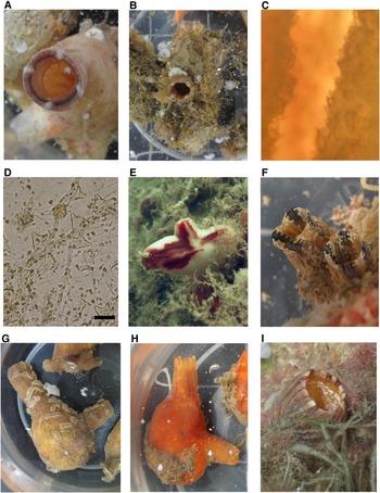
(Figures 3I & 4A)
Sites 7, 10, 15, 18, 21, 25, 26, 27, 29.
(A) Close-up of Polycarpa captiosa siphon; (B) Polycarpa olitoria?; (C) close-up of internal siphonal spines of P. olitoria?; (D) siphonal spines of P. olitoria? under light microscope; (E) Polycarpa papillata; (F) Styela canopus; (G) brown individual of Microcosmus exasperatus; (H) orange individual of M. exasperatus; (I) close-up of M. exasperatus siphon. Scale bar: D, 0.05 mm.

DIAGNOSIS
Animal is ovoid and often found attached on it posterior end. Tunic is tough, slightly lumpy and leathery but thin. When alive, the tunic is off-white underneath fouling; sometimes reddish-brown specimens were collected but the difference in colour is due to the presence of epibionts on the tunic. Atrial siphon opens at half the distance down the body. Both siphons are not very long; four white protrusions mark the lobes of the siphons and posterior to those in living specimens, is a ring of purple pigment. Body when removed from tunic is fleshy and opaque; transverse muscles are tightly packed and no longitudinal muscles could be observed, though the inner wall of the siphons is composed of tightly packed longitudinal muscles. In preservative, body is yellow with purple siphons, oral tentacles and dorsal tubercle. Branchial tentacles are simple and of three orders of length; 36 in a 7 cm animal, 40 in a 5.4 cm animal and 47 in a 7.5 cm individual. Dorsal tubercle is made up of multiple short slits presented on a raised cushion and fills the entire V-shaped prepharyngeal space. The neural ganglion is immediately posterior and projects in a small knob visible on the outside of the mantle between the siphons, as mentioned by Tokioka (Reference Tokioka1950). Dorsal lamina is smooth and entire; height increases near the oesophagus. There are four non-overlapping folds on each side of the branchial sac. Numbers of internal longitudinal vessels between folds mostly range from 6–8 and 17–19 on folds, with great variability throughout the sac. There are approximately 6–8 stigmata in each mesh. There are no parastigmatic vessels. The stomach is somewhat elongated with internal folds, and together with the U-shaped gut loop occupies only the posterior region of the body on the left. Pole of gut loop is at about the same level as anus, whose rim has very shallow lobes. Elliptical polycarps attached only by their proximal end are scattered along the mid-lateral region from prepharyngeal band to gut loop. In mature individuals with fully packed gonads, they tend to be more elongated. Large leaf-like endocarps pack the body wall and gut loop. Numerous slender fingerlike papillae line the inside of the atrial siphon but were not found in the oral siphon.
REMARKS
There are fewer longitudinal vessels per branchial fold on our specimens than reported by Tokioka (Reference Tokioka1950) and Monniot & Monniot (Reference Monniot and Monniot2008).
REFERENCES
Tokioka (Reference Tokioka1950), Millar (Reference Millar1975), Monniot & Monniot (Reference Monniot and Monniot1996, Reference Monniot and Monniot2001).
Polycarpa olitoria? (Sluiter, Reference Sluiter1890)
(Figure 4B–D)
Site 16 (1 specimen was attached to jetty piling, the other on P. papillata).
DIAGNOSIS
When alive, animal has a reddish-brown leathery tunic; red and slightly opalescent strips can be seen inside the siphons. The animal is laterally flattened and was attached to the substratum by its posterior end. The atrial siphon opens about halfway down the body. Hollow conical spines with basal flange are present on the external surface of the siphons and extend into the anterior region of the siphonal lining. Bladder cells are found around the regularly placed transparent spines. Average size of most spines is about 50 µm; they range from 40 to 70 µm. Body when removed from tunic is thin and flaccid. Circular and longitudinal muscles form a fine network that extends throughout the body. Both body and oral tentacles are yellow in preservatives. Fifty-seven simple tentacles of about equal length are crowded on the prepharyngeal band of the 4.5 cm tall specimen. Dorsal tubercle has a W slit with one end inrolled, in a swollen spongy cushion and fills the entire V-shaped prepharyngeal space. Dorsal lamina is smooth and entire. Branchial sac has four folds that do not overlap and the formula is: RE 7 (12) 5 (16) 7 (18) 8 (18) 8 DL 5 (14) 5 (18) 5 (17) 7 (15) 6 LE. There are primarily six stigmata in each mesh between folds, sometimes seven, and 7–8 stigmata per mesh closer to the endostyle. There are no atrial tentacles. The stomach folds do not extend to the posterior end of the stomach and are visible from the outside of the stomach; it and the gut loop occupy about one-third of the body wall on the left. The intestine forms an open loop at the posterior-ventral corner and bends at right angle to form the long rectum which opens near the atrial siphon. The anus has 16 long fingerlike lobes; a few of them bifurcated in one specimen. Elongate polycarps up to 4 mm in length are arranged in a single very irregular row down the centre of the body on both sides and are attached to the body wall by their entire length. The testes are difficult to count because they are nearly empty of sperm, but there may be about 4 or 5 with numerous small fingerlike branching. The sperm ducts join into a single duct distally that opens very close to the oviduct. Twenty polycarps were counted on the left and 32 on the right in the 4.5 cm animal. Endocarps are scattered on the body wall; there are about 16 slender ones crowded in the gut loop. A 4.3 cm animal missing the entire gut and branchial sac has 62 oral tentacles, similar siphonal spines, and 25 gonads on the left, 33 on the right.
REMARKS
There are a number of differences between our specimens and Kott's (Reference Kott1985). She recorded hollow conical siphonal spines measuring 80–130 µm, while the size of spines in our specimens ranged from 40 to 70 µm and they are similar in shape but not as narrowly pointed. The longitudinal muscles of our samples extend to the posterior end of the body, while in Kott's specimens they extend only halfway to the posterior end. There are a few more rectal lobes and more endocarps in the gut loop in our specimens. There are no other known Polycarpa species with this type of siphonal spines, which Monniot C. (Reference Monniot2002) considered a distinctive species character. Thus we provisionally place our specimens with P. olitoria with the understanding that it might be an undescribed but closely related species.
REFERENCE
Kott (Reference Kott1985).
Polycarpa papillata (Sluiter, Reference Sluiter1885)
(Figure 4E)
Sites 5, 9, 10, 11, 13, 14, 15, 16, 17, 21, 22, 23, 24, 25, 26, 27, 29, 31.
DIAGNOSIS
Animal is seldom found with fouling and its white and smooth but tough tunic is usually marked with irregular red stripes or patches in the anterior half, especially around the siphons. Animal is attached via its posterior end. The oral siphon is terminal, the atrial shorter and positioned at the anterior one-third of body and can either project sideways or upwards. Fine hair-like papillae are on the inner lining of the atrial siphon. Longitudinal and circular muscle bands extend throughout body, the latter overlaying the former. Branchial tentacles are simple with three orders of length: 70 in a 4.8 cm animal, 58 in a 5 cm animal and 49 in a 7 cm animal. Dorsal tubercle usually bears a large U-shaped slit with one horn rolling outwards and the other inwards in the V-shaped prepharyngeal area; in the largest specimen it has numerous slits. Dorsal lamina is a smooth membrane. Branchial sac has four folds that do not overlap. There are usually six internal longitudinal vessels between folds with fewer near the endostyle (range of 3–9) and about 13 on the folds (range of 6–18). The average number of stigmata per mesh is seven in the middle of the branchial sac. The stomach is short, with many internal folds, and the intestine forms a loop at the posterior-ventral corner. The anus rim has 16–24 long finger-like projections. The number of gonads varies with size (11 left, 15 right in a 5 cm animal, 20 left and 42 right in a 7 cm animal), are either globular or somewhat elongate, attached to, but not embedded in, the body wall along about half of their outer side or sometimes just at the proximal end, and scattered on both sides among numerous endocarps, but located only anterior to the gut loop on the left. The testis follicles are on the body wall side of each polycarp; the sperm ducts join to a single opening very close to the opening of the oviduct. Numerous endocarps are also located in the gut loop.
REMARKS
Our specimens differ in having more oral tentacles than reported by Monniot & Monniot (Reference Monniot and Monniot2001) and Tokioka (Reference Tokioka1952); they reported 16 and 14–21, respectively. Our specimens agree in all other characters. Monniot & Monniot (Reference Monniot and Monniot2001) corrected earlier misidentifications by themselves and by Kott (Reference Kott1985).
REFERENCES
Tokioka (Reference Tokioka1952, 1970), Kott (Reference Kott1985), Monniot C. (Reference Monniot1987b), Monniot & Monniot (Reference Monniot and Monniot2001).
Genus Styela Fleming, 1822
Styela canopus (Savigny, Reference Savigny1816)
(Figure 4F)
Sites 1, 15, 22 (attached to Pyura curvigona), 30.
DIAGNOSIS
Animals are attached at the posterior end and laterally flattened. Tunic is tough but usually thin, though the external surface is rough with swellings. Living animal is brown with distinctive siphons: white (slightly opalescent) and brown toothed bands form a striped pattern that can be seen externally and internally. Both siphons are placed very close together with the atrial at a 45 degree angle from the slightly more anteriorly placed oral siphon. Fan-shaped, flat tunic spines with a single, long pointed end on each side are present around the siphons and in the siphonal lining. Spines measure about 11 µm wide and 15–20 µm long inside the siphons; those outside the siphons are a bit longer. Circular muscles were obscured on the body wall and siphons. Longitudinal muscle bands are broad but not thick and extend from siphons to posterior end. Oral tentacles are simple, of three orders and well-spaced; 20 tentacles were counted in a single individual. There are also very fine and crowded hair-like tentacles on a short velum at the base of the atrial siphon, much finer and more numerous than the branchial tentacles.
Dorsal tubercle has a U slit on a raised cushion in the V-shaped prepharyngeal area. Dorsal lamina is smooth, single-membraned and entire. There are four low folds/side in the branchial sac; internal longitudinal vessels are very crowded on the folds. The branchial formula for a 2 cm tall specimen is: RE 5 (19) 4 (24) 7 (30) 8 (33) 10 DL 6 (28) 7 (27) 8 (27) 10 (21) 3 LE. There are 3–4 stigmata in each mesh and parastigmatic vessels are present. On the left side the stomach is elongate with many internal folds visible on the outside. The intestine makes a narrow loop at the posterior-ventral corner, then lies parallel to the long stomach and bends at a right angle to open at the atrial siphon. The shallow-lobed anus is anterior to pole of gut loop. There are two gonads each side of body. The tubular ovary is thick and slightly undulating with a single opening near the atrial aperture. Testis follicles in the shape of broad lobes often irregularly bifurcated are attached to the body wall and somewhat confined to the proximal end of the ovaries. Each lobe has a single duct that joins to a central vas deferens that runs along the ovary and opens near the oviducal opening. One gonad on the left side of body is placed inside the secondary gut loop adjacent to ascending rectum while the other curves above the pole of gut loop. Endocarps are present on body wall and inside the gut loop.
REFERENCES
Van Name (Reference Van Name1945 as Styela partita), Kott & Goodbody (Reference Kott1980), Kott (Reference Kott1985), Monniot & Monniot (Reference Monniot and Monniot1987).
Family PYURIDAE Hartmeyer, 1908
Genus Microcosmus Heller, 1877
Microcosmus exasperatus Heller, Reference Heller1878
(Figure 4G–I)
Sites 1, 4, 6, 7, 8, 9, 10, 11, 15, 17, 19, 21, 22, 24, 25, 26, 28, 29, 32.
DIAGNOSIS
The tunic is tough and leathery but thin, and individuals are usually attached at the posterior end. Various colours of living individuals have been collected: light- to dark-brown or reddish-orange. Both siphons are long with brown and white striping in brown individuals, while the siphonal lining is mottled with brown and white spots. When relaxed, the atrial siphon that opens mid way down the body is at a right angle to the terminal oral siphon. Gently curved but sharply pointed siphonal spines with open base and measuring about 40 µm long are present on both siphons on the external anterior region and in the siphonal lining. Internally there are four pockets about 1/3 down the branchial siphon, though usually (as in Kott, Reference Kott1985) these are described as being at the base of the siphon. The thin body wall adheres very tightly to the tunic and has well-developed musculature. Circular muscles are confined around the siphons. Thick longitudinal muscles in bundles of 3 or 4 extend from each siphon, criss-crossing to form a rather regular mesh extending to the posterior end of body. Branchial tentacles have two orders of branching; primary branches are slender and long while secondary branches are short. The dorsal tubercle has a large and raised U slit with both horns rolling inwards to form double spirals and occupies a V-shaped papillated prepharyngeal area. Dorsal lamina is smooth and entire. Branchial sac has nine folds on the right and eight on the left; those near the endostyle lower and may disappear towards the posterior end of the body. Branchial formula of a 5 cm tall, 4 cm wide specimen is: RE 2 (8) 4 (11) 3 (13) 6 (16) 5 (21) 3 (21) 3 (19) 3 (19) 3 (23) 3 DL 3 (19) 3 (20) 3 (20) 4 (22) 4 (17) 4 (15) 5 (14) 3 (2) 1 LE. There are about six stigmata in each mesh and parastigmatic vessels are present. The stomach is partially covered by the closely adherent lobes of the hepatic diverticula which are dull-yellow in preservation and made up of parallel lamellae. Gut loop is large, with a deep U form, and occupies almost the entire left side of body. Pole of gut loop is anterior to the smooth-rimmed anus. There is one gonad on each side divided into three broad lobes; the left gonad crosses the intestine and the most proximal gonadal lobe is inside the primary curve of the intestine.
REFERENCES
Van Name (Reference Van Name1945), Tokioka (Reference Tokioka1952), Kott (Reference Kott1985), Monniot C. (Reference Monniot2002), Monniot F. (Reference Monniot2010).
Genus Pyura Molina, 1782
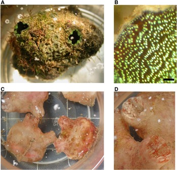
Pyura curvigona Tokioka, Reference Tokioka1950
(Figure 5A, B)
Sites 7, 15, 22.
(A) Pyura curvigona; (B) close-up of siphonal spines of P. curvigona; (C) Herdmania pallida; (D) close-up of H. pallida siphons. Scale bar: B, 0.25 mm.

DIAGNOSIS
This species has a tough, leathery tunic, brown beneath the epibionts. Both siphons are relatively short and directed anteriorly in a V pattern. Large spines 0.25 mm long are present in the siphonal lining and along the external edge of the siphons and consist of a long central needle with a flat rounded iridescent scale positioned at the mid-point of the needle. The body wall is thin and translucent, revealing the thick and well-developed musculature. Circular muscles are restricted to siphons; longitudinal muscle bands from each siphon extend downward, criss-crossing on the body wall to form a rather regular mesh and ramifying ventrally. Branchial tentacles are of two orders of branching. Dorsal tubercle has a raised U slit with inrolled horns and occupies the entire V-shaped prepharyngeal groove. The dorsal languets are long and finger-like. The branchial sac has six non-overlapping folds/side; the formula is: RE 7 (14) 7 (15) 6 (18) 6 (17) 5 (17) 7 (12) 4 DL 4 (13) 7 (17) 7 (18) 6 (19) 5 (16) 6 (9) 6 LE. There are 6–7 stigmata in each mesh and parastigmatic vessels are present. The stomach is elongate with an olive-brown lobed hepatic diverticulum attached loosely at its posterior end. The gut loop is large, taking up the entire body wall on the left; it makes a deep U curve. The rectum, swollen with mud, ends in the shallow lobed anus. There is one somewhat U- or J-shaped gonad on each side of the body, divided into multiple rounded ovotestes attached to the body wall, with a common oviduct and vas deferens. The left gonad is entirely enclosed within the gut loop. Endocarps project from the stomach, gut loop and each rounded ovotestis.
REFERENCES
Tokioka (Reference Tokioka1950), Millar (Reference Millar1975), Kott (Reference Kott1985).
Genus Herdmania Lahille, 1888
Herdmania pallida (Heller, Reference Heller1878)
(Figure 5C, D)
Sites 1, 2, 3, 4, 7, 8, 9, 10, 11, 15, 19, 21, 22, 24, 25, 26, 27, 28, 29, 31, 33.
DIAGNOSIS
In life the tunic is light-pink and slightly translucent but usually thick; in mature individuals, bright-red or pink gonads can be seen through the body wall when the animal is removed from its tunic. Tiny calcareous pointed spicules (40–150 µm long) form in the tunic blood vessels similar to those in Herdmania momus (Lambert & Lambert, Reference Lambert and Lambert1987) and project from the tunic, with the rounded spiny ball-shaped proximal end firmly anchored in the tunic. A red web-like pattern lines the insides of the siphons; sometimes white iridescent bands lining the ridges of the siphon lobes are also present. When relaxed the animal swells into a rounded sac attached at its posterior end, and its siphons are directed anteriorly in a V pattern. Even after preservation, the tunic is relatively soft. Muscles are concentrated anteriorly. Circular muscles are found only in the siphons while thick longitudinal muscles extend posteriorly from the siphons to form a mesh-like network. No muscles were observed in the lower half of the body, beyond the level of the gonads. A velum with small shovel-like projections was found only at the base of the atrial siphon. Oral tentacles are large with relatively short secondary branching. The dorsal tubercle is a fleshy cushion with a U slit with horns rolling inwards. Dorsal languets are tusk-like. There are eight folds on each side of the branchial sac that tend to overlap closer to the dorsal region. In a 6 cm tall, 5 cm wide specimen, the branchial formula is: RE 1 (11) 1 (15) 2 (12) 2 (16) 1 (14) 2 (19) 2 (17) 2 (12) 1 DL 2 (14) 1 (20) 2 (17) 1 (19) 1 (17) 2 (15) 1 (14) 1 (8) 1 LE. There are 8–11 stigmata in each mesh although some meshes between folds are flat with no stigmata, especially when these meshes lie above thick transverse vessels. Parastigmatic vessels are present. Longitudinal vessels on folds terminate as finger-like projections around the oesophagus. The hepatic diverticulum is a compact mass that adheres closely to the stomach and does not project freely into atrial cavity. The gut loop is large and occupies the entire left body wall. It makes an open loop with the pole just posterior to the base of the oral siphon and terminates in a smooth-rimmed anus. There is one long gonad on each side of the body; the left gonad is entirely enclosed within the gut loop. Each gonad consists of a slightly sinuous ovary lined along the sides by branched testis follicles. Multiple ducts from the testis follicles join to a central vas deferens on the body wall surface of the ovary. There is a single large opening of the oviduct and close to it a separate, smaller single opening of the vas deferens. Both openings are simple and without hoods or ornaments (in H. momus there are multiple openings of the testis and the oviducal opening has a large lobulate collar (Monniot C., Reference Monniot2002)). Long (0.8–1 mm) barbed needle-like calcareous spicules are distributed all over the body including the branchial sac.
REFERENCES
Kott (Reference Kott2002), Nishikawa (Reference Nishikawa2002), Monniot C. (Reference Kott2002).
RESULTS
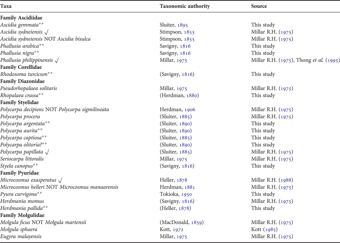
Twelve new records of solitary ascidians in Singapore waters were recorded in this study: Ascidia gemmata, Phallusia arabica, Phallusia nigra, Rhodosoma turcicum, Rhopalaea crassa, Polycarpa argentata, Polycarpa aurita, Polycarpa captiosa, Polycarpa olitoria?, Styela canopus, Pyura curvigona and Herdmania pallida (Table 1). Tamilselvi et al. (Reference Tamilselvi, Sivakumar, Jaffar Ali and Thilaga2011) cited Kott (Reference Kott2005b) and erroneously reported Herdmania pallida as native to Singapore. However, Kott (Reference Kott2005b) did not state this species as being documented in Singapore.
Checklist of solitary ascidians reported from Singapore waters. New records are indicated with double asterisks (**). Species that were previously recorded and also encountered in this study are marked with a check (√).

The most commonly encountered species on artificial substrates was Herdmania pallida which occurred in 21 out of 33 of the collection sites visited, from the north-east coast to the western tip of Singapore. Microcosmus exasperatus, the second most common ascidian (19 sites) has a similar distribution pattern to H. pallida. These two species are often found in abundance on fish nets and navigational buoys.
Polycarpa papillata and Phallusia arabica were also common and widely distributed around Singapore, occurring for the most part in similar areas, though the latter was not found along the western coast. Phallusia nigra, P. philippinensis, Ascidia gemmata and A. sydneiensis were found in 12–13 out of 33 sites. Phallusia nigra and A. gemmata were mainly distributed from the eastern to western coast and were absent from the north-eastern region. Ascidia sydneiensis had a wide distribution similar to H. pallida and M. exasperatus although it was not as common as those two. Phallusia philippinensis and Polycarpa captiosa (9 sites), like Phallusia arabica, were found only from the eastern tip to the south-east region of the port area.
Rhopalaea crassa (8 sites) were observed mainly from sites near the southern islands along the Singapore Straits. Styela canopus was only encountered four times but was found from far up in the north-eastern area to the south-eastern port area. Pyura curvigona (3 sites) was found further offshore along the Singapore Straits; Polycarpa aurita was only recorded at two sites located offshore to the south-east and at the western coast. A single specimen of Rhodosoma turcicum was collected from the port area while Polycarpa argentata and P. olitoria? were found only at the southern islands.
DISCUSSION
The worldwide geographical range of the species described in this study is given in Table 2. Of the 16 species of ascidians identified, only four (Ascidia sydneiensis, Phallusia philippinensis, Polycarpa papillata and Microcosmus exasperatus) were previously reported. Ascidia sydneiensis, Phallusia nigra, Styela canopus and M. exasperatus were reported by Lambert (Reference Lambert2003) and Jaffar Ali et al. (Reference Jaffar Ali, Sivakumar and Tamilselvi2009) as non-indigenous to their area of studies and potentially Indo-Pacific as a whole. While most of the ascidians encountered have been recorded within South-East Asian waters, there are no previous records of P. nigra, suggesting that it may be new to the region.
Worldwide geographical range of identified solitary ascidians in Singapore waters (this study). Type locality data were obtained from Kott (Reference Kott2005) and Monniot F. & C. (Reference Monniot and Monniot2001).

The new records substantially improve the biodiversity information for Singapore. Unlike earlier works, this study focused on fauna found on man-made coastal infrastructure, to obtain baseline information of the status and presence of potential marine pest species that occur in Singapore's Port waters. Carlton (Reference Carlton1996) defined cryptogenic species as not demonstrably native or introduced, due to unavailability of historical records in an area for the species. The lack of historical baseline surveys and biodiversity records for the South China Sea and poorly resolved taxonomy of many tropical groups are major impediments to defining the invasive status of different species.
Based on the geographical range consolidated from published literature (Table 2), 15 species appear to be common in South-East Asia, and have been described in the Philippines, Indonesia and now, Singapore. The exception is Phallusia nigra, not previously reported for the South China Sea. The closest documentation of this species is in Ginowan Port Marina on Okinawa Island (Hirose, Reference Hirose1999), but recent unpublished DNA sequencing indicates these specimens may be P. philippinensis (B.J. Swalla, personal communication). The type locality for P. nigra is in the Red Sea but is listed as an alien species on the Levantine coast of Turkey (Cinar et al., Reference Çinar, Bilecenoglu, Öztürk and Can2006), considered non-indigenous in the Mediterranean Sea (Izquierdo-Munoz et al., Reference Izquierdo-Muñoz, Díaz-Valdés and Ramos-Esplá2009; Kondilatos et al., Reference Kondilatos, Corsini-Foka and Pancucci-Papadopoulou2010), Guam (Lambert, Reference Lambert2003) and Hawaii (DeFelice et al., Reference DeFelice, Eldredge, Carlton, Eldredge and Smith2001), and considered invasive in India (Jaffar Ali et al., Reference Jaffar Ali, Sivakumar and Tamilselvi2009). It is a common and widespread species in the Caribbean Sea and Brazil coast. Based on these records, the current distribution for P. nigra appears cosmopolitan across the tropics.
Marins et al. (Reference Marins, Novaes, Rocha and Junqueira2010) compared colonization of experimental subtidal plates placed in port and natural environments in tropical Angra dos Reis, Brazil and showed that only introduced ascidian species were found in the port areas. Their study indicated the positive correlation between non-indigenous species with eutrophic conditions and artificial substrates. A similar correlation was found by Lambert (Reference Lambert2003) in Guam, with little overlap in species between artificial and natural benthic substrates. Ascidia sydneiensis, Phallusia nigra, Styela canopus and Microcosmus exasperatus are widely distributed in the Indo-Pacific (see Table 2), and were also listed by Marins et al. (Reference Marins, Novaes, Rocha and Junqueira2010). Given that most of the localities surveyed lie along major shipping lanes to and out of Singapore Port, it is not inconceivable that these animals may be introduced and associated with human mediated activities.
As there is a lack of local historical data, it was not possible to determine conclusively which species are native. Rhopalaea crassa, first recorded in this survey, is common in coral reef habitat at Heron Island, Australia (Kott, Reference Kott1985) and occurs in natural habitats in many locations in the tropical West Pacific (Table 2). As it was found from several artificial substrates in the southern islands of Singapore (this study), it is possible that they may have been recruited from coral reefs in the vicinity of the buoys; further studies are planned. Some other possible native species include A. gemmata, all the Polycarpas and Pyura curvigona, which have been previously reported from within the South-East Asia region. Ascidia gemmata, Polycarpus papillata and P. captiosa are relatively common on buoys and jetty pilings as well. Lambert (Reference Lambert2005) notes that many ascidian species are ‘pre-adapted' to this type of protected artificial habitat in harbours, and in many regions native species are common in both types of habitats. Nevertheless, without hard data we conservatively assign most of the species described in this paper as cryptogenic in Singapore.
Increased sampling efforts and the addition of DNA information, will be useful to resolve many of these questions. As there have been few reports of invasive species in the South China Sea, Hutchings et al. (Reference Hutchings, Hilliard and Coles2002) hypothesized that tropical communities were more resistant to marine introductions than temperate communities owing to their higher species diversity and greater community complexity (e.g. fewer unoccupied niches). However, he was referring to natural benthic communities. Given the poor knowledge about marine biodiversity for most of South-East Asia, the apparent lack of marine introductions may simply reflect insufficient documentation or lack of historical baseline data. The findings from this study highlight the value and urgent need for more surveys, to improve upon existing marine biodiversity baseline information, and mitigate further invasive species incursions into Singapore and the South China Sea.
ACKNOWLEDGEMENTS
The authors acknowledge the staff of the Hydrographic Department of the Maritime Port Authority of Singapore in coordinating the sampling of navigational buoys. Many thanks to staff of the Tropical Marine Science Institute, including Dr Tan Koh Siang, Ms Joyce Ong, Mr Lim Chin Sing and Mr Yeo Wei, for their assistance with the surveys, collection of specimens and guidance in development of this manuscript. Special thanks to Mr Su Shih Wei, National Sun-Yat-sen University, Taiwan for his assistance and valuable comments. We are grateful to the referees, whose careful reading and comments on the manuscript greatly improved it and enabled us to correct some errors and include additional information.
FINANCIAL SUPPORT
The authors acknowledge the generous funding support from the Coastal and Marine Branch of the National Parks Board of Singapore (NUS WBS R347-000-121-490). S.C.S. Lee acknowledges support from the Agency for Science and Engineering Research Council ‘Innovative Marine Antifouling Solutions’ research programme, Grant number 102-166-0102.