Introduction
A transmeatal approach is widely used for the surgical treatment of small epitympanic cholesteatomas. This inevitably makes it necessary to create a defect of the bony scutum to enable sufficient visualisation of the epitympanum. Sufficient overview of the surgical field is essential for complete removal of pathology. However, it is also relevant to aim for the smallest defect possible because a scutum defect relates to recurrence of retraction pockets and cholesteatoma.Reference Hinohira, Yanagihara and Gyo1–Reference Sanna, Zini, Gamoletti, Delogu, Scandellari and Russo9 To achieve adequate visualisation, a given extent of tissue trauma is needed when using linear view modalities, such as the microscope. The wide-angle view of the endoscope on the other hand is potentially less invasive while providing a better overview.Reference Pollak10
Within the field of otology, a trend towards minimally invasive surgery is present. In other surgical specialties, minimally invasive surgery has been associated with faster healing and better post-operative quality of life.Reference Chittawar P, Franik, Pouwer and Farquhar11,Reference Schwenk, Haase, Neudecker and Müller12 Benefits of endoscopic ear surgery that have been postulated are reduction of residual disease in cholesteatoma surgery,Reference Badr-el-Dine13–Reference Tarabichi22 shorter time of surgeryReference Plodpai and Paje23–Reference Huang, Ho, Wang, Chien and Wang25 and better cost effectiveness.Reference Bennett, Wanna, Francis, Murfee, O'Connell and Haynes19,Reference Patel, Mohammadi and Jufas26,Reference Tseng, Lai, Wu, Yuan and Ding27 Imai et al. states that the transmeatal endoscopic approach is minimally invasive, given the limited scutum defect for resection of cholesteatoma.Reference Imai, Nishiike, Oshima, Tanaka, Tsuruta and Tomiyama28 However, up to now, no volumetric information is available concerning invasiveness for transmeatal visualisation of the middle ear.
In this computed tomography (CT) based study, we used advanced three-dimensional (3D) imaging technology to quantify the bony scutum defect needed to visualise a defined area in the epitympanum and attic, using a microscopic and endoscopic transmeatal approach. We hypothesised that an endoscopic approach requires less bone removal compared with a microscopic approach. We expected the area of microscopic resection to overlap the endoscopic resection area. We therefore aimed to compare both methods within the same ear and rule out the effect of inter-ear variability.
Materials and methods
In this study, human cadaveric heads were used. Specimens with a fracture through the ear canal were excluded.
Groups and surgery
Two groups were defined to compare the resected amount of bone between the endoscopic and microscopic approach. In group 1, which consisted of 10 heads with 20 ears, middle-ear landmarks were visualised endoscopically. Following a baseline CT scan, bone of the scutum and posterior bony ear canal was resected with a curette or drill under endoscopic view (length: 175 mm, outer diameter: 4.0 mm, angle: 0 degrees, Richard Wolf, Knittlingen, Germany). Middle-ear landmarks to be visualised were: anteriorly, the anterior and superior border of the malleus head; superiorly, the tegmen; and posteriorly, the antrum or start of mastoidal trabecular air cells and the posterior border of the lateral semicircular canal.
For adequate visualisation of these landmarks, the incus was resected. Bone dust was removed by rinsing with water and suctioning. Afterwards a post-endoscopic CT scan was done. To evaluate if more bone resection was needed via the microscopic approach, all endoscopically operated ears in group 1 were subsequently approached by microscope (type: Opmi 9, Carl Zeiss Gmbh, Oberkochen, Germany). This group is identified as the group 1 microscopic approach following endoscopy. Any additional amount of bone was resected transmeatally through an ear speculum (type: Hartman (Olympus, Hamburg, Germany), diameter: 5.0 mm) until all landmarks were visualised. Afterwards, another CT scan was done: the post-microscopy after endoscopy CT scan. The location of resected bone was evaluated visually during the procedure.
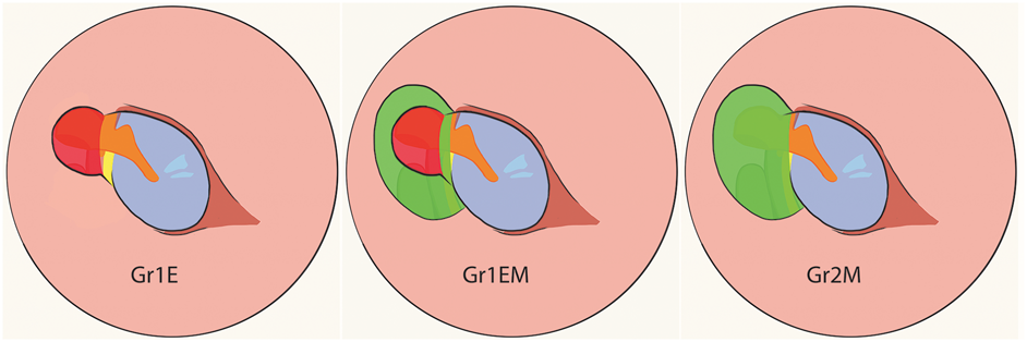
In order to establish whether the endoscopic and subsequent microscopic approach removed more bone than a solely microscopic approach, a second group was added. This group, the microscopic group (group 2 microscopic approach), consisted of four heads with eight ears. After a baseline pre-operative CT scan, middle-ear landmarks were visualised microscopically through an ear speculum similar to that performed in the group 1 microscopic approach following endoscopy group. Following this procedure, a post-microscopy CT scan was done. Figure 1 shows a right ear illustration of the transmeatal view of resected regions of the scutum for all (sub-)groups. All surgical steps were performed by the senior author (M de Wolf).
Right ear illustration of the transmeatal view of resected regions of the scutum for all
(sub-)groups. Gr1E = group 1 endoscopic approach; Gr1EM = group 1 microscopic approach following endoscopy; Gr2M = group 2 microscopic approach

Quantifying the resected volume
All CT scans were performed on a Siemens Somatom Force CT scanner (Siemens Gmbh, Erlangen, Germany). Image data were reconstructed into volume images with voxel spacing of 0.45 × 0.45 × 0.45 mm.
Differences in the amount of resected bone between the approaches were evaluated using custom-made software.Reference Dobbe, Strackee, Schreurs, Jonges, Carelsen and Vroemen29 All CT scan images per specimen were aligned by image segmentation and registration. To this end, the available part of the skull in the first image was segmented and registered to the subsequent images, yielding inter-image positioning matrices. The inverse of such matrix was used for image alignment. A 3D image of the resected bone volume was created by subtracting the image intensities on a voxel-by-voxel basis. Three subtraction images were created. Two of these were made by subtracting the post-endoscopy or -microscopy CT scan from their baseline CT scan (group 1 endoscopic approach, group 2 microscopic approach). This subtraction image clearly distinguished the resected incus from the resected bone and allowed manual segmentation of the resected bone voxels in the 3D selection using a painting tool. The third subtraction image was made by subtraction of the post-microscopy after endoscopy CT scan from the post-endoscopy CT scan (group 1 microscopic approach following endoscopy). This image visualised the additional bone resection for subsequent segmentation and volume quantification.
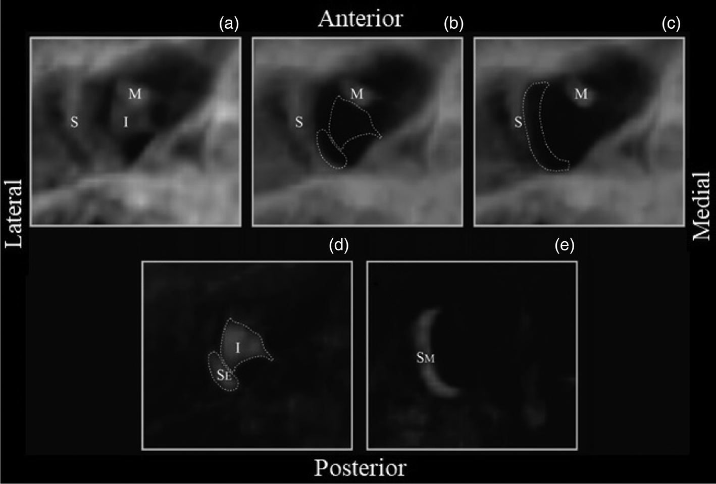
An example of a digitally reconstructed radiograph of the baseline, post-endoscopic, post-microscopic and subtraction images of a right ear is shown in Figure 2. The sum of the selected voxels represents the resected volume of bone and was expressed in millimetres cubed. The median of resected bone is reported with its 25th and 75th percentile.
Right ear example from group 1 visualised using a digitally reconstructed radiograph of the selected volume of interest (15 × 14 × 14 mm), showing an inferior view of a bony ear canal and middle ear. (a) Baseline situation, (b) situation after endoscopic visualisation with removal of the incus and part of the scutum, (c) situation after microscopic visualisation with additional removal of the scutum, (d) subtracted image (post-endoscopy from baseline) showing the resected incus and part of the scutum and (e) subtracted image (post-microscopy from post-endoscopy) showing the additionally resected scutum. S = scutum, I = incus, M = malleus, SE = endoscopically resected part of scutum, SM = extra microscopically resected scutum

Data analysis
Z-values for skewness and kurtosis were evaluated to analyse normality of the data (data were considered normally distributed if –1.96 < z < 1.96). To evaluate correlation between the measured volumes of ears within one head, the Spearman's rank correlation coefficient between the left and right ears was determined.
Resected volumes of the group 1 endoscopic approach were compared with the group 1 microscopic approach following endoscopy and the group 2 microscopic approach. The amount of resected bone in the group 1 microscopic approach following endoscopy was compared with the group 2 microscopic approach. The resected volume in the group 1 endoscopic approach was expressed as a percentage of the total resected volume after microscopic visualisation.
The authors assert that all procedures contributing to this work comply with the ethical standards of the relevant national and institutional guidelines on human experimentation and with the Helsinki Declaration of 1975, as revised in 2008. Approval from the institutional review board was not necessary.
Results
One right ear from group 1 was excluded from analysis because noise due to displacement of defrosted water and soft tissue made it impossible to reliably select voxels representing resected bone.
Data were not normally distributed in all groups. The Spearman's rank correlation coefficient between left and right ears showed a low correlation (rs = 0.251; p = 0.259; median right ears = 9.88 mm3 and median left ears = 8.28 mm3).
All predetermined landmarks were visualised successfully using the endoscopic and microscopic approach after removal of the incus and sufficient part of the scutum. During the dissection, the endoscopically resected part of the scutum lay inside the area of resected bone for microscopic visualisation.
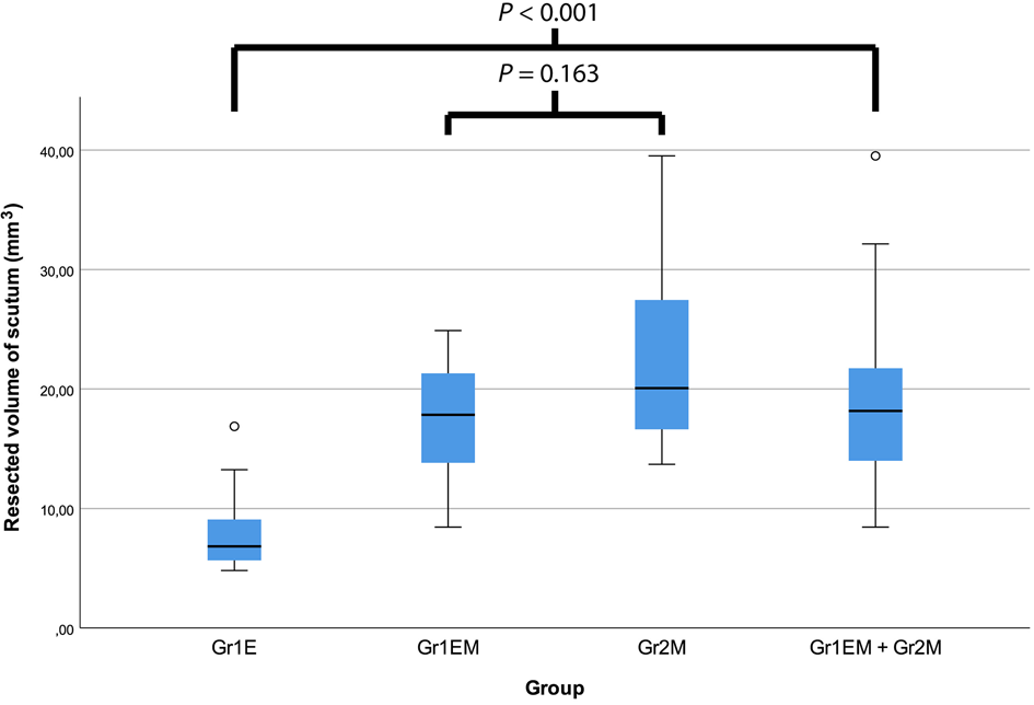
In the group 1 endoscopic approach, a median of 6.84 mm3 (25th percentile: 5.66, 75th percentile: 9.61 mm3) bone was resected. No statistically significant difference (Mann–Whitney U test, p = 0.163, U = 49.000) of resected bone was found between the group 1 microscopic approach following endoscopy (median 17.84; 25th percentile: 13.78, 75th percentile: 21.47 mm3) and the group 2 microscopic approach (median 20.08; 25th percentile: 15.86, 75th percentile: 29.80 mm3). Since there is no statistically significant difference between these 2 groups, they were combined to form 1 microscopic evaluation group (group 1 microscopic approach following endoscopy + group 2 microscopic approach: median 18.16; 25th percentile: 13.89, 75th percentile: 22.00 mm3). The difference between the group 1 endoscopic approach and group 1 microscopic approach following endoscopy plus group 2 microscopic approach is statistically significant (Mann–Whitney U test, p < 0.001, U = 18.000). By using an endoscope, 38 per cent (6.84 / 18.16 mm3) of the amount of bone for microscopic visualisation had to be resected. Figure 3 shows a boxplot representing resected volumes of bone from the endoscopic and microscopic groups.
Boxplot of resected volumes of bone from the scutum per (sub-)group. Statistical analysis of differences between groups was done by Mann–Whitney U test. Gr1E = group 1 endoscopic approach; Gr1EM = group 1 microscopic approach following endoscopy; Gr2M = group 2 microscopic approach

Discussion
This cadaveric study compared volumes of resected bone from the scutum to visualise middle-ear landmarks transmeatally by endoscope and microscope. State of the art, custom-made 3D imaging technology was used to evaluate resected volumes of bone in three dimensions.Reference Dobbe, Strackee, Schreurs, Jonges, Carelsen and Vroemen29 Since there was a low correlation between left and right ears, all ears were interpreted as independent. During the dissection, the endoscopically resected part of the scutum was overlapped by the area of resected bone in the microscopic group. The finding that there was no statistically significant difference between the group 1 microscopic approach following endoscopy and the group 2 microscopic approach confirms this observation.
Therefore, both methods were compared within the same ear and the effect of inter-ear variability on resected volumes could be ruled out. However, despite the inter-ear variability, there was no significant difference found between resected volumes in the group 1 microscopic approach following endoscopy and group 2 microscopic approach. So, these groups were combined to create one larger group for comparison with the group 1 endoscopic approach.
• Small epitympanic cholesteatomas are often resected through the ear canal
• Iatrogenic scutum defects relate to recurrence of retraction pockets and cholesteatoma
• The endoscope allows adequate visualisation of the middle ear
• Endoscopic visualisation of middle-ear landmarks preserves more of the bony scutum in comparison to the microscope
A statistically significant difference of resected volumes between the endoscopic and microscopic approach was found. To visualise middle-ear landmarks, the endoscopic approach preserves a larger part of the scutum compared with the microscopic approach.
To minimise intra- and inter-observer variability, all procedures, including identifying the middle-ear landmarks, were performed by one surgeon. Quantification of resected bone with the 3D-imaging software was done by the first author. This study established that the endoscopic approach is less invasive compared with the microscopic approach despite inter-scan and the aforementioned variabilities. Our results strengthen the conclusion of Imai et al, who state that the endoscopic approach allows minimally invasive transmeatal removal of cholesteatoma. They used two-dimensional imaging technology by measuring templates for reconstruction of the scutum after endoscopic transmeatal resection of cholesteatoma (median, 37.3; minimum, 14.7, maximum, 68.4 mm2).Reference Imai, Nishiike, Oshima, Tanaka, Tsuruta and Tomiyama28 In our study, the aim was to transmeatally visualise predetermined landmarks. Imai et al. performed a retrospective analysis of endoscopically treated cholesteatoma cases. These two studies can therefore not be compared. In the Japanese study there was no microscopic control group. Notably, there is a large difference between the size of the smallest (14.7 mm2) and largest (68.4 mm2) created scutum defect. The authors explained this by pointing out a correlation between the size of the cholesteatoma and the post-surgery scutum defect (correlation coefficient, R2 = 0.617).
We believe that the position of the external ear canal in relation to the horizontal semicircular canal also plays an important part in posterior visualisation. When the external ear canal, in relation to the horizontal semicircular canal, is positioned relatively anteriorly, large parts of the bony ear canal have to be removed for adequate view in the posterior direction. In our study, for three ears, large amounts of bone had to be endoscopically removed (group 1 endoscopic approach) with hardly any additional resection for microscopic visualisation (group 1 microscopic approach following endoscopy): 27, 31 and 21 per cent, respectively. In these cases, the endoscopic advantage regarding preservation of the bony scutum is limited. In the study performed by Imai et al., this might also have played a role for cases that required a large scutum defect, next to the size of the cholesteatoma.Reference Imai, Nishiike, Oshima, Tanaka, Tsuruta and Tomiyama28 A future study might investigate the possibility to create a predictive model to select patients who are suitable for an endoscopic transmeatal approach.
Since the 1980s, it has been known that a scutum defect plays a role in the recurrence of retraction pockets after canal wall up mastoidectomy. Additionally, the importance of scutal defect reconstruction to prevent recurrence has been stressed.Reference Sanna, Zini, Gamoletti, Delogu, Scandellari and Russo9 Many studies strengthen this statement.Reference Hinohira, Yanagihara and Gyo1–Reference Sakai, Shinkawa, Miyake and Fujii8 It is plausible that smaller iatrogenic scutum defects may reduce the risk of recurrence of pathology. To our knowledge, only one study (Bae et al.) has compared results of cholesteatoma surgery after an endoscopic and microscopic transmeatal approach.Reference Bae, Kang and Chung30 In the microscopic group, a Lempert incision was made and no mastoidectomy was performed. Neither residual, nor recurrent disease was reported for both groups. Limitations of this study were a short follow-up time (19.75 vs 41.05 months) and small groups (10 vs 10 patients). Whether the reduced invasiveness of the endoscopic transmeatal approach will actually decrease recurrences compared with a microscopic transmeatal approach has to be evaluated in larger, prospective, long-term follow-up studies.
Multiple studies have compared the outcomes of a transmeatal endoscopic with a retroauricular microscopic approach with mastoidectomy for cholesteatoma cases.Reference Hunter, Zuniga, Sweeney, Bertrand, Wanna and Haynes20,Reference Marchioni, Soloperto, Rubini, Villari, Genovese and Artioli21,Reference Presutti, Anschuetz, Rubini, Ruberto, Alicandri-Ciufelli and Dematte31,Reference Magliulo and Iannella32 Although their results favour the endoscopic transmeatal approach, the studies also have their limitations. Interpretation is difficult because of heterogeneity of the study groups and short follow-up time. Despite the promising usefulness of the endoscope, high-quality studies are needed to evaluate its position in relation to canal wall up mastoidectomy with obliteration as a surgical modality in management of primary cholesteatoma.Reference Hellingman, Geerse, de Wolf, Ebbens and van Spronsen33
Conclusion
The results of this study show that an endoscopic transmeatal approach preserves more of the bony scutum than a microscopic transmeatal approach to acquire visualisation of the same middle-ear landmarks and antrum. Long-term follow-up studies of endoscopically treated epitympanic ear pathology will have to show if preservation of larger parts of the scutum plays a role in preventing disease recurrence.
Acknowledgements
Completion of this article was made possible by a generous contribution of the Heinsius-Houbolt Foundation