INTRODUCTION
Zoos and public aquaria experience an increasing interest of their visitors in living coral reef exhibits. In order to improve the skills of zoos and aquaria to grow and maintain stony corals (Scleractinia), a consortium consisting of several zoos and public aquaria, four specialized research institutions and a coral culture company executed research and innovation activities on the following topics:
1. Sexual and asexual breeding of corals in captivity, including techniques for propagation, feeding and induction of natural coral colony morphogenesis.
2. Coral husbandry: development of generic bioassays to evaluate biotic and abiotic husbandry parameters and to monitor coral health, elaboration of methods for identification and treatment of coral diseases and optimization of transport and acclimation procedures.
These activities were executed under the umbrella of a European research project entitled CORALZOO, which was executed for the benefit of the European Association of Zoos and Aquaria (EAZA). CORALZOO started on 1 June 2005 and ended on 31 August 2009. This review summarizes the main achievements of the project.
PREPARING BROODSTOCK: CORAL PROPAGATION
The first step in coral culture is to acquire coral materials for breeding. This so-called broodstock can be obtained in two ways:
1. Asexual reproduction (cloning); this is done by the fragmentation of parent colonies into smaller pieces that will grow out to adult colonies.
2. Sexual reproduction, leading to the formation of planula larvae, each of which has a different genetic background.
Whereas cloning is useful for efficient breeding of genotypes that have proven to be suitable for aquarium use, sexual reproduction is needed to maintain genetic diversity among captive coral populations, thus increasing the resilience of these populations. Both approaches have been studied intensively during the CORALZOO project.
Asexual reproduction (fragmentation)
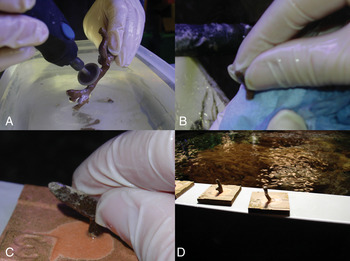
Asexual propagation or ‘fragmentation’ is a simple method for obtaining large numbers of coral colonies (Borneman, Reference Borneman2001; Calfo, Reference Calfo2001). New coral colonies are obtained by cutting a mother colony into smaller pieces (Figure 1) and, if necessary, by attaching these fragments to a solid support. It makes use of the remarkable regeneration capabilities of corals: even a single, isolated polyp or a fragment that contains few polyps can grow out to a new adult colony. Hence, one adult coral colony can be cut into numerous small fragments that will all form healthy, new colonies.
An experimental procedure for fragmentation (example: Montipora digitata). (A) Fragments being cut from colony using the modelling saw; (B) drying the newly cut end of the fragment; (C) gently holding the fragment until a bond is formed; (D) fragments adhered to the tiles awaiting placement back in the tank after the glue has set. Photographs by Andrew McLeod.

A generic, two-step procedure was defined for optimizing protocols for asexual reproduction of a given coral species by fragmentation. In the first step, growth kinetics and variability among individuals of the species studied are determined. Information on growth kinetics (Osinga et al., Reference Osinga, Schutter, Griffioen, Wijffels, Verreth, Shafir, Henard, Taruffi, Lavorano and Gili2011) can be used to determine the best fragment size for effective asexual propagation. The obtained data on variability can be used to define sample sizes for further (second step) optimization experiments, such as comparative studies on fragmentation methods (e.g. using tweezers, knives, electric cutters and saws), support materials (plastics, ceramic tiles, etc) and methods to attach fragments to their supports (superglue, two-component epoxy and nylon threads). Following this procedure, protocols describing the best practises for fragmentation of Montipora digitata (Dana 1846), Galaxea fascicularis (Linnaeus 1767) and Stylophora pistillata (Esper 1797) were elaborated (Leewis et al., Reference Leewis, Wijgerde, Laterveer and Osinga2009). For M. digitata, an optimal sample size of 20 to 30 replicates was determined, which was used to evaluate the effect of fragment size, orientation and origin (i.e. growth tips versus middle sections) on growth of fragments that were glued onto ceramic tiles using cyanoacrylate glue. Coral tips grew faster (90% weight increase in ten weeks) after initial fragmentation than all the other areas of the colony where fragments had been taken from. 1 cm rings taken from the middle of the branches grew faster than 2 cm growth rings taken from the same areas. Fragments taken from the middle sections of the colony branch that had been cut longitudinally grew faster if they were approximately 2 cm in length compared to 1 cm in length.
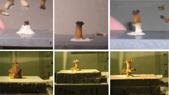
Optimal fragment size for the culture of G. fascicularis was deduced from growth kinetics. Specific growth rates of this species were found to exhibit a decreasing trend in time (Schutter et al., Reference Schutter, Crocker, Paijmans, Janse, Osinga, Verreth and Wijffels2010). Hence, small fragments will have the highest productivity. The relatively large size of the polyps of G. fascicularis enables the production of single-polyp nubbins. The most suitable method to produce large series of viable fragments of this species is by removing single polyps from a parent colony using tweezers and by attaching the single polyps to small plastic plates using two-component epoxy. The position of the polyp in the epoxy determines the subsequent degree of attachment of the developing colony onto the support (Figure 2). When the epoxy is not completely covering the dead skeletal part of the initial polyp, the colony will grow like an umbrella that is attached to the support only through the initial polyp. This type of attachment can be convenient if the newly formed colony has to be removed from its substrate and positioned in a display tank. By pushing the polyp deeper into the epoxy, the newly formed polyps will all attach to the substrate.
Development of colonies of Galaxea fascicularis (lower row of photographs) after inserting the initial polyps in three different ways into two-component epoxy (upper row of photographs). Left: dead skeleton and part of living tissue covered in epoxy; middle: dead skeleton completely covered in epoxy; right: dead skeleton partially covered in epoxy. Partial coverage of the dead skeletal material by epoxy resulted in an umbrella-like shape. Photographs by Max Janse.

Procedures to prepare large numbers of S. pistillata fragments were outlined by Shafir et al. (Reference Shafir, Van Rijn and Rinkevich2006), who used small fragments (approximately 10 polyps per fragment) that were glued on plastic pins. Within the first few years of their development, S. pistillata colonies grow exponentially following first order kinetics. This implies that for this species, fragment size is not influencing subsequent growth performance.
A list of common (but not-optimized) techniques for more than one hundred coral species was compiled based on information obtained from participating aquaria (Leewis et al., Reference Leewis, Wijgerde, Laterveer and Osinga2009). Shafir & Rinkevich (Reference Shafir, Rinkevich, Leewis and Janse2008) further described procedures for propagation of branching coral species.
Sexual propagation
In CORALZOO, research on sexual propagation has focused on the concept to raise spat obtained from natural populations under controlled laboratory conditions (Petersen et al., Reference Petersen, Laterveer, Van Bergen, Hatta, Hebbinghaus, Janse, Jones, Richter, Ziegler, Visser and Schuhmacher2006; Amar et al., Reference Amar, Chadwick and Rinkevich2008a). This approach has been studied concurrent with analogous research carried out in another project to which many of the CORALZOO partners participate (SECORE—www.secore.org).
CORALZOO activities focused on the branching species S. pistillata. The reproductive cycle of this species in the Red Sea has been studied extensively (see Amar et al., Reference Amar, Chadwick and Rinkevich2008a and references therein). Based on these observations, a procedure for controlled production of sexually derived propagules was elaborated by bringing captive bred, reproductive colonies of S. pistillata into a sea-based nursery, where spawning followed the natural cycle. The procedures for collection, settlement and subsequent handling of the planula larvae were established (Leewis et al., Reference Leewis, Wijgerde, Laterveer and Osinga2009; Amar & Rinkevich, Reference Amar and Rinkevich2007; Amar et al., Reference Amar, Chadwick and Rinkevich2008a). Larvae of S. pistillata were collected by positioning plankton nets overnight over gravid colonies. Collected larvae were allowed to settle on transparent, waterproof paper (Mailers paper) that was placed in petri dishes filled with natural seawater. Some larvae metamorphosed without attachment, hanging upside-down on the water surface. These free-floating primary polyps were picked up and glued to Mailers paper. The primary polyps on the Mailers papers holding the primary polyps were glued onto glass slides and transferred to running seawater aquaria for subsequent culture for three months. Survival of the collected larvae was 60–68%, of which 30% settled. Survival during the three-month culture phase was 80%. Hence, from a total of 1315 collected planula larvae, approximately 200 new colonies were obtained for outgrow in the sea-based hatchery. Comparable results have been reported for other coral species (Petersen et al., Reference Petersen, Laterveer, Van Bergen, Hatta, Hebbinghaus, Janse, Jones, Richter, Ziegler, Visser and Schuhmacher2006), showing the validity of the concept.
‘TOGETHER WE ARE STRONG’—SETTLEMENT IN AGGREGATES
A new approach was investigated that deals with aggregates of settling larvae (chimeras). Coral larvae of many species are usually found to settle in aggregates (e.g. Duerden, Reference Duerden1902; Hidaka, Reference Hidaka1985; Raymundo & Maypap, Reference Raymundo and Maypab2004), and in the past, scientists tried to separate between the aggregated spats. Aggregated settlement of kin larvae in sessile marine invertebrates may result in a complex array of compatible and incompatible allogeneic responses within each assemblage. Each such aggregate can, therefore, be considered as a distinct self-organizing biological entity representing adaptations that have evolved to maximize the potential benefits of gregarious settlement. However, only sparse information exists on the selective forces and ecological consequences of allogeneic coalescence.
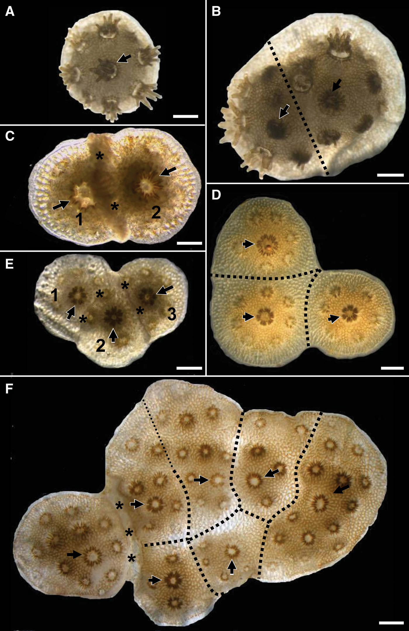
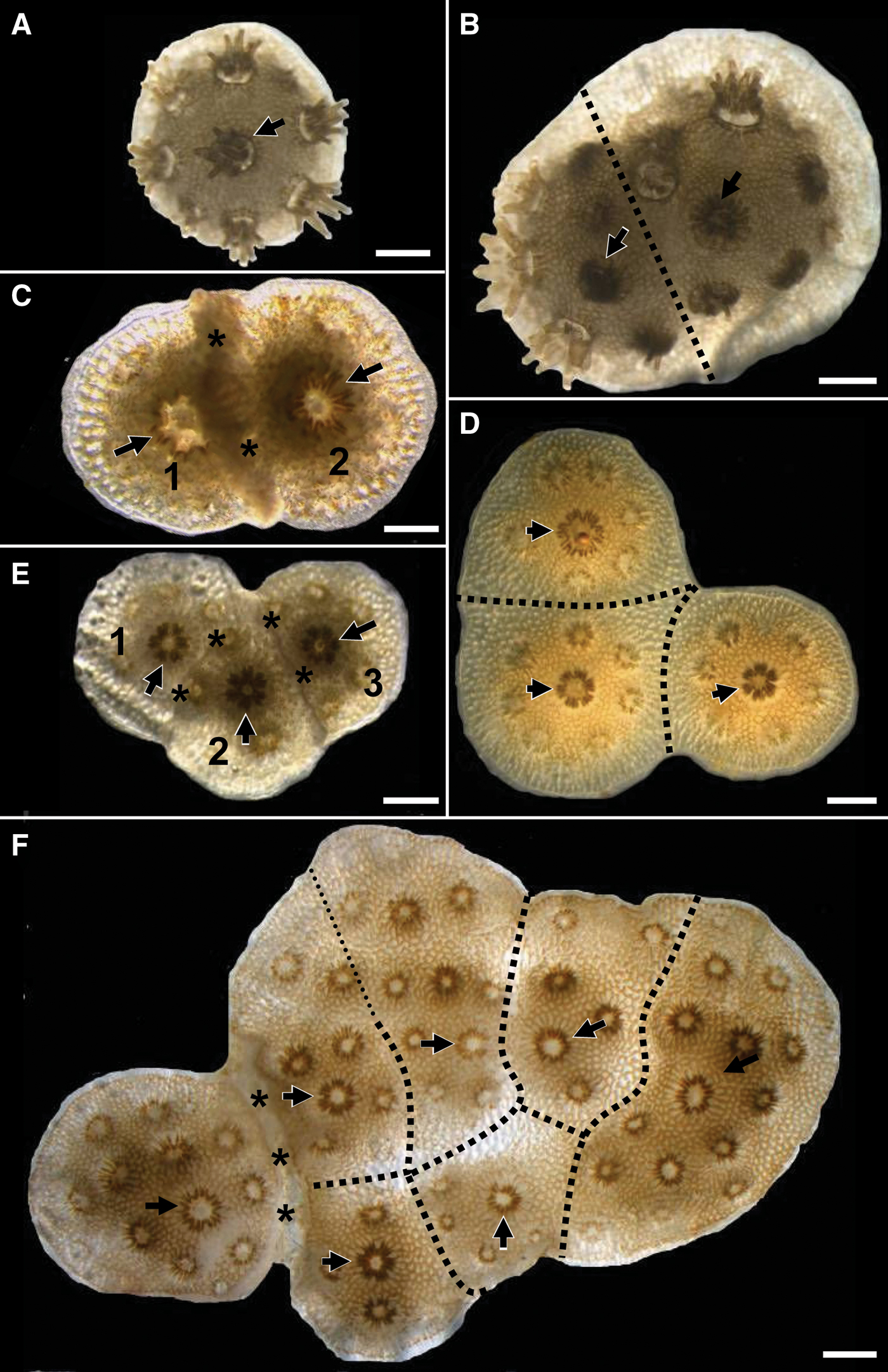
In CORALZOO, under controlled laboratory settings the consequences of aggregated settlement of kin larvae of S. pistillata were studied (Amar et al., Reference Amar, Chadwick and Rinkevich2008b). When spat came into contact, they either fused, establishing a chimera, or rejected one another. A one-year study on growth and survivorship of 544 settled S. pistillata genotypes revealed six types of biological entities (Figure 3): single genotypes (SG); bi-chimeras; bi-rejecting genotypes; tri-chimeras; three-rejecting genotypes; and multi-partner entities (MP) consisting of 7.5 ± 2.6 partners. In general, MP exhibited the highest survivorship rate and SG the lowest. These results bring us to suggest that the ‘group level’ of kin aggregates may serve as a ubiquitous legitimate selection entity in the evolution of a sessile mode of life in marine organisms. Therefore, handling the whole aggregate as a single unit is a novel approach for successful handling of settled larvae.
Types of biological entities in young colonies of Stylophora pistillata. (A) Single genotype; (B) bi-chimera; (C) bi-rejecting genotype (colony no. 2 has begun to overgrow colony no. 1); (D) tri-chimera; (E) entity with three rejecting genotypes numbered 1, 2 and 3. Clear borderlines demarcate each genotype; (F) aggregated colony composed of seven genotypes (multi-partner entity). Asterisks indicate rejecting areas. Thick dotted lines depict presumed borders between genotypes. Thin dotted lines indicate no clear border between genotypes. Arrows point to the sites of founder polyps in each genotype. Scale bars = 1 mm. Reproduced from Amar et al. (Reference Amar, Chadwick and Rinkevich2008b).
SIGNS OF INBREEDING?
A study of the fusion and rejection of planulae larvae (formation of kin-aggregates and chimera between two or more genetic individuals) on S. pistillata during two successive breeding seasons in the natural waters of Eilat (Israel), revealed signs of inbreeding within this natural population (Amar & Rinkevich, Reference Amar and Rinkevich2010). Such signs were not observed in a comparative study that was executed a decade ago. The study demonstrates that the immunoresponse of corals in genetically small populations such as aquarium populations may change over time, potentially leading to reduced population fitness. These findings support the strategy to obtain spat from natural population for breeding purposes in aquaria.
Identification of corals—tagging
The identification and administration of aquarium corals (including new coral recruits obtained from induced or spontaneous sexual reproduction) is an important issue, for which no suitable technology was available up till now. The possibility to use amplified fragment length polymorphism (AFLP: Vos et al., Reference Vos, Hogers, Bleeker, Reijans, Van de Lee, Hornes, Friters, Pot, Paleman, Kuiper and Zabeau1995) as a genetic fingerprinting technique to discriminate genetic individuals (in analogy to molecular fingerprinting for forensic investigations) was successfully explored for wild spat from S. pistillata obtained during two successive reproductive seasons in Eilat (Israel, Red Sea) (Amar et al., Reference Amar, Douek, Rabinowitz and Rinkevich2008c). The method is not only suitable to identify genetic individuals but it can also be used to relate offspring to parents (population genetics). Another tagging method that was successfully tested on corals during the project is the use of passive integrated transponders (PIT: Whitfield Gibbons & Andrews, Reference Whitfield Gibbons and Andrews2004). Such transponders can be attached to a coral colony using cyanoacrylate glue or two-component epoxy (Leewis et al., Reference Leewis, Wijgerde, Laterveer and Osinga2009). The transponders are passive, but can be activated by a hand-held reader that scans the code of the tagged individual. AFLP fingerprinting combined with PIT tagging provides a set of complementary tools for effective administration of aquarium populations.
REARING CORALS—CULTURE AND HUSBANDRY
When the rearing (also termed: breeding) of corals is concerned, two aspects need to be considered:
— the growth rate of the corals, which determines the quantity of corals that an aquarium system of a given size can produce per unit of time (the volumetric productivity); and
— the growth form (shape) of the corals, which largely determines their value as ornamental animals (natural growth forms being preferred by spectators and aquarium hobbyists).
Both aspects were studied within CORALZOO in order to provide a rationale for further improvement of breeding techniques (i.e. production of corals with a nice, natural-looking growth form at a low production cost). Results are presented in the next two subsections.
Factors controlling coral growth
CORALZOO focused on zooxanthellate stony corals, these are the corals that live in symbiosis with unicellular algae called zooxanthellae. In order to optimize the breeding regimes for zooxanthellate corals, it should be understood how growth (including both skeletal growth and growth of the organic tissue) is controlled in these organisms (see review by Osinga et al., Reference Osinga, Schutter, Griffioen, Wijffels, Verreth, Shafir, Henard, Taruffi, Lavorano and Gili2011). Three major processes drive the growth of zooxanthellate corals:
1. photosynthesis (the translocation of photosynthetically acquired organic carbon from symbiotic zooxanthellae to the coral host);
2. heterotrophic feeding (grazing on zooplankton, phytoplankton, bacteria and dissolved organic matter);
3. calcification (the formation of a calcium carbonate skeleton).
For these processes, the following resources are needed: for photosynthesis: light, carbon dioxide and inorganic nutrients; for heterotrophic feeding: organic food; and for calcification: Ca2+, CO2−3 and metabolic energy.
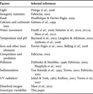
Several other factors have been reported to influence coral growth, either positively or negatively (Table 1). Within CORALZOO, research concentrated mainly on the factors light, water movement, nutrition and genotype, since these factors are likely to be determining the cost-efficiency of coral culture to a large extent.
Overview of factors that have been reported in relation to coral growth.

*, this factor will not play a role in aquaria that use artificial lighting.
LIGHT
At the start of the project, it was assumed that light is the growth-limiting factor for most corals in artificially illuminated aquaria. Hence, a first experiment was designed to measure the effect of different photon flux densities on the growth (measured as the increase in buoyant weight) of G. fascicularis when cultured under artificial light.
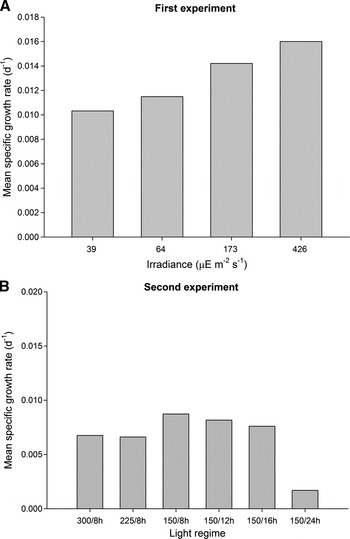
Indeed, a positive correlation between photon flux density (PFD) and growth was found in this experiment (Schutter et al., Reference Schutter, Van Velthoven, Janse, Osinga, Janssen, Wijffels and Verreth2008; Figure 4A). In a comparable experiment, that was designed to study effects of length of day on growth of G. fascicularis, no differences between treatments (both different lengths of day and different PFDs) were found (Schutter et al., Reference Schutter, Van der Ven, Janse, Verreth, Wijffels and Osinga2011b; Figure 4B). Specific growth rates were much lower than in the first experiment. This indicates that under common aquarium conditions, coral growth may in many cases not be light-limited. Notwithstanding these conflicting data, it was unambiguously demonstrated that G. fascicularis cannot be grown under a regime of 24 hours illumination per day: corals grown under this regime bleached and died after 7 weeks in culture (Schutter et al., Reference Schutter, Van der Ven, Janse, Verreth, Wijffels and Osinga2011b).
Effects of different irradiance regimes on the specific growth rate of Galaxea fascicularis. (A) Effects of increasing irradiance; (B) effects of increasing irradiance and length of the day. On the X-axis, the regimes are shown, for example: 150/8h refers to a regime where the corals receive 150 µE m−2 s−1 during 8 hours per day.

WATER MOVEMENT
Clones of this species were grown under four different hydrodynamic regimes (average flow velocities of 0, 10, 20 and 25 cm/s) under low light, to test the effect of water movement on the growth (measured as the increase in buoyant weight) of G. fascicularis (Schutter et al., Reference Schutter, Crocker, Paijmans, Janse, Osinga, Verreth and Wijffels2010). The corals cultured under the highest flow rate grew fastest, despite the fact that they had the lowest photosynthesis to respiration ratio. Corals grown at high flow appear to be more efficient in the utilization of resources, probably because the high flow keeps these organisms more free from fouling (Fabricius, Reference Fabricius2005; Box & Mumby, Reference Box and Mumby2007). It was also shown that corals grew significantly slower without active movement of the surrounding water.
NUTRITION
Feeding promotes coral growth and increases their resistance (Ferrier-Pagès et al., Reference Ferrier-Pagès, Gattuso, Dallot and Jaubert2000, Reference Ferrier-Pagès, Witting, Tambutté and Sebens2003; Bongiorni et al., Reference Bongiorni, Shafir and Rinkevich2003). Hence, fed corals are happy corals. Following upon this new dogma, considerable effort was put by members of the CORALZOO team into studies on coral nutrition. The next subsections describe studies on the digestive physiology of a model coral (S. pistillata), feed optimization for two coral species (S. pistillata and Pocillopora damicornis Linnaeus 1758) and the testing of two alternative strategies for feeding: feeding with inorganic nutrients and feeding with dry fish food. In addition, a simulation tool was developed that shows the effect of system design on the fate of planktonic food added to the aquarium system (Leewis et al., Reference Leewis, Wijgerde, Laterveer and Osinga2009). This tool can help to design an optimal strategy to administer food to coral breeding systems.
How coral process food—a study on the digestive system of S. pistillata
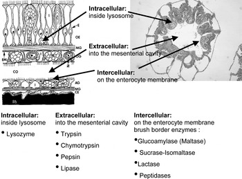
An extensive histological study was done to characterize the cellular structure of the polyps of S. pistillata. The gut and digestive apparatus of this species were shown to be more complex than previously assumed. The polyp's digestive system along the oral–aboral axis is divided into several segments. Each of these segments can be termed as a separate compartment, consisting of different cell types and their distribution, implying for unalike digestive functions along the polyp's gastric cavity. We found an extensive actinopharynx loaded with nematocysts through a third of the polyp's length. We detected two kinds of gastric filaments with a different anatomic location containing different cell types. The digestion system of corals may comprise three unalike compartments of activity (Figure 5), each representing a different digestion pathway:
1. extracellular digestion—through secreted enzymes into the digestion cavity;
2. intercellular digestion type ‘a’—through lysosomes; and
3. intercellular digestion type ‘b’—on the digestive system cell walls membranes.
Three potential digestion pathways in Stylophora pistillata. There is one extracellular pathway whereby enzymes are secreted into the mesenterial cavity and two intracellular pathways: digestion inside lysozomes and digestion through enzymes located on the cell membranes of specialized gut cells.

Almost nothing is known about these digestion pathways in corals. Therefore, the digestive system of S. pistillata was further characterized on an enzymatic level. Using the in situ hybridization technique, we detected specific gastric areas which are producing the digestive enzyme chymotrypsin. Furthermore, it was demonstrated for the first time that a cnidarian may elicit a brush border enzymes pathway of digestion. In mammals and other vertebrates, brush border enzymes are located on the apical side membrane of the intestine cells, where they break (among others) carbohydrates into mono-sugars and peptides into amino acids.
Effect of different feeds on the growth of P. damicornis
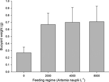
The best practises for feeding this species with planktonic food were defined (composition and concentration) based upon four years of nutrition experiments with P. damicornis. Improvement of growth (compared to corals that were not additionally fed) was found with live Instar I nauplii of the brine shrimp Artemia (Lavorano et al., Reference Lavorano, Gili, Muti, Taruffi, Corsino, Gnone, Leewis and Janse2008), the rotifer Brachionis sp. and the marine diatom Tetraselmis suecica. The latter finding is in contrast to the generally prevailing idea that stony corals do not eat phytoplankton, but is in good agreement with the discovery described above that stony corals possess brush border enzymes. Feeding with the microalgae Nannochloropsis sp. did not promote growth in P. damicornis. For each of the feeds that were found useful, an optimal concentration was determined (based on daily batch feeding, see for example Figure 6).
Effect of different concentrations of Artemia nauplii (end concentrations of Artemia nauplii per litre of seawater in the tank after feeding—daily batches) on the growth of nubbins of Pocillopora damicornis, measured as the buoyant weight after four months. Error bars indicate standard deviations (N = 216).

Also, mixed diets were tested. These did not further improve growth when compared to the best single-species diet, but were found to be more attractive in a cost–benefit analysis. Artemia is the best food to maximize productivity at the lowest cost. From an environmental point of view, however, large-scale usage of Artemia can be questioned, since this product is obtained through harvesting natural resources.
Daytime feeding appeared to be advantageous over nighttime feeding, despite the observations in nature that suggest that most stony corals are nighttime feeders. This shows that stony corals are opportunistic feeders, which start to consume whenever food is available.
Effect of different feeds on the growth of S. pistillata
In analogy to the work on P. damicornis described above, a series of feeding tests was done to optimize the diet for S. pistillata. Here, feeding did not show pronounced effects on coral growth, most likely because this study was done with natural seawater from an area that is relatively rich in organic matter. The study revealed the importance of genotypic variation as a factor determining coral growth: variability among genotypes was in all cases much larger than variability among treatments. It was therefore decided to further study this genotype factor (see subsection 3.1.4).
Corals as plants?—nutrition through inorganic enrichment
A study was done to investigate if the need for heterotrophic feeding (being a source of nitrogen) can be reduced by adding inorganic nutrients (nitrate and ammonia) to a coral culture. In agreement with the literature (e.g. Stambler et al., Reference Stambler, Popper, Dubinsky and Stimson1991; Marubini & Davies, Reference Marubini and Davies1996; Marubini & Thake, Reference Marubini and Thake1999; Ferrier-Pagès et al., Reference Ferrier-Pagès, Gattuso, Dallot and Jaubert2000, Reference Ferrier-Pagès, Schoelzke, Jaubert, Muscatine and Hoegh-Guldberg2001; Langdon & Atkinson, Reference Langdon and Atkinson2005), no beneficial effect of the nutrient additions was found. The high nutrient loadings did not negatively affect coral performance either, this is in contrast to most papers cited above.
Corals as fish?—nutrition through dry fish food additions
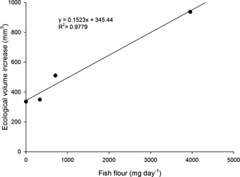
Providing dry food to corals will reduce labour costs when compared to supplying live planktonic food (no need for preparation of live feeds). Because previous studies indicated that batch feeding with dry fish food harmed the corals, a new system was designed that continuously provides dry fish food. This system was tested on a culture system in which P. damicornis was grown. Growth was good and showed a positive correlation with food concentration (Figure 7). Hence, this approach can be recommended for use in coral breeding systems.
Correlation between growth of Pocillopora damicornis (increase in ecological volume in 122 days) and amount of dry feed added.

GENOTYPE: A PAN-CORALZOO EXPERIMENT
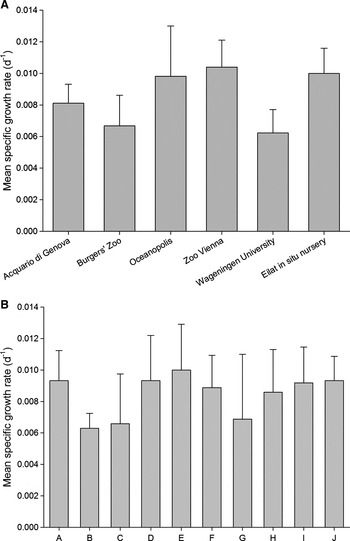
A series of more than one thousand nubbins obtained from ten genetically different colonies of S. pistillata was shipped to seven CORALZOO partners in Europe in spring 2007. Growth (measured as the increase in ecological volume) and survival of these nubbins have been monitored for more than two years on six of the seven originally selected locations. Average survivorship of all nubbins was 70%, but showed considerable variation among location and genotype. Survival was better in the aquaria than in the sea-based nursery, revealing the potential importance of public aquaria as a modern ‘Noah's ark’ for corals. The variability in growth after two years was also large, both between locations and between genotypes (Figure 8), which leads to the following conclusions:
1. Differences in breeding efficiency between the breeding centres are considerable, further optimization is possible in many cases.
2. Genotypic variability is an important factor to take into consideration when starting a breeding programme for a coral species.
Results of the pan-CORALZOO nubbin experiment with Stylophora pistillata. Upper graph: average specific growth rates for all genotypes at the different experimental locations; lower graph: average specific growth rates for all locations per genotype.

Another result of this study is the availability of genetically different materials of S. pistillata in EAZA-associated breeding centres, which is a good starting point for a breeding programme on this species.
INTERACTIONS
Several experiments that had been done in the first years of the project pointed towards potential interactive effects of factors that potentially influence coral growth (see for example Figures 4 & 6). Interactions are defined as the effect that one factor exerts on the effect of another factor. We expected to find interactions between light and water flow and between light and nutrition. Experiments were designed to study these potential interactions.
Light and water movement
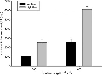
The interactive effect of light and water movement on coral growth (measured as the increase in buoyant weight) was studied using G. fascicularis as a model species (Schutter et al., Reference Schutter, Kranenbarg, Wijffels, Verreth and Osinga2011a). A significant positive interaction was found (Figure 9), but no concurrent increase in photosynthetic activity could be detected. Most likely, water movement modulates the efficiency of light utilization by removing oxygen, thus reducing inhibition of photosynthesis by photorespiration (Mass et al., Reference Mass, Genin, Shavit, Grinstein and Tchernov2010; Schutter et al., Reference Schutter, Kranenbarg, Wijffels, Verreth and Osinga2011a).
Average increase in buoyant weight for four groups of nine genetically identical colonies of Galaxea fascicularis during a 275 days culturing period under different combinations of light and water movement: intermediate light/low flow; intermediate light/high flow; high light/low flow; and high light/high flow (2-factorial design). Reproduced from Schutter et al. (Reference Schutter, Kranenbarg, Wijffels, Verreth and Osinga2011a).

Light and nutrition
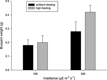
In analogy to the combined study on light and water movement, the interactive effects of light and food supply on coral growth (measured as the increase in buoyant weight) were studied using Pocillopora damicornis as a model species. Also here an interaction was found (Osinga et al., Reference Osinga, Schutter, Griffioen, Wijffels, Verreth, Shafir, Henard, Taruffi, Lavorano and Gili2011; Figure 10), high feeding only promoted growth under the highest irradiance level applied. This shows that adding food can be more than just giving the corals an additional source of energy: feeding is likely to have an effect on the efficiency of stony corals to utilize light for growth. It is therefore recommended to optimize feeding concurrent with the optimization of light when designing a culture regime for maximal volumetric productivity of coral materials.
Interactive effect of light and food concentration on the growth of Pocillopora damicornis. Reproduced from Osinga et al. (2011).

Morphology
CHARACTERIZATION OF PATTERN FORMATION IN BRANCHING CORALS
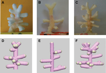
Analyses of the branching and sprouting patterns of growing colonies of S. pistillata indicate how these processes lead to an overall morphology of this species (Figure 11). Analyses revealed plastic morphometric characters at branch level, and predetermined morphometric traits at colony level. Therefore, under the experimental manipulations of this study, phenotypic plasticity in S. pistillata appears to be related to branch level of organization, whereas colony traits are controlled by predetermined genetic architectural rules (Shaish et al., Reference Shaish, Abelson and Rinkevich2006). A second study confirmed that colony astogeny in S. pistillata is a regulated process expressed through genetically programmed events and not directly related to simple energy trade-off principles or to environmental conditions (Shaish et al., Reference Shaish, Abelson and Rinkevich2007).
Representative photographs illustrating the typical colonial architectures developed from isolated branches of six Stylophora pistillata genotypes. Reproduced from Shaish et al. (2007).

A study was conducted to find the importance of branch tips (polarity) to coral colony astogeny and to address the possible existence of apical dominance, a well-documented phenomenon in the plant world, in shaping coral architectures (Shaish & Rinkevich, Reference Shaish and Rinkevich2009). Branches of S. pistillata were positioned in three morphometric settings: vertically growing branches (control); horizontally growing branches with an intact growth tip; and horizontally growing branches with the growth tips cut off. The branches were allowed to grow for one year and were used in subsequent morphometric analyses (of 23 morphological parameters such as branch length, etc) to assess the importance of branch polarity and apical dominance as dictators of colony astogeny. Changing branch orientation affected the way morphology developed, the complexity of the branching system (represented by numbers of branch generations), and the development of new branches originating from branch tip or along the branch. No evidence for apical dominance was found.
MODELLING CORAL GROWTH
Literature on modelling coral morphology was reviewed in order to define the best approach to transfer this biological process into mathematical data. Until now, the modelling approach described by Merks et al. (Reference Merks, Hoekstra, Kaandorp and Sloot2004) yielded the closest morphological resemblance between modelled corals and real corals. Their modelling approach was based upon the availability of a growth-limiting nutrient and the influence of water flow on the distribution of this nutrient among the coral branches. The limitation of the model by Merks et al. (Reference Merks, Hoekstra, Kaandorp and Sloot2004) is that it can only be applied to branching corals, and that it does not take into account genetic drivers of astogeny. Within CORALZOO, a new mathematical model was developed to describe morphogenesis in S. pistillata. The main concept of the new model is that concentrations of signalling molecules govern the occurrence of morphogenetic events such as sprouting and branching. These molecules can either be nutrients available in the water surrounding the coral, morphogenetic agents produced by the coral itself, or photosynthetically derived products in the coral. Growth was assumed only to take place at the growth tips and the growth rate per growth tip was assumed to be constant. The experimental studies on pattern formation described above were used to validate the model. The model outcomes were compared both qualitatively and quantitatively to different genotypes. Overall, the model performed very well in these comparisons.
In silico simulation of coral growth
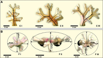
A computer simulation application was made that visualizes the modelled growth of S. pistillata. The visualization enables a direct comparison of the modelled growth and the experimental data described above (Figure 12). The simulation platform can also generate short animations showing the morphogenetic development of a growing colony of S. pistillata. The simulation platform can be used for educational purposes in public aquaria.
Comparison of simulations and experiments for three genotypes of Stylophora pistillata. Picture by Michael Kuecken.

CORAL HEALTH
Health bioassay
Stress imposed on coral colonies in captivity and in the wild, represents a wide array of causes and impacts. Therefore, it is difficult to quantitatively evaluate stress imposed on a coral colony, and to define the parameters to quantify it. In aquaria, stress is usually ‘monitored’ by visual observations of the outer appearance of corals. In CORALZOO, it was aimed to develop a more quantitative, generic health assay. Many organisms produce heat shock proteins (chaperone proteins) as a response to multiple stressors. Hence, expression patterns of the gene encoding for heat shock proteins may be used to quantify stress. We evaluated the use of HSP70 transcript analysis as a ubiquitous bioassay tool for coral health (a quantitative polymerase chain reaction assay), by applying a series of stressors to the model species S. pistillata (of which the HSP70 gene has been cloned and sequenced) and subsequent analysis of the HSP70 gene expression pattern. Monitoring the expression of the HSP70 gene turned out not to be viable as a generic tool for health monitoring in S. pistillata. The response of the corals varied between genotype and was not consistent over time. As an alternative, an existing generic bioassay for detecting DNA damage (the COMET assay) was tested, again on the model species S. pistillata. It was shown that this assay can be effectively used to detect signs of stress in aquarium corals caused by genotoxic agents such as UV-radiation.
Coral diseases
Phenomena that cause tissue regression, bleaching and deterioration of tissue are usually referred to as coral diseases. Since it is in most cases unknown whether the disease-like phenomena are caused by pathogenic agents, it is suggested to refer to these disease-like phenomena as syndromes rather than as diseases. In CORALZOO, work on coral syndromes comprised four successive steps:
1. Description of the problem—identification of the most frequently occurring syndromes among aquarium corals;
2. Identification of potential pathogens associated with those syndromes;
3. Development of diagnostic tools for those pathogens; and
4. Development of treatments for coral affected by pathogen-induced syndromes.
IDENTIFICATION OF THE MOST FREQUENTLY OCCURRING DISEASES AMONG AQUARIUM CORALS
A large body of information on captive coral syndromes was acquired using a dual-based strategy. One approach has been based on the retrieval of new information from the curators of the aquaria involved in the project. In this regard, CORALZOO have provided the unique opportunity to document, in an unprecedented way, the real entity of the coral syndrome problem in public European aquaria. This information has been collected by distributing to all contractors an ad hoc interview, which contained specifically-designed questions about past records of coral syndromes in their aquaria, affected species and environmental parameters of the tanks hosting the corals. The second approach has been devoted to reviewing the existing information about syndromes of stony corals held in captivity in private aquaria. As most of such literature is represented by non-refereed, grey literature, our search has been conducted on the web, by screening websites, hobbyists' forums and magazines. The screening has been conducted on selected sites, over ~469,000 hits on the Google web search engine, retrieved after typing the words ‘coral’, ‘disease’ and ‘aquarium’. Most of this information has been obtained from public popular forums, where home aquarists continuously report the appearance of diseases and/or abnormalities occurring in their private displays. The results from these two approaches were combined with those obtained from a ‘monitoring programme’, which was launched from the beginning of the CORALZOO project and has lasted for two consecutive years (i.e. June 2005–May 2007). All partners involved in this monitoring programme recorded the occurrence of coral syndromes within their displays. For each record, they prepared relevant samples for subsequent analysis and (when needed) disease classification.
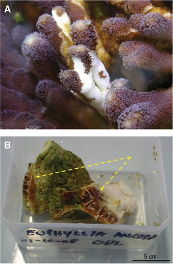
The survey revealed that two coral syndrome types occur most frequently in aquaria: white syndromes (Figure 13A) comprised nearly 70% of the records, brown jelly syndrome (BJS; Figure 13B) being second in occurrence frequency. ‘White syndromes’ are here defined as ‘a disease-like syndrome characterized by the presence of severe tissue loss from the coral’, while ‘brown jelly syndrome’ is characterized by tissue death and massive mucus production. The ‘white syndrome’ records displayed extremely variable rates of lesion progression, as well as patterns of tissue loss; this definition also includes what is commonly referred to, by most professional and home aquarists, as rapid tissue necrosis (RTN) or shut down reaction (SDR). The complete result of the survey will be published elsewhere.
(A) (left): white syndrome affected on Stylophora sp; (B) (right): brown jelly syndrome affected colony of Euphyllia ancora. Photographs by Dominique Barthelemy (A) and Elsa Santos (B).

AGENTS CAUSING DISEASES AND SYNDROMES
Many of the coral samples showing white syndromes had large proportions of pathogenic Vibrio species (Vibrio harveyi, and few other Vibrio strains) associated with their tissue. The pathogenic role of V. harveyi in aquarium corals was convincingly demonstrated (Luna et al., Reference Luna, Biavasco and Danovaro2007, Reference Luna, Bongiorni, Gili, Biavasco and Danovaro2010); it is the main causative agent of RTN and most likely is involved in other white syndromes as well. Fungi are the main suspects with regard to causation of BJS.
DIAGNOSIS AND TREATMENT OF WHITE SYNDROMES
A diagnostic kit was completed that allows both diagnosis of RTN in corals and enumeration of the pathogens in the aquarium water (preventive monitoring). The kit was distributed for testing among the CORALZOO aquarium partners.
A conceptual framework (Figure 14) describes the recommended procedure on how to handle species when a coral is suspected to suffer from white syndromes. A best practice for treatment of white syndromes with antibiotics was elaborated. Affected corals should be treated with 5–10 mg l−1 of tetracycline in secluded quarantine tanks.
Conceptual framework for antibiotic treatment of ‘white syndrome’ affected aquarium stony corals.

Pests: metazoan parasites
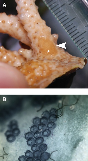
Different flatworms (phylum Platyhelminthes) are known to live together with Scleractinia. Some types of flatworms, for example species of the Convolutidae, are commensal towards Scleractinia and can become a nuisance when occurring in large numbers. A parasitic polyclad worm (Aplidioplana sp.) is known to eat tissue specifically from acroporid corals (Nosratpour, Reference Nosratpour, Leewis and Janse2008). Studies were done to evaluate the use of ivomectin to treat infestations of corals by a parasitic flatworm, Waminoa sp. (Figure 15). The results from these experiments allowed identification of the best procedure (2 mg ivomectin/l in a 5 hour incubation) to efficaciously treat these common infestations in acroporid corals (see also Leewis et al., Reference Leewis, Wijgerde, Laterveer and Osinga2009 for the detailed protocol).
(A) Polyclad flatworm on an Acropora sp. The camouflage is magnificent and the tissue of the coral is clearly removed by the flatworm; (B) eggs of the Acropora-associated flatworms. Photographs by Max Janse.

TRANSPORT AND ACCLIMATION OF CORALS
One of the general suggestions that we can make after four years of CORALZOO, is that a breeding programme may be most efficient when each participating breeding centre is concentrating on one or few target species. This emphasizes the need for good procedures to transport corals from one institution to the other, including best practises for the acclimation of transported corals to their new environment. Although literature on these topics is scarce (Petersen et al., Reference Petersen, Laterveer, Van Bergen and Kuenen2004, Reference Petersen, Hatta, Laterveer and Van Bergen2005; Delbeek, Reference Delbeek, Leewis and Janse2008), a substantial amount of undocumented information on transportation and acclimation exists among aquarists. Therefore, a database was set up (Leewis et al., Reference Leewis, Wijgerde, Laterveer and Osinga2009) that contains data (a short description of the transportation technique used, species concerned, number of individuals transported and percentage survival) on a large number of registered transportations carried out during (and prior to) the project. The data revealed that survival was in most cases nearly 100%, particularly when wet (submerged) transportation was applied. Since submerged transportation is expensive (water being the main constituent of the transported package), CORALZOO particularly focused on strategies to reduce transportation costs.
Comparative studies on coral transportation methods
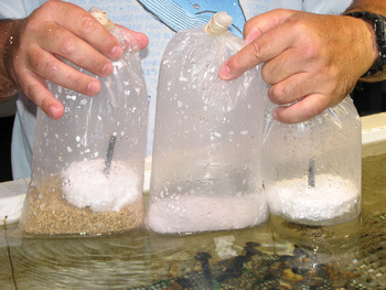
A classical wet transportation technique for coral colonies (corals submerged in seawater by putting them upside down on a floating piece of Styrofoam™) was compared to a dry transportation method using wet tissue paper and a dry transportation method using vermiculite (Figure 16). Three coral species (Acropora sp., S. pistillata and P. damicornis) were subjected to simulated transport conditions. Transportation in wet tissue paper was found to be a useful alternative for wet transportation for S. pistillata and P. damicornis. It reduces the weight of the shipment five- to tenfold without a significant change in survival rates. The use of vermiculite is not recommended: survival was lower for all species, while shipment weight is higher compared to the wet tissue method.
Three transportation methods, vermiculite (bag at left), tissue paper (bag in the middle) and wet (bag at right).

In addition, a semi-dry transportation method using bubble plastic and a minimal amount of seawater was tested for the transport of large colonies. The method was suitable for the species tested, Echinopora lamellosa (Esper 1791) and Acropora sp.
A simulated transport experiment was done that focused on the effects of light and temperature on transportation success. There seemed to be a correlation between survival rate and temperature decline during transportation. Tests at a constant ambient temperature of 25°C tended to confirm this hypothesis. Addition of light to the animals during transport had no effect on survival and is therefore not considered useful. Based on these and other CORALZOO data, a calculating tool for designing wet transports was elaborated.
Acclimation
Light upshocks are generally considered as one of the largest potential risks with respect to acclimation of corals. A tool for light acclimation was developed that uses sheets to dim the aquarium lights. In this way, a gradual increase in photon flux density can be achieved.
The role of water movement in acclimation is often neglected. Therefore, the effect of rapid changes in both light and water movement regimes on coral survival was tested on G. fascicularis. Under high light, a downshock in flow velocity was found to be potentially detrimental to larger sized corals (partial necrosis). Changes in hydrodynamic regimes should be seriously taken into consideration when repositioning corals, both among tanks and within tanks.
THE CORALZOO BOOK OF PROTOCOLS
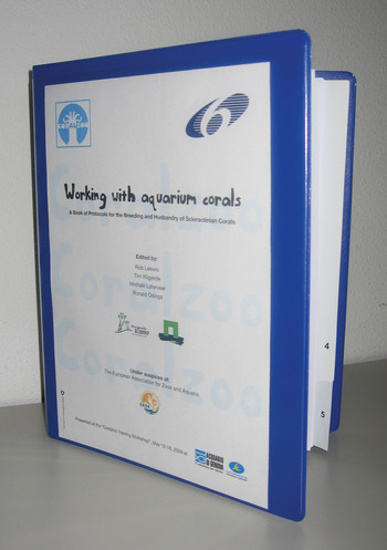
The main deliverable of CORALZOO was a Book of Protocols (within this paper referred to as Leewis et al., Reference Leewis, Wijgerde, Laterveer and Osinga2009), which provides guidelines and tools to establish a breeding programme for stony corals. The CORALZOO Book of Protocols (Figure 17) contains 70 user-protocols accompanied by introducing texts and general guidelines on the major categories of topics (the Book is publicly disseminated through the CORALZOO website www.coralzoo.org)
The CORALZOO Book of Protocols. We have chosen a loose leaflet system, so that protocols can be added and modified. The book contains over 70 user-protocols distributed over four main sections: Determination of Coral Size; Broodstock Production; Culture & Husbandry; and Transport & Acclimation.

CONCLUDING REMARKS
CORALZOO successfully achieved its major aims:
1. A toolkit and information package was delivered that provides a blueprint for a breeding programme for stony corals in EAZA-associated zoos and aquaria.
2. The developed tools and background information have been described in a small business-friendly format in the CORALZOO Book of Protocols, which is disseminated through the CORALZOO website (www.coralzoo.org).
A series of new insights in coral biology were generated through the project, which will be useful for both aquarists and coral scientists:
1. When growth of corals is concerned, interactions between factors controlling growth are important. Both feeding and water movement were shown to enhance the capability of corals to channel photosynthetic activities by the symbiotic zooxanthellae into coral growth. Optimization of growth should therefore preferably be based upon multifactorial experiments that include at least light, feeding and water movement as variables, whereby other potentially limiting factors such as calcium and carbonate should continuously be provided in excess.
2. Fed corals are happy corals: experiments on different coral species with different feeds showed that in most cases, feeding promoted growth. Furthermore, it was unambiguously demonstrated that corals can digest phytoplankton and benefit from phytoplankton feeding.
3. Genotype matters: the influence of genotypic variability on growth is often larger than effects of external factors. Therefore, comparative studies on generic biological principles are better studied using a single genotype, whereas optimization studies should always take genotypic variability into account.
4. In aquaria, the frequency of occurrence of diseases is completely different from the patterns observed in the sea. White syndromes comprise 70% of the disease record in aquaria. The marine pathogen V. harveyi was identified as the main agent causing white syndromes in aquaria.
Not all studies that were done within CORALZOO provided conclusive results. The main recommendations for further studies are listed below:
1. Control on sexual reproduction in captivity was not achieved. The alternative approach (rearing naturally generated spat under controlled conditions) is useful and probably even preferred with respect to maintaining a high genetic diversity. Nevertheless, for completely captive breeding of endangered species, more studies on the mechanisms that control sexual reproduction are needed.
2. The effects of variations in the length of day on coral growth need further investigation.
3. The role of trace elements in coral growth could not be studied within CORALZOO due to technical limitations, but is an important gap in the current knowledge on coral culture.
Another important consideration after four years of CORALZOO is that the coupling of the research infrastructure of the scientific partners to the facilities and technological knowledge present in public aquaria proved to be an efficient strategy to generate new knowledge. It enabled the execution of novel types of coral studies, such as long term growth experiments under controlled conditions and the demonstration of causal relationships between pathogens and coral diseases. In conclusion, CORALZOO provides a clear example of:
— the potency of zoos and public aquaria as partners in science;
— the added value of collaboration between scientists and aquarists; and
— the stimulating role that funding agencies such as the European Commission can play to develop aquarium science and technology.
As such, CORALZOO can serve as a role model for future collaborations between scientists and aquarists.
ACKNOWLEDGEMENT
The project CORALZOO was funded by the FP6 programme of the European Community, grant number 012547.