Introduction
Since its first description by Guild Reference Guild1 in 1941, a number of papers from Europe and the United States have reported on glomus tumours. However, there have been few reports of the condition in non-Caucasians. Only 88 cases (46 tympanicum tumours, 31 jugulare tumours and 11 unknowns) have been reported in Japan.
Glomus tumours sometimes secrete adrenalin and noradrenalin. The tympanicum tumour accounts for approximately half of such hormone-secreting tumours. Functioning tumours account for 1 to 3 per cent and are known to prompt peri-operative episodes of abnormal hypertension.Reference Schwaber, Glasscock, Nissen, Jackson and Smith2 Since paroxysmal hypersecretion of catecholamine occasionally occurs during tumour extraction, careful examination and management prior to tumour extraction are important. Endovascular embolisation is frequently performed pre-operatively in order to decrease catecholamine secretion and to reduce bleeding during the operation.
We describe the case of a catecholamine-producing tympanicum glomus tumour in a 73-year-old woman. Pre-operative endovascular embolisation and potassium titanyl phosphate (KTP) laser were used to remove the tumour, with minimal bleeding. We also discuss the treatment of catecholamine-producing glomus tumours.
Case report
A 73-year-old woman presented to the otorhinolaryngology department of Shiga University of Medical Science complaining of hearing impairment and left ear pain. The patient had a history of hypertension, angina pectoris and cerebral infarction.
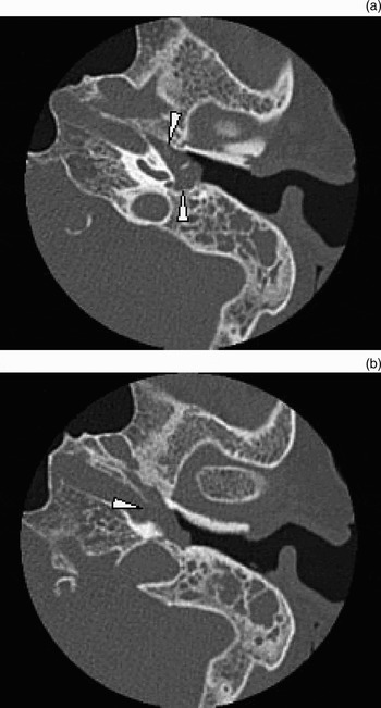
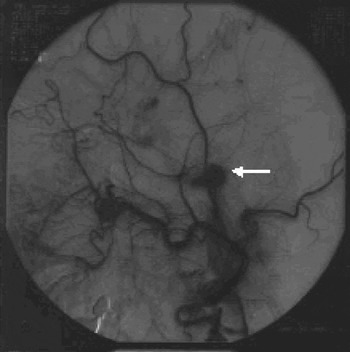
Physical examination revealed a red, pulsating swelling in the left tympanic membrane (Figure 1). Audiography showed a combined hearing loss on the left side. Computed tomography demonstrated a soft tissue mass filling the entire middle-ear cavity and a partial osteolytic lesion in the internal carotid artery. Bone was preserved between the hypotympanum and the jugular bulb (Figure 2). Angiographic examination revealed a densely contrasting tumour, which had feeding vessels from the ascending pharyngeal artery, a branch of the external carotid artery (Figure 3).
Operating microscope photograph showing a red, pulsating swelling in the left tympanic membrane.

Axial computed tomography showing (a) a soft tissue opacity (arrowheads) filling the entire middle-ear cavity and (b) a partial osteolytic lesion (arrowhead) in the internal carotid artery.

Angiographic image showing densely contrasting tumour with a feeding vessel from the ascending pharyngeal artery, a branch of the external carotid artery (arrow).

Laboratory data showed the following serum concentrations (shown with their normal ranges in parentheses): adrenaline, 64 (≤170 pg/ml); noradrenalin, 1850 (150–570 pg/ml); and dopamine, 40 (≤30 pg/ml); the urine VMA concentration was 8.6 (1.4–4.9 mg/day). The patient's blood pressure was 130/75 mmHg and her heart rate was 55 beats/minute.
According to Fisch's classification,Reference Fisch3 we diagnosed a class A glomus tympanicum tumour, which in this case was catecholamine-producing.
Clinical course
Endovascular embolisation was completed a day before the main surgical procedure, by placing a coil into the ascending pharyngeal artery (the tumour feeding vessel) and injecting Spongel (Astellas Pharma Inc., Tokyo, Japan). Antihypertensive drugs were on hand in case of an abnormal hypertensive episode during embolisation; however, there was no elevation of blood pressure. The serum noradrenalin concentration had normalised by the following day, when the main surgical procedure was planned.
At operation, the red, pulsating tumour was found to have pervaded the entire middle-ear cavity and continued into the eustachian tube. The internal carotid artery was partially exposed. The tumour was removed entirely, in piecemeal fashion, using a KTP laser. The incus and malleus head were removed with the tumour, and the ossicular chain was reconstructed with a piece of auricular cartilage positioned between the stapes and the tympanic membrane. The amount of bleeding during the operation was 30 ml. Abnormal hypertension did not develop during the operation.
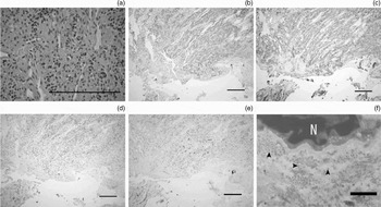
Histopathological examination of the resected tumour showed a proliferation of cells bearing circular nuclei and rich cystid, surrounded by interstitium with abundant capillary vessels. Immunohistological examination demonstrated positive staining of cytoplasm for neuron specific enolase (NSE) and synaptophysin, and of supporting cells for S-100. Chromogranin was partially positive (Figure 4a to e). Ultrastructurally, many electron-dense, neurosecretory granules were observed around the tumour cell nuclei (Figure 4f). The histological diagnosis was a catecholamine-producing glomus tumour.
(a) Photomicrograph showing proliferation of cells bearing circular nuclei and rich cystid, surrounded by interstitium with abundant capillary vessels (H&E). Photomicrographs taken after immunological staining demonstrate positive staining of cytoplasm for (b) neuron specific enolase (NSE), (c) synaptophysin and (d) chromogranin, and (e) positive reaction of supporting cells for S-100. (f) Transmission electron micrograph showing many electron-dense, neurosecretory granules (arrowheads) around a tumour cell nucleus. N = nucleus. (a) to (e) bars = 200 µm; (f) bar = 1 µm

The post-operative course was uneventful. The concentrations of serum noradrenalin and urine VMA normalised after the operation. Audiography revealed improvement of the left-sided hearing loss. No tumour recurrence was noted over two years' follow up.
Discussion
Glomus tumours are benign lesions originating from the adventitia of the jugular bulb. They proliferate circumferentially and may cause bone destruction. The histopathology of glomus tumours resembles that of the carotid body, and they occasionally secrete catecholamines. Since the first description by Parkinson,Reference Parkinson4 23 cases of catecholamine-secreting glomus tumour have been reported, including two cases in Japan. The treatment of glomus tumours should be determined by their age, size and histological type. According to Fisch's classification, complete surgical resection should be primarily considered for tumours confined to the tympanic cavity, and total resection can be achieved by a procedure based on tympanoplasty.
Recently, pre-operative treatment with concomitant embolisation has been used with increasing frequency, not only for functional tumours but also for glomus tumours in general. Pre-operative embolisation may prevent a hypertensive crisis caused by catecholamine release during the main procedure. However, the development of a hypertensive crisis has been also reported during such embolisation procedures, and antihypertensive drugs should thus be prepared not only for the main surgical procedure but also for the endovascular embolisation procedure. Embolisation is also effective for reducing tumour bleeding during surgery and for decreasing tumour size. In our case, pre-operative embolisation was useful not only for normalising the serum catecholamine level, but also for minimising peri-operative bleeding.
In order to ensure successful surgical resection of a glomus tympanicum tumour, it is important to establish an adequate operative field and to assess the relationship between the tumour and the surrounding tissues. We used a KTP laser to reduce the tumour size and then to resect it. Tumour separation and extraction procedures were simplified by reducing the tumour size, without significant bleeding. One of the features of the KTP laser is a higher absorbing capacity for haemoglobin in tissues with abundant vessels, resulting in more effective haemostasis, coagulation and evaporation. The tissue penetration of the KTP laser is only 1 to 2 mm, which is useful for avoiding associated injuries in the tympanic cavity. The use of a hyperbolic electrosurgical knife and a neodymium-yttrium-aluminium-garnet laser have also been reported for resection of glomus tumours. However, the hyperbolic electrosurgical knife blade must be wiped after each ablation, and the neodymium-yttrium-aluminium-garnet laser has deeper tissue penetration, compared with the KTP laser.
• Glomus tumours sometimes secrete adrenaline and noradrenalin; tympanicum tumours account for approximately half of such hormone-secreting tumours
• In the reported case, the serum noradrenalin concentration normalised after embolisation of the tumour feeding vessel from the ascending pharyngeal artery
• Potassium titanyl phosphate laser was also useful for complete resection of the tumour
Surgical resection is the primary treatment choice for small glomus tumours with limited localisation, such as Fisch's classification classes A and B. However, for larger intracranial tumours, such as Fisch's classification classes C and D, radiation therapy has been reported to be effective.Reference Springate and Weichelbaum5 However, for the small number of cases of catecholamine-producing glomus tumours reported so far, there has been no specific therapeutic policy. A different therapeutic approach is required for functional tumours, compared with non-functional tumours, in order to control catecholamine secretion as well as to treat the tumour itself. It has been reported that radiation therapy alone does not reduce catecholamine production, and that the chief cells, which produce adrenalin, survive even after irradiation.Reference Schwaber, Glasscock, Nissen, Jackson and Smith2 For this reason, surgical resection, even if partial, is preferred for large catecholamine-producing glomus tumours, along with concomitant use of vascular embolisation to reduce catecholamine secretion. However, strict control of blood pressure is required when handling giant catecholamine-producing tumours.
Conclusion
We report a case of catecholamine-producing glomus tympanicum tumour. The serum noradrenalin concentration normalised following embolisation of the tumour feeding vessel from the ascending pharyngeal artery. Pre-operative embolisation is useful, not only for preventing a hypertensive crisis but also for reducing peri-operative bleeding. The KTP laser is also useful for complete resection of the tumour.