Transcatheter interventional therapy, which necessarily exposes children to ionising radiation during the procedure, has gained increasing popularity in management of CHD. Compared to adults, children are potentially at a greater risk of radiation-induced stochastic injuries resulting from the greater radiation sensitivity of their tissues, along with the longer lifespan ahead of them to accumulate further exposures or to develop potential neoplasms.Reference Justino1–Reference Walsh, Noga and Rutledge3 As the stochastic effects of radiation are unpredictable and threshold independent, there is no definite safe radiation dose.Reference Ait-Ali, Andreassi, Foffa, Spadoni, Vano and Picano2, Reference Kamiya, Ozasa and Akiba4, Reference Song, Liu and Zhang5
Hence, the principle of as low as reasonably achievable has been strongly recommended by the Society for Pediatric Radiology and the International Commission on Radiologic Protection to minimise radiation exposure as low as reasonably achievable while maintaining acceptable imaging quality for diagnostic and therapeutic purpose.Reference Justino1, Reference Strauss and Kaste6–Reference Khong, Ringertz and Donoghue8 Radiation dose during interventional cardiac procedure greatly depends on the operator’s knowledge, training, and tactics of radiation protection.Reference Justino1, Reference Strauss and Kaste6, Reference Hernanz-Schulman, Goske, Bercha and Strauss9–Reference Aldoss, Patel, Harris and Divekar13
The main goal of this study was to investigate how much radiation reduction could be achieved during the most common paediatric interventional procedures by using a modified radiation safety protocol with efforts to increase the awareness and refine the tactics of the operators.
Materials and methods
The study period included the 2 years of initial default protocol from January, 2014 to December, 2015 (Standard group) and another 2 years from January, 2016 to December, 2017 (Low-dose group) after implementation of the modified radiation safety protocol. Radiation dose data were extracted from database of the catheterisation in our institution, involving the following five most common interventional procedures: atrial septal defect closure; patent ductus arteriosus closure; perimembranous ventricular septal defect closure; pulmonary valvuloplasty; and supraventricular tachycardia ablation, including atrioventricular reentrant tachycardia and atrioventricular nodal reentrant tachycardia. The ablation procedures were performed without application of three-dimensional mapping. Patients who underwent two or more procedures simultaneously were excluded.
All the procedures were performed using a flat-panel biplane angiography system (AXIOM TrtisdBA DSA, Siemens, Germany). The study was approved by Ethics Committee of our institution (2017-162) and written consents were obtained from guardians of the patients.
Radiation metrics
Our equipment is capable of reporting radiation dose by the end of procedure. Total fluoroscopy time (minutes) and total air Kerma (mGy) were recorded for all patients. However, dose area product (μGy.m2) was only recorded prospectively and thus available for the Low-dose group.
Radiation reduction approaches
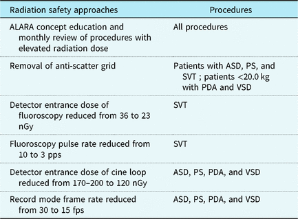
The modified radiation safety protocol was introduced to all the staffs involved in the catheterisation procedure (Table 1). All procedures included in this study were carried out by two paediatric interventional cardiologists with over 10 years of experience. When performing procedure with the modified protocol, once operators felt image quality was unacceptable, they could easily switch back to the initial default protocol by one button.
Details of the modified radiation safety protocol.

ALARA = As Low As Reasonably Achievable; ASD = atrial septal defect; PDA = patent ductus arteriosus; PS = pulmonary valvular stenosis; SVT=supraventricular tachycardia; VSD = ventricular septal defect.
Statistical analysis
Patients’ characteristics and radiation dosages were presented as median with range for continuous variables. Comparisons between two groups were conducted using Independent-sample t-test or Mann–Whitney U-test for normally or not normally distributed continuous variables, respectively. Kruskal–Wallis test was applied for multiple-group comparisons. Statistical analysis was performed by SPSS Version 23. A p value of <0.05 was considered statistically significant.
Results
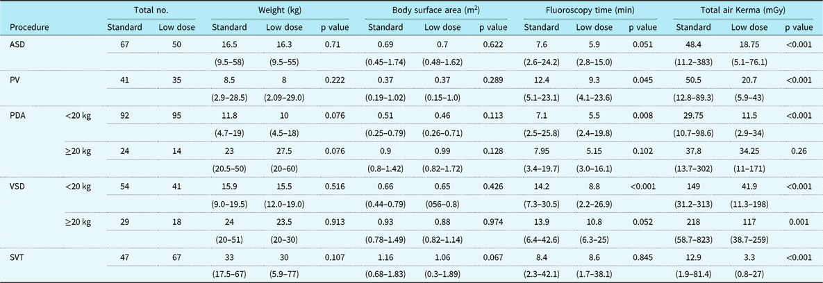
A total of 674 cases were enrolled, which included 117, 225, 142, 76, and 114 cases of transcatheter closure of atrial septal defect, patent ductus arteriosus, ventricular septal defect, pulmonary valvuloplasty, and supraventricular tachycardia ablation, respectively. Patients’ characteristics and radiation doses of both groups from the procedural types are listed in Table 2. There was no difference in patients’ weight and body surface area between Standard group and Low-dose group in any procedural type.
Patient details and radiation dose of both groups.

ASD = atrial septal defect; PDA = patent ductus arteriosus; PV = pulmonary valvuloplasty; SVT=supraventricular tachycardia; VSD = ventricular septal defect.
Both operators were able to perform the procedures using the modified radiation safety protocol. Switching to the initial default protocol was not necessary in any case.
Comparison of radiation dosage between two groups
Median fluoroscopy time was 12.4, 7.1, and 14.2 minutes for pulmonary valvuloplasty, patent ductus arteriosus <20 kg, and ventricular septal defect <20 kg in Standard group, respectively, which significantly decreased to 9.3, 5.5, and 8.8 minutes in Low-dose group (p < 0.05). There was no statistical difference in fluoroscopy time between two groups in procedural type of atrial septal defect, patent ductus arteriosus ≧20 kg, ventricular septal defect ≧20 kg, and supraventricular tachycardia.
Median air Kerma was 48.4, 50.5, 29.75, 149, 218, and 12.9 mGy for atrial septal defect, pulmonary valvuloplasty, patent ductus arteriosus <20 kg, ventricular septal defect <20 kg, ventricular septal defect ≧20 kg, and supraventricular tachycardia in Standard group, respectively, which significantly decreased to 18.75, 20.7, 11.5, 41.9, 117, and 3.3 mGy in Low-dose group (p < 0.05). This represents a reduction in dose to each patient between 46 and 74%. There was no difference in air Kerma between two groups in the procedure of patent ductus arteriosus ≧20 kg (37.8 versus 34.25 mGy) (p = 0.26) (Table 2).
Comparison of radiation dosage among different procedures
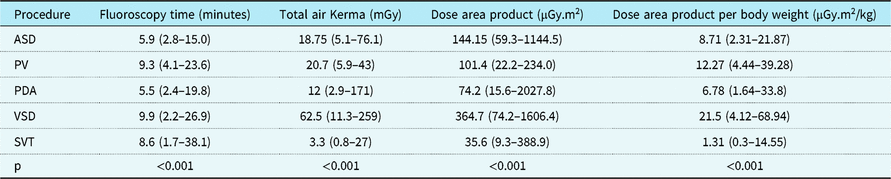
The radiation dose levels of all five procedures in Low-dose group, including fluoroscopy time, air Kerma, dose area product, and dose area product per body weight (dose area product normalised to body weight in kilogram), were compared (Table 3).
Radiation exposure measures by procedure type in Low-dose group.

ASD=atrial septal defect; PDA=patent ductus arteriosus; PV=pulmonary valvuloplasty; SVT=supraventricular tachycardia; VSD=ventricular septal defect.
The fluoroscopy time of the procedure of patent ductus arteriosus closure was significantly shorter than that of the ventricular septal defect closure, pulmonary valvuloplasty, and supraventricular tachycardia ablation (p < 0.001), without significant difference with that of atrial septal defect closure.
The air Kerma was highest in the procedure of ventricular septal defect closure and lowest in supraventricular tachycardia ablation (62.5 versus 3.3 mGy); so was the dose area product value (364.7 versus 35.6 μGy.m2).
The median dose area product per body weight value was 21.5, 12.27, 8.71, 6.78, and 1.31 μGy.m2/kg for ventricular septal defect closure, pulmonary valvuloplasty, atrial septal defect closure, patent ductus arteriosus closure, and supraventricular tachycardia ablation cohort, respectively, from the maximum to the minimum. The dose area product per body weight was highest in the procedure of ventricular septal defect closure, which was not significantly different with pulmonary valvuloplasty but much higher than that of any other three procedures (p < 0.001). The dose area product per body weight in supraventricular tachycardia ablation was significantly lower than that of either of the other four procedures (p < 0.001).
Discussion
In the present study, we adopted a modified radiation safety protocol in paediatric interventional procedures and compared the radiation dose level with previous data. Despite the same equipment and operators, a significant reduction in radiation exposure was achieved for all types of the procedures after the implementation of our modified protocol. The result underscores that increasing awareness and training of techniques of the operators and relevant staffs are the key factors to improve radiation protection. Dose reduction is a team effort and every member managing the angiography equipment plays an important role.
All the cases in Low-dose group were accomplished by using the modified radiation safety protocol with a tendency of shorter fluoroscopy time. It indicated that the low-dose strategy did not increase the difficulty of performing the procedures; meanwhile, it might reduce fluoroscopy time by avoidance of unnecessary radiation exposure during the procedure attributed to as low as reasonably achievable concept education. Apparently, the influence of other factors on fluoroscopy time could not be ruled out in the present study, including the complexity of procedures and the skill of operators.
Compared with the initial default protocol, our modified protocol resulted in the reduction of air Kerma values by 46–74% with the exception of patent ductus arteriosus ≧20 kg cohort. Removal of anti-scatter grid for patients lower than 20 kg, as well as patients of any weight for atrial septal defect closure, pulmonary valvuloplasty, and supraventricular tachycardia ablation, was one major component of our modified protocol. It was shown by previous paediatric studies that the anti-scatter grid increased radiation dose with limited improvement in image quality in patients under 20 kg.Reference Onnasch, Schemm and Kramer14–Reference Osei, Hayman, Sutton and Pass18 From an operator standpoint, given the relatively lower image quality required for atrial septal defect closure, pulmonary valvuloplasty, and supraventricular tachycardia ablation, removal of grid for patient over 20 kg created the reduced but acceptable image quality for the procedures in the current study. The strategies of decreasing the detector entrance doses, pulse rate for fluoroscopy, and frame rate for cine-digital acquisition also contributed to a significant reduction in radiation dose.Reference McFadden, Hughes, Mooney and Winder16, Reference Lamers, Moran, Torgeson and Hokanson19–Reference Gossett, Sammet, Agrawal, Rychlik and Wax24 There was no significant reduction in radiation dose for the patients of patent ductus arteriosus ≧20 kg due to less times of image acquisition required during procedure in comparison to that of ventricular septal defect ≧20 kg.
Of the three metrics recorded, fluoroscopy time is not a good measure of dose received by the patient, as the dose with any given fluoroscopy time can be greatly variable, depending on the size of the patient, and the equipment settings. Total air Kerma is a good predictor of deterministic effects by estimating the radiation exposure at the interventional reference point.Reference Cevallos, Armstrong and Glatz25, Reference Kwon, Little and Miller26 Dose area product is the best indicator of the risk of stochastic effects, representing the products of radiation dose and exposed area.Reference Cevallos, Armstrong and Glatz25, Reference Stecker, Balter and Towbin27, Reference Ghelani, Glatz and David28 The dose area product per body weight is regarded as a necessary standardised adjustment to facilitate direct comparison of radiation dose level among different procedures.Reference Cevallos, Armstrong and Glatz25, Reference Onnasch, Schroder, Fischer and Kramer29 Among the five procedures in Low-dose group, the dose area product per body weight value for ventricular septal defect closure (21.5 μGy.m2/kg) was highest along with longest total fluoroscopy time, due to complexity of the procedure. In contrast, the dose area product per body weight dose for supraventricular tachycardia ablation is lowest (1.31 μGy.m2/kg) in spite of similar total fluoroscopy time. The much lower dose level was attributed to the radiation settings with least entrance detector dose and fluoroscopy rate and removal of anti-scatter grid during the procedure of supraventricular tachycardia ablation. It indicates that fluoroscopy time is not a good measure of radiation exposure to patient.
In comparison to radiation dose benchmarks established in 2017,Reference Cevallos, Armstrong and Glatz25 the median dose area product per body weight values in the present study for atrial septal defect and patent ductus arteriosus closure and pulmonary valvuloplasty are lower than those data (8.7 and 34 μGy.m2/kg, 6.8 and 37 μGy.m2/kg, 12.3, and 53 μGy.m2/kg, respectively), supporting the effectiveness of our modified protocol. Radiation dose benchmark of ventricular septal defect closure is not available, as transcatheter closure of perimembranous ventricular septal defect closure is still strictly restricted in United States of America. Therefore, our data could be regarded as a preliminary supplement to the reference for quality measurement and comparative assessment among institutions.
The present study has several limitations. Firstly, the values of dose area product were not recorded for initial baseline. Secondly, 3D navigation system has not been applied in supraventricular tachycardia ablation in our institution, which should lead to a further reduction of radiation exposure in electrophysiology interventions.Reference Bourier, Reents and Ammar-Busch30–Reference Pass, Gates, Gellis, Nappo and Ceresnak32 Finally, the equipment (AXIOM TrtisdBA) used in the present study is old generation, and new generation equipment may further result in substantial radiation dose reduction.Reference Osei, Hayman, Sutton and Pass18, Reference Lamers, Moran, Torgeson and Hokanson19, Reference Patel, Ganley, Zhu, Rome, Shah and Glatz22, Reference Bourier, Reents and Ammar-Busch30
Our study has provided a feasible radiation safety protocol with specific settings on a case-by-case basis, which is able to substantially decrease radiation exposure during paediatric interventional cardiac catheterisation. Increasing awareness and adequate training of a practical radiation dose reduction program are essential to improve radiation protection for children, no matter what equipment is used.
Author ORCIDs
Lin Wu, 0000-0001-6737-4646
Acknowledgements
None.
Financial support
This study was funded by the National Key Research and Development Program (2016YFC1000500) and Innovation Project for Clinical Science and Technology Development (SHDC12016125).
Conflicts of interest
None.
Ethical standards
The authors assert that all procedures contributing to this work comply with the Helsinki Declaration of 1975, as revised in 2008, and has been approved by the Institutional Ethics Committee of Children’s Hospital of Fudan University (2017-162).