I. INTRODUCTION
It is perhaps astonishing to have synthesized, by the standard ceramic procedure of co-firing oxides, an ~90% pure very complex oxide crystal type containing Al, Ca, Fe, Mn, Ti, and Zr, with an enormous face-centered cubic (FCC) unit cell with a = 39.269 Å units. It is a member of the pyrochlore–murataite modular polysomatic family (Urusov et al., Reference Urusov, Organova, Karimova, Yudintsev and Ewing2007) with an eightfold supercell of a fluorite-type basic sub-cell, viz. M8 (i.e. containing 8 × 8 × 8 = 512 basic sub-cells, about 5250 atoms and 40 distinct symmetry cation positions (Laverov et al., Reference Laverov, Yudintsev, Stefanovsky, Omel'yaneko and Nikonov2006, Reference Laverov, Urusov, Krivovichev, Pakhomova, Stefanovsky and Yudintsev2011a)).
The first member of this group, now dubbed M3 (nearly) isotypic with the natural mineral, murataite (Adams et al., Reference Adams, Botinelly, Sharp and Robinson1974), was discovered during research on radwaste disposal, in a mixture with many cations [PDF 00-036-0138 (ICDD, 1986)]. It was recognized as a 3 × 3 × 3 fluorite supercell (structurally very similar to the mineral murataite, M, but with a surprisingly very different elemental content) (Morgan and Ryerson, Reference Morgan and Ryerson1982; Morgan et al., Reference Morgan, Harker and Flintoff1984). The detailed crystal structure was published much later (Pakhomova et al., Reference Pakhomova, Krivovichev, Yudintsev and Stefanovsky2013).
The well-known 2 × 2 × 2 cousin is pyrochlore, Py, also frequently considered for radwaste disposal. Subsequently in a superior research effort in Russia, aimed specifically at radwaste disposal, it was discovered that there were a series of modular polytypes between Py and M, such as M5, M7, M8, in addition to M3 (Laverov et al., Reference Laverov, Yudintsev, Omel'yanenko, Nikonov and Stefanovskii1999); other packing variants are probably yet, likely soon, to be revealed. Moon tranquillityite found during the Apollo 11 mission (Gatehouse et al., Reference Gatehouse, Grey, Lovering and Wark1977), was initially amorphous, but now appears plausibly to be one of these. The regions connecting the fluorite-type sub-cells can vary in fine detail.
The new polytypes, identified by very careful electron diffraction, were first achieved by melt methods; these produced good-sized crystals but with zoning (a downside for single-crystal XRD but a plus for radwaste entombment). The polytypes are most routinely made by simple mixed oxide firings, whereupon, the X-ray powder diffraction (XRPD) method is the method of choice for simplicity and speediness in monitoring exactly how a day-to-day synthesis program may be proceeding, bearing in mind the many variables, of about six cation content ratios, the temperatures and times of syntheses and so forth.
Very large unit cells turn out to be much more difficult to analyze with advanced powder methods, such as a Rietveld method; analysis on the largest oxide unit cell attempted so far (only three cations), was published as La3Ti5Al15O37 (Morris et al., Reference Morris, Owen, Stalick and Cheetham1994). It was later corrected to the originally proposed LaTi2Al9O19 (Morgan, Reference Morgan1984) with the aid of the subsequent single-crystal determination of isomorphous SrTi3Al8O19 (Strunk and Müller-Buschbaum, Reference Strunk and Müller-Buschbaum1993) and checked (as always required today) with bond–valence sums (Kasunič et al., Reference Kasunič, Meden, Škapin, Suvorov and Golobič2011). The unit cell considered here, with six or more cations in a mixed oxide, is much larger by volume, in fact about three times larger, than an inorganic one previously referred to as “a giant unit cell” (Aliev et al., Reference Aliev, Kovrugin, Colmont, Terryn, Huve, Siidra, Krivovichev and Mentre2014).
The natural mineral, an M3 (Ercit and Hawthorne, Reference Ercit and Hawthorne1995) is known to contain many defects that include mixed polytypic layers and this probably applies to the “reconstituted-by-heating” tranquillityite (Gatehouse et al., Reference Gatehouse, Grey, Lovering and Wark1977). In addition, the main diffraction peaks of the supercells, derived from the similar basic sub-cells, means, close overlaps occur for all the polytypes at all the strongest peaks (Laverov et al., Reference Laverov, Yudintsev, Stefanovskii, Omel'yanenko and Nikonov2011b). Therefore, for quick analysis, we need accurate XRPD patterns especially emphasizing the lowest angle lines where these complex polytypes are most readily distinguishable.
The first possible FCC line/peak of M8, the (111), would be expected at about 2θ = 3°, so special steps must be taken to enable searching at unusually low 2θ angles by masking the background – mimicking a small-angle X-ray scattering technique. Other modular types, with lower symmetry, could produce even lower angle lines. We present here the XRPD for M8, found to be FCC as predicted (Laverov et al., Reference Laverov, Urusov, Krivovichev, Pakhomova, Stefanovsky and Yudintsev2011a), exemplifying the ease of indexing and unit-supercell confirmation. Quote: “From the crystal chemical point of view, the murataite series are the most complex mixed oxides known to date” (Laverov et al., Reference Laverov, Urusov, Krivovichev, Pakhomova, Stefanovsky and Yudintsev2011a).
II. EXPERIMENTAL
Commercial α-Al2O3 (99.99% purity, Taimei Chemicals Co. Ltd., Saitama, Japan), TiO2 anatase (99% purity, Kojundo Chemical Laboratory Co. Ltd.), CaO (99.9% purity, Kojundo Chemical Laboratory Co. Ltd.), MnO2 (99% purity, Kojundo Chemical Laboratory Co. Ltd.), ZrO2 (98% purity, Kojundo Chemical Laboratory Co. Ltd.) and α-Fe2O3 (99.9% purity, Wako Pure Chemical Industries Ltd.) powders were used as starting materials. The starting powders, α-Al2O3 : CaO : TiO2 : MnO2 : α-Fe2O3 : ZrO2 = 6.4 : 9.6 : 37.7 : 7.1 : 17.1 : 22.1 in wt.% (viz. 5.8 : 16.0 : 43.9 : 7.6 : 10.0 : 16.7 in mol.%), were mixed by planetary ball-milling with ZrO2 media for 30 min (acceleration: 4g). The powder mixtures were sieved through a 150-mesh screen. Cylindrical pellet (diameter of 15 mm) was prepared by the uniaxial cold isostatic pressing (CIP) of the mixed oxides at 200 MPa for 10 min. The green sample was then sintered at 1300 °C for 6 h in air in an alumina crucible coated with Pt foil.
Transmission electron microscope–selected area electron diffraction pattern (TEM–SAED) of the crushed sample was observed by aberration-corrected transmission electron microscopy (JEOL, JEM-2200FS, operated at 200 kV), in order to discover the superstructure of this particular murataite. The atomic composition on a polished section of the bulk sample was measured by scanning electron microscopy (Hitachi TM3000, operated at 15 kV) equipped with energy dispersive X-ray spectrometry (SEM–EDS).
The phases in the pulverized sample were analyzed by the XRPD (Rigaku Multiflex counter diffractometer with graphite-monochromatized CuK α1α2 radiation (λ CuKα1 = 1.540 56 Å, λ CuKα2 = 1.544 39 Å), operated at 40 kV and 40 mA, 27 °C), using a reflection-free single-crystal silicon stage. The slit sizes of divergence slit, scatter slit, and receiving slit were 1°, 1°, and 0.15 mm, respectively. Very slow step scans (step: 0.01°) were done with the scan time of 70 s step−1 in a range of 2θ = 2–10° with judicious masking of the incident beam and background forward scatter. A normal scan (viz. without masking) was done with the scan time of 30 s step−1 in a range of 2θ = 9–50°, and with the scan time of 20 s step−1 in a range of 2θ = 50–150° with the background removed. Kα 2 lines were removed by the Rachinger algorithm.
The peak search was carried out with peak search algorithm based on the Savitzky–Golay method in powder pattern integrated analysis software (JADE7, RIGAKU, Japan). A fully automatic peak search software, however, has the possibility of failure in detecting the weak peaks; the murataite phase contains large numbers of weak peaks from the supercell (i.e. superlattice reflections); these supercell peaks were manually searched. All indices and the relative intensities were manually calculated without using any specific software.
III. RESULTS AND DISCUSSION
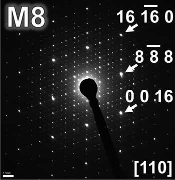
TEM–SAED (Figure 1) revealed that the murataite phase had an 8 × 8 × 8 (M8) superstructure. Indexing for all peaks of the murataite phase were carried out mostly manually, securely aware of this.
SAED pattern of M8 in the (1 1 0)-plane of the reciprocal lattice.

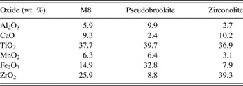
SEM–EDS analyses (Table I) measured the atomic ratio of each element. The pseudobrookite (P)-type phase, containing much Al, Ti, and Fe elements, agrees well with the conventional formula. We concur that Mn in the starting material is an essential condition for synthetic M formation (Laverov et al., Reference Laverov, Yudintsev, Omel'yanenko, Nikonov and Stefanovskii1999), where it may reside in trigonal bipyramids [as it often prefers, e.g. in YMnO3, as Mn3+ (Aken et al., Reference Aken, Meetsma and Pastra2001)]. Moreover, Mn is known also to aid in the formation of tranquillityite (Gatehouse et al., Reference Gatehouse, Grey, Lovering and Wark1977), another reason that we think that it has similarities in this extended polytypic group. M8 has 2 α-Keggin groups with an AlO4 tetrahedron at its center (Laverov et al., Reference Laverov, Urusov, Krivovichev, Pakhomova, Stefanovsky and Yudintsev2011a), resulting in the higher Al contents in M8 than in zirconolite (Z).
Chemical compositions (EDS) of the coexisting phases in the sintered sample.

In Table I, Z's composition is quite like M8 but with a little less Al, and P's composition is quite similar to M8 but with more iron and less zirconium. Reaction appears to be incomplete because the contents of the Z and P sum to about the same composition as the M8. With six elements, it is almost certain that, at 1300 °C, a liquid formed from which, on cooling, Z and P crystallized and which facilitated the good crystallization of M8 as micrographic observations indicated (to be published elsewhere).
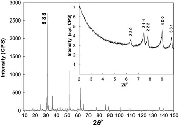
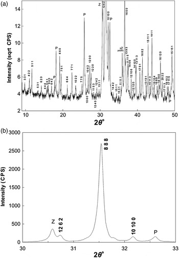
Figures 2 and 3 and Table I revealed that the resultant phases after sintering were about 90% major M8, about 5% minor (Fe,Al,Mg)2(Ti,Zr)O5 P, and about 5% minor (Ca,Mn)ZrTi2O7−2M Z [PDF 00-041-1432 and 00-034-0167 (ICDD, 1991 and 1984)]. In the present work, the XRPD pattern was collected in three distinct 2θ ranges, with specific experimental conditions in each of them.
X-ray diffraction pattern on a linear scale exemplifies how very weak are the superlattice lines, dominated by the sub-cell lines. The low angle, 2θ = 2–10°, is shown inset on a square root scale. Advantage accrues at low angles where intensities are slightly enhanced by the (1 + cos2 θ) term in the usual scattering equation.

Expanded section of Figure 2. (a) Better to emphasize the many low-intensity peaks from the superlattice on a square root scale. (b) In the region of (8 8 8), all peaks are M8 with minor impurities: P, pseudobrookite and Z, zirconolite; there are no visible peaks belonging to M3 or M5.

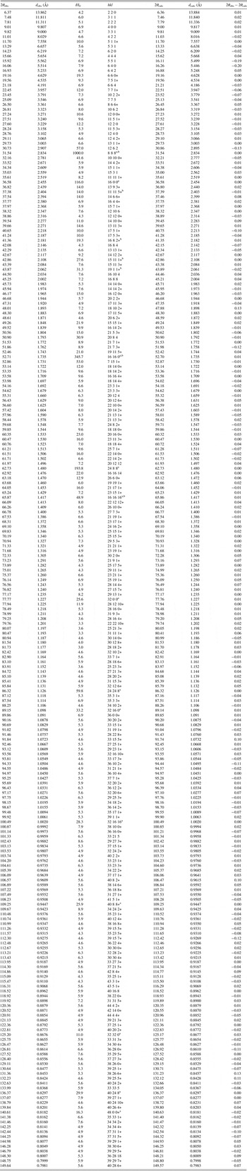
The XRPD indexing (Table II) confirms that M8 is FCC, with (h k l) all odd or all even. Low-angle lines are well visible but with the noticeable absence of the (1 1 1), for which there seems not to be any systematic reason, and we do not believe that it is smothered by the background. M8 conforms also to the extra condition (h 0 0) where h = 4n only, consequently, and especially noticeable, leading to the absence of the (2 0 0). This allows for four space groups (SGs), Fd
 $\bar 3$
(203), F4132 (210), Fd
 $\bar 3$
m (227), and Fd3c (228). Three of these SGs have (h k 0) where h + k = 4n only, but we do find the (6 4 0) and (12 2 0), without overlaps, to be present, thus leaving only F4132 (210), not the same SG of F4
 $\bar 3$
m (216) as for M3.
Detailed list of X-ray diffraction peaks of M8 sample. There are a total of 239, mostly very weak FCC lines, from the supercell, 13 stronger FCC lines come from the fluorite-type basic sub-cell. In the case of many overlapping values of h 2 + k 2 + l 2, we choose, for simplicity, to show only the one with the lowest value of l (marked with the “+” symbol).

a 13 stronger FCC lines come from the fluorite-type basic sub-cell.
b Several of the stronger lines agree with all the indexing of stronger spots in the SAED patterns (Laverov et al., Reference Laverov, Urusov, Krivovichev, Pakhomova, Stefanovsky and Yudintsev2011a).
The lattice constant was calculated using only the strong certain reflections at high angles [(32 0 0), (24 24 8), (32 16 0), (32 16 16), (40 8 8), (32 32 0), (40 24 8), and (48 0 0)] – where all the (h k l) are divisible by 8, i.e. those from the FCC basic sub-cell. The lattice constant, a, of the M8 superstructure is 39.269 ± 0.001 Å, calculated using a Nelson–Riley extrapolation.
Here there is no rare earth, so those elements are not mandatory but can play a role as they do in the work of Laverov et al. The full range of elements that will enter these structures is to be determined but appears to be most of the periodic table excepting, perhaps, the very largest ions – making them especially interesting for long-term radwaste sequestration.
IV. SUPPLEMENTAL COMMENT
There is considerable risk when not observing possible low-angle very weak peaks, especially as we move to these more complex materials. One particularly “good” example hampered the (maybe overspecialized) applied luminescent/fluorescent community; these researchers published well over 70 papers that maintained that YBO3, and its isotypes, were “vaterites” (isotypic with a form of CaCO3) and/or had hexagonal symmetry – this was actually the strong basic sub-cell with B hardly contributing to the intensities. Low-angle very weak peaks would have revealed the larger monoclinic two-layer pseudowollastonite cell, isotypic with CaSiO3 [pseudowollastonites are notorious for having polytypic complexity (Morgan et al., Reference Morgan, Carrol and Lange1977; Yang and Prewitt, Reference Yang and Prewitt1999)]; unawareness of the true unit cells, or intergrowths thereof, quite possibly might partly explain some variable luminescence results. Single-crystal work was necessary, coming to the rescue (Lin et al., Reference Lin, Sheptyakov, Wang and Allenspach2004; Pitscheider et al., Reference Pitscheider, Kaindl, Oeckler and Huppertz2011) even though the powder technique had, indeed, much earlier flagged the problem (Morgan et al., Reference Morgan, Carrol and Lange1977). Although Lin et al. identified the correct monoclinic cell, they were unaware that it is a known pseudowollastonite type.
For routine “fingerprinting”, slow scans only from ~2° to 15–20° may be sufficient. Higher intensity sources would be beneficial, but may not be available on a day-to-day basis. A rotating stage would also help as so many planes must orient in the diffraction mode.
This approach should be applied to M5 and M7 (M3 already done) so as to provide the engineering synthesizer a speedier monitoring to gauge progress in his/her efforts with so many possible cations. Likely other families of very complex polytypes may be discovered more quickly as ceramics style efforts pursue evermore complex cationic mixes for novel/exotic physical properties.
V. CONCLUSION
-
(1) M8 has been synthesized remarkably pure by the simple standard ceramic procedure of co-firing oxides. Its XRPD contains more than the 239 surely identified lines.
-
(2) The M8 superstructure was first observed by TEM–SAED enabling the analysis by the powder method with some confidence. The calculated lattice constant, a, of this M8 superstructure is 39.269(1) Å; it is most likely the largest inorganic mixed-oxide unit cell ever studied by somewhat conventional powder diffraction.
-
(3) Systematic extinctions of all (h 0 0), where h ≠ 4n, and the existence of some (h k 0) peaks, where h + k ≠ 4n, suggests it belongs in the SG, F4132 (210), contrasting with an earlier suggestion that the SG might be F4
 $\bar 3$
m (216). Some random varying site occupancies of the cations would, in any case, make assigning an ideal SG impossible.

SUPPLEMENTARY MATERIAL
To view supplementary material for this article, please visit http://dx.doi.org/10.1017/S0885715615000913
ACKNOWLEDGEMENTS
The authors thank the National Institute for Material Science (NIMS) for the use of SEM–EDS. We also appreciate to the Institute for Chemical Research, Kyoto University for the help of TEM–SAED analysis. Detailed work of these techniques will be published later.