Introduction
The nematode Gnathostoma turgidum has been reported in eight Mexican states, including Sinaloa (Pérez-Álvarez et al., Reference Pérez-Álvarez, García-Prieto, Osorio-Sarabia, Lamothe-Argumedo and León-Règanon2008; Díaz-Camacho et al., Reference Díaz-Camacho, Willms, Rendón-Maldonado, de la Cruz Otero, Delgado-Vargas, Léon-Règanon and Nawa2009), which has been described as an endemic area of human gnathostomosis with more than 10,000 estimated human cases. Gnathostoma binucleatum, a related species, has been reported to be the causative agent of the human disease (Almeyda-Ártigas et al., Reference Almeyda-Ártigas, Bargues and Mas-Coma2000). Both Gnathostoma species have been reported in the endemic area, suggesting that they co-inhabit the same location. The prevalence of G. turgidum, an annual parasite of Didelphis virginiana, is highest during May and June (Nawa et al., Reference Nawa, de la Cruz-Otero, Zazueta-Ramos, Bojórquez-Contreras, Sicairos-Félix, Campista-León, Torres-Montoya, Sánchez-Gonzales, Guzmán-Loreto, Delgado-Vargas and Díaz-Camacho2009). It has been reported that advanced third-stage larvae (A3L) localize to the liver of infected animals from February to April, and subsequently adult worms are located in the stomach from April to July (Díaz-Camacho et al., Reference Díaz-Camacho, Delgado-Vargas, Willms, de la Cruz-Otero, Rendón-Maldonado, Robert-Guerrero, Antuna-Bizarro and Nawa2010). The beginning and end of synchronous G. turgidum egg expulsion in the faeces of infected D. virginiana, as well as the expulsion of adult worms from May to September by spontaneous curing, have been reported (Torres-Montoya et al., Reference Torres-Montoya, Galaviz-Rentería, Castillo-Ureta, López-Moreno, Nawa, Bojórquez-Contreras, Sánchez-Gonzales, Díaz-Camacho, Rocha-Tirado and Rendón-Maldonado2014). However, the histopathological changes of D. virginiana caused by infection with G. turgidum are unknown. Although G. binucleatum is the reported causative agent of human gnathostomosis, the histopathology of lesions has been poorly studied, because it is difficult to obtain samples of the parasite from infected humans, due to its migratory behaviour. Experimental infection with G. binucleatum in dogs has been reported; however, stomach histopathology has not been studied adequately (Álvarez-Guerrero et al., Reference Álvarez-Guerrero, Muñoz-Guzmán and Alba-Hurtado2012). In addition, it is not clear whether G. turgidum is also involved in human gnathostomosis. As such, further studies are needed to achieve a better understanding of the life cycle of the parasite and to describe the pathological changes associated with natural host–parasite interactions. Such studies could further clarify the progression of human infections caused by G. binucleatum. In this report, we studied the histopathology of the liver and stomach of wild D. virginiana infected with G. turgidum.
Materials and methods
Three opossums suspected to be infected with G. turgidum were captured in an endemic area of Sinaloa (Torres-Montoya et al., Reference Torres-Montoya, Galaviz-Rentería, Castillo-Ureta, López-Moreno, Nawa, Bojórquez-Contreras, Sánchez-Gonzales, Díaz-Camacho, Rocha-Tirado and Rendón-Maldonado2014). All of them were positive for Gnathostoma, based on stool examination. Three opossums captured from a non-endemic area of G. turgidum in Navolato, Sinaloa (24°45′55″N, 107°42′7″W) were used as a negative control. Experimental and control specimens were registered as (T1–T3) and (TC1–TC3), respectively. Opossums were transported to the lab and kept under natural conditions of humidity, temperature and light in 50-cm2 cages, without access to external food. They were fed with commercial dog food and had access to water ad libitum. Animals were sacrificed in March (T1), May (T2) and December (T3) to follow the process of infection and spontaneous curing. Opossums were sedated with 10 mg/kg of intramuscularly administered tiletamine-zolazepam (Zoletil50, Virac, Guadalajara, Jalisco, México). After applying an intra-cardiac dosage of 50 ml potassium chloride (KCl) to sacrifice the opossums, necropsies were performed to collect liver and stomach samples. Samples were fixed with 10% formalin–phosphate-buffered saline (PBS), processed in paraffin wax, and stained with haematoxylin–eosin (HE) and Masson's trichrome (Marcos et al., Reference Marcos, Yi, Machicado, Andrade, Smalvides, Sanchez and Terashima2007). Histopathology was performed using an optical microscope (PrimoStar, Zeiss, Oberkochen, Germany).
Results and discussion
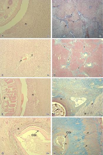
Histopathology of the liver collected in March (T1) showed fibrous connective tissue, hepatocyte necrosis and haemorrhage, with polymorphonuclear cells surrounding the parasitic structure in the liver parenchyma (fig. 1A). Periportal fibrosis and formation of collagen septa between the portal structures were observed (fig. 1B). In animals T2 and T3, inflammatory infiltration of macrophages and periportal hyperplasia were observed, along with some evident haemorrhagic areas (fig. 1C). Histopathology showed a thickening of the collagen fibres and the presence of inflammatory cells inside the fibrotic areas (fig. 1D). Stomach histopathology in T1 showed normal histology. Anchylostoma spp. and Turgida turgida adults were also observed; however, nodule lesions in stomachs caused by these nematodes have not been described previously. Stomach histopathology of T2 showed an adult worm between the lamina propria and muscular tissue surrounded by apparent necrosis (fig. 1E), along with collagen fibres in the muscular layer (fig. 1F). In T3, a nodule in the stomach caused by the parasite was observed, as well as necrosis in the periphery of the lesion, inflammatory infiltrate and infiltration of the muscular layer with possible thickening of the collagen fibres (fig. 1G, H).
Liver histopathology of D. virginiana infected with G. turgidum. (A) Liver damage of T1, sacrificed in March, suggests necrosis (n) around the cuticular surface of the parasite (p) (stain HE). (B) Liver of T1 with formation of collagen septa (*) between the periportal structures (arrow) (stain Masson). (C) Representative image of livers of T2 and T3, similar damage in both, with periportal fibrosis (f) in portal vein (arrow) (stain HE). (D) Representative image of livers from opossums T2 and T3, where thickening in the collagen septa (*) is shown (stain Masson). (E) Stomach histopathology of T2, necrosis (n) in the periphery of the lesion caused by the parasite (p) (stain HE). (F) Section of T2 stomach with a parasite (p), necrosis (n) in the periphery of the nodular lesion and infiltration of collagen fibres (arrow) in the muscle layer (stain Masson). (G) Stomach of T3, necrosis (n) in the periphery of the nodular lesion and apparent remains of cuticle from an adult G. turgidum worm (CGt) (stain HE). (H) Stomach of T3, apparent cuticular surface of parasite (CGt) in the nodular lesion with necrosis (n) over the periphery, and apparent thickening of collagen fibres in the muscular layer (arrows).

In the current study, the establishment of collagen septa in the liver and stomach of opossums infected with G. turgidum was observed. Even though the fibrogenic process in the stomach has not yet been described for this type of infection, it has been shown that in bovine liver infections with Fasciola hepatica, fibrosis is observed in at least two-thirds of the liver parenchyma (Raadsma et al., Reference Raadsma, Kingsford, Suharyanta, Spithill and Piedrafita2007). Moreover, it seems that the degree of fibrosis is correlated with the parasite load in other helminth infections, suggesting that establishment, and even partial reversibility, of fibrosis is directly proportional to the number of parasites and degree of damage (Marcos et al., Reference Marcos, Yi, Machicado, Andrade, Smalvides, Sanchez and Terashima2007). In the present study, the beginning of collagen septa formation was observed, and interconnection of the septa in liver tissue increased with infection time. Collagen septa were observed after 7 months, when the parasite had left the liver or had been expelled by the opossum, as has been described previously in other animal models (Friedman. Reference Friedman2008). However, more studies are needed to verify that G. turgidum causes fibrosis, and to determine if the state of fibrosis depends on parasite load.
Stomach histopathology revealed similar pathology as that caused by other nematodes, such as Spirocerca lupi in dogs (Dvir et al., Reference Dvir, Clift and Williams2010), suggesting that remains of G. turgidum may cause a chronic inflammatory response, and fibrosis proceeds even after the parasite has been expelled. Further immunological studies are necessary to verify that this stimulation is caused by antigenic remains of the parasite, and to determine how these factors contribute to the chronic inflammatory process.
Acknowledgements
The authors wish to acknowledge to local hunters from Tecualilla, Escuinapa, Sinaloa, who worked as volunteers to capture the opossums.
Financial support
This work was supported by institutional funds (PROFAPI2012/144).
Conflict of interest
None.
Ethical standards
Animals used in this article were captured with permission from the Mexican Wildlife Animal authorities (SEMARNAT, 02197/12) and were maintained following guidelines from the Bioethical Animal Committee of the Universidad Autónoma de Sinaloa.