Introduction
Benign osteoblastoma is a rare tumour, constituting just 1 per cent of all bone tumours and 3 per cent of all benign bone tumours.Reference McLeod, Dahlin and Beabout1 It is defined as a vascular, osteoid- and bone-forming, benign neoplasm of bone characterised by the presence of numerous osteoblasts.Reference McLeod, Dahlin and Beabout1, Reference Matsumoto, Kakita and Fukuma2 Around 60 per cent of cases occur in the long bones and spine. Kroon et al. described the clinical features of 98 cases of osteoblastoma: 33 per cent originated in the spine, 26 per cent in long tubular bones, 26 per cent in the bones of the hands or feet, and 14 per cent in other sites (including the pelvis, ribs, skull, scapula and clavicle).Reference Kroon and Schurmans3 Other, rarer sites have been described, including the temporal bone, ethmoidal sinus, frontal bone and orbits.Reference Matsumoto, Kakita and Fukuma2, Reference Ungkanont, Chanyavanich, Benjarasamerote and Tantinikorn4–Reference Parodi, Iustulin and Isola7
Involvement of the nasal cavity is rare, with only 10 previous reports in the English-language literature. The commonest site of origin was the ethmoid sinus, with extension to involve the nasal cavity. Four cases originated from the nasal cavity: two from the nasal bones, one from the middle turbinate periosteum and one from the perpendicular plate of the ethmoid bone.Reference Sooknundun, Kacker and Kapila11–Reference Imai, Tsujiguchi, Toda, Sung, Tajima and Sakamoto14
In this paper, we present the second report of osteoblastoma originating from the bony nasal septum.
We also present the results of a literature review, performed by searching the English-language literature using the PubMed database, using the search terms ‘osteoblastoma’, ‘nasal cavity’, ‘nasal septum’ and ‘nose’.
Case report
A 45-year-old man presented to the ENT out-patient clinic with a six-month history of recurrent, right-sided epistaxis associated with right-sided nasal obstruction. There was no previous history of epistaxis. The patient was otherwise fit and well, and took no regular medication.
Anterior rhinoscopy revealed a friable, polypoidal mass completely filling the right nasal cavity, which bled on contact.
A provisional diagnosis of haemangioma was made, and the patient was commenced on oral steroids and topical antibiotics. We requested an urgent computed tomography (CT) scan of the sinuses, and a staging CT of the neck. A biopsy was not taken in clinic due to the risk of bleeding.
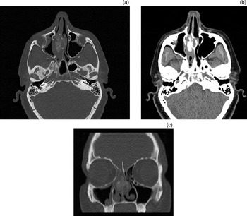
The sinus CT scan showed a 2.2 × 1.3 × 2.5 cm mass centred on the bony nasal septum and extending mainly into the right nasal vault, but with some extension into the left vault (Figure 1). The mass was noted to have a significant osseous component in continuity with the nasal septum. The mass occluded the right osteomeatal unit, with resultant mucosal thickening throughout the right maxillary sinus.
Axial (a, b) and coronal (c) computed tomography sinus scans, showing a mass centred on the bony nasal septum and extending into the right nasal vault. The mass obstructs the right osteomeatal complex, with associated maxillary sinus disease.

The staging CT of the neck showed that the neck, lungs and upper abdominal viscera were clear of disease.
The patient was scheduled for urgent functional endoscopic sinus surgery with lesion biopsy.
Under anaesthesia, a large, expansive lesion was seen to arise directly from the right bony nasal septum. The lesion was removed along with most of the bony nasal septum, using a chisel. A middle meatal antrostomy and uncinectomy were performed to treat the maxillary sinus disease.
Post-operatively, the patient was kept in hospital overnight as there was a small amount of bleeding.
On macroscopic inspection, the mass was grey-brown and measured 3.2 × 2.5 × 1.2 cm, with a haemorrhagic cut surface. There were some firm areas the consistency of bone.
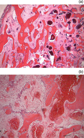
Microscopic inspection showed an expansile lesion composed of several nidi of well formed and anastomosing trabeculae of osteoid and woven bone, rimmed by osteoblasts and a few osteoclasts (Figure 2). The background consisted of a richly vascular, loose, fibrous stroma. There was no osteoblastic atypia, and mitoses were few. At the periphery of the lesion, the bony trabeculae gradually merged with, but did not permeate, the adjacent tissues, where present (although assessment was limited due to the fragmented nature of the specimen).
Photomicrographs of the lesion, showing (a) a general overview of lesion (H&E; ×40), and (b) the centre of the lesion (H&E; ×100). The latter view shows bone trabeculae with peripheral osteoblastic rimming, and background fibrous and vascular stroma.

A diagnosis of osteoblastoma arising from the bony nasal septum was made.
At six-month follow up, the patient was well and free of recurrence.
Discussion
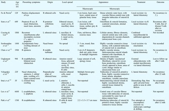
Osteoblastoma typically occurs in the long bones and spine, but, rarely, can occur in sites including the nasal cavity, as described above. We identified 10 previously reported cases, in the English-language literature, of osteoblastoma involving the nasal cavity (see Table I).
Osteoblastoma involving nasal cavity: reported cases

Y = years; F = female; R = right; mth = months; L = left; M = male; A-P = anteroposterior
Ninety per cent of osteoblastoma cases (at any site) occur in patients in their second and third decades, although the reported age range extends from three to 72 years.Reference McLeod, Dahlin and Beabout1 Patients with nasal cavity osteoblastoma have an equally broad age range, from three to 69 years (based on English-language literature reports) (see Table I). Our literature review identified six male and four female patients.
Presenting signs and symptoms depend on the site and extension of the tumour. Of the 10 cases identified by our literature review, exophthalmos or proptosis was the most common presenting symptom or sign, occurring in six cases; in all these cases, proptosis was related to lateral extension of the tumour from the ethmoid cells. Nasal obstruction and epistaxis were also common symptoms, occurring in three and two of the 10 cases, respectively. Two patients were completely asymptomatic, complaining only of an external bony swelling. Interestingly, none of the 10 patients complained of pain.
In all 10 cases, the osteoblastoma was found to either originate from or extend into the nasal cavity. Five cases originated from the ethmoid cells and extended to involve the nasal cavity. Four cases originated from the nasal cavity: one from the middle turbinate, two from the nasal bones and one from the perpendicular plate of the ethmoid. One case originated from the posterior cribriform plate.
Histologically, osteoblastoma can be difficult to distinguish from other bone lesions, including osteoid osteoma, low grade osteosarcoma and aneurysmal bone cyst. Osteoid osteomas can often be identical histologically to osteoblastomas. Often, the only distinguishing factor is size: osteoid osteomas are usually smaller than 2 cm due to their limited growth potential.Reference McLeod, Dahlin and Beabout1 Osteoblastomas also typically have broader, longer and less densely packed trabeculae than osteoid osteomas. In our literature review, all lesions were at least 3 cm in diameter, in cases with reported size. In addition, osteoblastomas tend to lack the ‘halo’ of sclerotic bone normally associated with osteoid osteomas.Reference Kroon and Schurmans3 None of our 10 identified cases had bone pain as a presenting symptom. This is in contrast with osteoid osteomas, which often produce severe pain relieved by non-steroidal anti-inflammatory drugs.
Some osteoblastomas can exhibit histological features suggestive of malignancy, such as low-grade osteosarcoma. In osteoblastomas occurring in the nasal septum, it is important to make this distinction. It is difficult, if not impossible, to histologically differentiate between osteoblastoma and osteosarcoma if the edge of the lesion cannot be clearly visualised. Clinical and radiological correlation is therefore essential. Features that can aid the discrimination of benign osteoblastoma from osteosarcoma include the absence of anaplasia, the lack of permeation at the lesional edge, the absence of ‘trapping’ of host lamellar bone by tumour bone, and the absence of cartilage production.Reference Kroon and Schurmans3
Computed tomography is the best modality for investigating osteoblastoma, and demonstrates the origin and local extension of the tumour. Typically, an expansile lesion is seen, with remodelling of adjacent bone. It is usually well circumscribed, without bone destruction, and with large, discrete areas of bone density; alternatively, it may have a mixed osseous and fibrous appearance.Reference Velegrakis, Prokopakis, Papadakis, Karampekios, Koutsoubi and Helidonis13
Magnetic resonance imaging adds little to the investigation of this lesion, as it typically overestimates the extent of the tumour due to the surrounding extensive inflammatory reaction.Reference Papaioannou, Sebire and McHugh15
Treatment of osteoblastoma is via complete surgical excision.
In the present case, the osteoblastoma involved only the nasal cavity, without extension to surrounding areas, and was therefore removed by simple endoscopic curettage.
• Osteoblastomas are rare tumours constituting 1 per cent of all bone tumours
• They rarely involve the nasal cavity; the reported case is only the second involving the bony nasal septum
• Histologically, osteoblastomas can be difficult to distinguish from osteoid osteomas, osteosarcomas and aneurysmal bone cysts
• Treatment is via complete excision
• Recurrence has not been reported after complete excision, although long-term follow up is advised for patients without clear surgical margins
In the cases identified by our literature search, the most common approach to excision was via a lateral rhinotomy approach. This was employed in five of the reported cases.Reference Ungkanont, Chanyavanich, Benjarasamerote and Tantinikorn4, Reference Fu and Perzin8, Reference Som, Bellot, Blitzer, Som and Geller9, Reference Chen, Weinberg, Simpson and Tschang12, Reference Velegrakis, Prokopakis, Papadakis, Karampekios, Koutsoubi and Helidonis13 This approach allows full exposure of the ethmoid sinus and the lateral wall of the nasal cavity. However, since the late 1990s this approach has generally been superseded by the endoscopic approach, as tumours which can be resected via lateral rhinotomy can just as easily be resected endoscopically. In one case, a lateral rhinotomy approach was combined with a medial maxillectomy to allow access to a tumour extending from the right ethmoid sinus to the anterior cranial fossa, right orbit, right maxillary sinus and sphenoid sinuses.Reference Ungkanont, Chanyavanich, Benjarasamerote and Tantinikorn4 In 1985, Coscina et al. used a combined transethmoidal and subfrontal approach to facilitate excision of a tumour extending from the left ethmoid sinus to the left frontal sinus and orbit.Reference Coscina and Lee10 Imai et al. used a dismasking flap to expose a tumour arising from the left nasal bones.Reference Imai, Tsujiguchi, Toda, Sung, Tajima and Sakamoto14 Following resection, the defects of the nose and orbit were reconstructed using split calvarial bone grafts.
The patients identified by our literature search were generally followed up for between five months and three years. Follow up was not documented in three of the cases.
There were two case of recurrence following an initial incomplete excision: one occurred seven years after the original excision, the other nine months after. In the latter case, the patient was disease-free for a further nine months following re-excision. Of the four cases of osteoblastoma originating from the nasal cavity, all were disease-free after follow up of between 8 months and three years.Reference Sooknundun, Kacker and Kapila11–Reference Imai, Tsujiguchi, Toda, Sung, Tajima and Sakamoto14 Osteoblastomas of all sites has a surprisingly high recurrence rate, 9.8 per cent; however, recurrence after complete en-bloc excision has not been reported.Reference Jackson16 Recurrence can occur up to nine years after the initial excision; therefore, complete excision followed by long-term follow up is recommended. Radiotherapy does not prevent recurrence, and indeed may possibly be associated with late sarcomatous change.Reference Jackson16 In cases of sinonasal osteoblastoma, recurrence should be treated with further excision.