Introduction
Cystic echinococcosis (CE) caused by the metacestode stage of Echinococcus granulosus is one of the most important helminth zoonoses in the world; it affects both humans and domestic livestock (Thompson, Reference Thompson2008). The disease is endemic in Argentina and highly endemic in the province of Neuquén, located in the Argentinean Patagonia region (Pierangeli et al., Reference Pierangeli, Soriano, Roccia, Giménez, Lazzarini, Grenóvero, Menestrina and Basualdo2007).
Considerable genetic and phenotypic variation has been demonstrated in E. granulosus, and to date ten distinct genotypes (G1–G10) have been identified using molecular tools (Bowles et al., 1992; Lavikainen et al., Reference Lavikainen, Lehtinen, Meri, Hirvelä-Koski and Meri2003). This categorization follows very closely the pattern of strain variation described, based on the morphological and biological characteristics (McManus & Thompson, Reference McManus and Thompson2003). Through an analysis of complete mitochondrial genomes, it has recently been proposed that E. granulosus sensu lato (E. granulosus sl) may be considered a species complex maintained in distinct cycles of transmission, comprising E. granulosus sensu stricto (E. granulosus ss) (genotypes G1–G3), E. equinus (G4), E. ortleppi (G5) and E. canadensis (G6–G10) (Nakao et al., Reference Nakao, McManus, Schantz, Craig and Ito2007). These species/genotypes can differ in several characteristics, such as morphology, growth and development rates in the definitive hosts or infectivity to intermediate hosts, all of which have implications for control strategies. Therefore, in endemic areas where several species of livestock could be infected with E. granulosus sl, it is important to know whether each harbours a different species/genotype and if there is the possibility of interaction between cycles (Thompson, Reference Thompson2008). A variety of criteria has been used to characterize isolates of Echinococcus (Thompson & McManus, Reference Thompson, McManus, Eckert, Gemmell, Meslin and Pawlowski2001). Although DNA sequencing is undoubtedly the most accurate tool to achieve this purpose, large-scale sequence-based studies are expensive and, therefore, less affordable for many research laboratories in endemic areas (Harandi et al., Reference Harandi, Hajialilo and Shokouhi2012). The use of simpler and more economical methods to distinguish between species could be of practical value in field epidemiological studies. Characterization of E. granulosus strains or variants has been carried out by different analytical methods, such as morphology, physiology or biochemistry, with variable results. Although the use of larval rostellar hook morphometry as a sole criterion for differentiation has been questioned, because hook dimensions can be influenced by host or environmental factors (Thompson & Lymbery, Reference Thompson and Lymbery1988; Hobbs et al., Reference Hobbs, Lymbery and Thompson1990), more recent studies have shown a strong correlation between hook morphometric results and molecular genotyping (Ponce Gordo & Cuesta Bandera, Reference Ponce Gordo and Cuesta Bandera1997; Harandi et al., Reference Harandi, Hobbs, Adams, Mobedi, Morgan-Ryan and Thompson2002; Ahmadi, Reference Ahmadi2004; Ahmadi & Dalimi, Reference Ahmadi and Dalimi2006; Latif et al., Reference Latif, Tanveer, Maqbool, Siddiqi, Kyaw-Tanner and Traub2010). However, validation of larval hook morphometry as a suitable method to characterize E. granulosus isolates is still lacking.
In a previous molecular survey carried out in the province of Neuquén, the occurrence of E. granulosus ss G1, E. canadensis G6 and E. canadensis G7 was identified in fertile cysts – with protoscoleces (PSCs) – from sheep, goats and pigs, respectively (Soriano et al., Reference Soriano, Pierangeli, Pianciola, Mazzeo, Lazzarini, Saiz, Kossman, Bergagna, Chartier and Basualdo2010). In a study carried out thereafter in similar locations, five fertile cysts from cattle were also characterized as E. canadensis G6 (authors' unpublished data). It must be pointed out that in Neuquén, although sheep, goats and cattle are infected with different species/genotypes of E. granulosus sl, fertile cysts seem to be produced only by a particular genotype in each species of intermediate host. Using the same samples analysed in those studies, the objectives of the present work have been to morphologically characterize the isolates, to evaluate which morphometric parameter is more useful to differentiate between species/genotypes of E. granulosus sl, and to see if a cut-off value based on such a parameter can be defined.
Materials and methods
Collection and examination of cysts
The province of Neuquén is located in the north of the Argentinean Patagonia region, extending from 36°39′ to 41°01′S latitude and 60°00′ to 71°58′W longitude. CE is highly endemic in this province, where mean annual incidence rates of 24.4 per 100,000 inhabitants for the total population and 9.7 per 100,000 in children aged 0–14 years were reported over the period 1995–2004 (Pierangeli et al., Reference Pierangeli, Soriano, Roccia, Giménez, Lazzarini, Grenóvero, Menestrina and Basualdo2007). In rural areas the main economic activity is raising livestock for subsistence, under a transhumant model of production. Goats are the most common livestock but sheep and cattle are also raised. Pigs are mostly raised in urban areas. A prospective, descriptive and analytical study design was carried out.
Parasite material consisted of hepatic and pulmonary cysts from naturally infected sheep, goats, cattle and pigs slaughtered in abattoirs throughout the Province of Neuquén. Ten randomly selected fertile cysts from each intermediate host species (goats and pigs) and all the fertile cysts obtained from sheep (n= 11) and cattle (n= 5) were included in the study. All the samples were collected between June 2005 and October 2010.
Each individual cyst was considered as a sample or isolate. In no case were several cysts pooled and processed jointly. Protoscoleces were aspirated from each cyst, washed three times with sterile saline and stored in 10% buffered-formalin at room temperature for morphometric analysis.
Hook morphometry
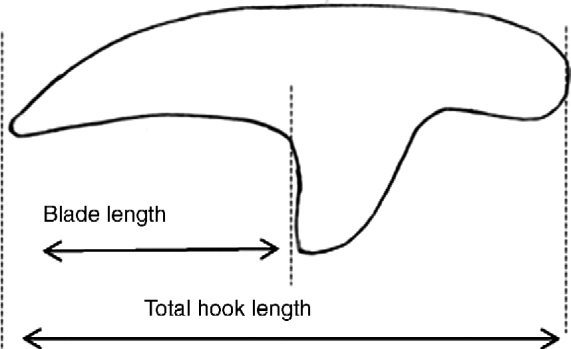
Protoscoleces were mounted in polyvinyl lactophenol on a glass slide, and sufficient pressure was applied to the cover slip in order to flatten but not damage the hooks (Ponce Gordo & Cuesta Bandera, Reference Ponce Gordo and Cuesta Bandera1997; Ahmadi, Reference Ahmadi2004; Latif et al., Reference Latif, Tanveer, Maqbool, Siddiqi, Kyaw-Tanner and Traub2010). The hook parameters were measured according to Hobbs et al. (Reference Hobbs, Lymbery and Thompson1990). Measurements of the total length of large (LTL) and small (STL) hooks as well as the blade length of large (LBL) and small (SBL) hooks were made using a calibrated eyepiece micrometer under oil immersion (fig. 1). Morphometric data were collected from three large and three small hooks per rostellum from each of six PSCs for each E. canadensis isolate (goat, pig and cattle isolates). In order to analyse an equivalent number of data from E. granulosus ss isolates, four large and four small hooks per rostellum from each of ten PSCs for each sheep isolate were measured.
Measurements (μm) of total hook and blade lengths of larval rostellar hooks of E. granulosus sensu lato.

Molecular characterization
All the isolates included in this research had already been characterized genetically in previous studies by amplification and sequencing of a 450 base pair fragment of the mitochondrial cytochrome c oxidase subunit 1 (cox 1) gene (Soriano et al., Reference Soriano, Pierangeli, Pianciola, Mazzeo, Lazzarini, Saiz, Kossman, Bergagna, Chartier and Basualdo2010; authors' unpublished data). The distribution of species/genotypes found was: E. granulosus ss G1 (sheep isolates); E. canadensis G6 (goat and cattle isolates) and E. canadensis G7 (pig isolates). The identified sequences were registered in GenBank database under the accession numbers GU980908, GU980913, JN176932 and GU980914.
Data analysis
Data analysis was carried out using SPSS statistical software (SPSS 17.0; SPSS Inc., Chicago, Illinois, USA). To evaluate which morphometric parameter is more useful to differentiate between hosts and/or species/genotypes, data were subjected to a multivariate analysis (ANOVA) with Tukey's post-hoc test. Differences were considered statistically significant when P< 0.05.
The diagnostic performance of morphometry as a test to differentiate between species of E. granulosus sl was evaluated using genetic characterization as gold standard. For analytical purposes, E. canadensis (G6 and G7) isolates were considered ‘positive’ and E. granulosus ss G1 isolates were considered ‘negative’. Receiver operating characteristic (ROC) analysis was applied to determine the optimum cut-off value and the area under the curve (AUC) was used to evaluate the global diagnostic performance of the test. Diagnostic parameters were calculated as follows: sensitivity (SENS) = TP × 100/(TP + FN); specificity (SPEC) = TN × 100/(TN + FP); positive predictive value (PPV) = TP × 100/(TP + FP); negative predictive value (NPV) = TN × 100/(TN + FN); diagnostic efficiency (DE) = (TP + TN) × 100/(TP + FP + TN + FN), where: TP = true positive; TN = true negative; FP = false positive and FN = false negative.
Results
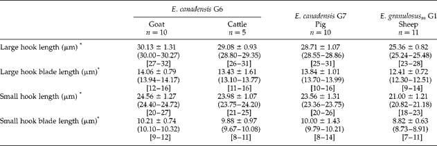
The first step in this study was to characterize the morphometric components of larval rostellar hooks from different intermediate hosts and from the different species/genotypes of E. granulosus sl present in Neuquén. Overall, 1780 hooks of 36 isolates were measured and analysed, and the results are shown in table 1.
Rostellar hook characteristics of protoscoleces of Echinococcus canadensis (E. canadensis) G6 and G7 and of Echinococcus granulosus sensu stricto (E. granulosus ss) G1 collected from hydatid cysts in intermediate hosts from the province of Neuquén.
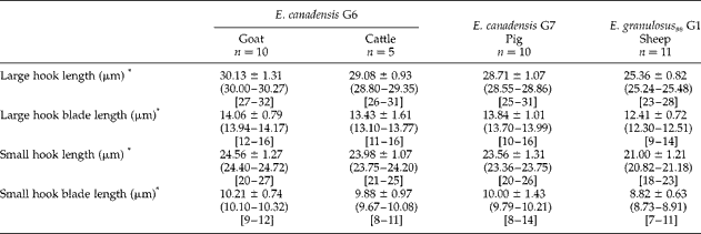
*All values are shown as mean ± SD; (95% CI) and [range]; n= number of cysts examined.
The second step was to assess which hook parameter is more useful to differentiate between hosts and/or species/genotypes. Data were subjected to multivariate analysis (one-way ANOVA), and highly significant differences were observed for all the parameters and among all the intermediate hosts. The measurements of LTL showed the strongest difference between groups: LTL, F= 735.24; LBL, F= 97.91; STL, F= 280.13; and SBL, F= 68.91. However, a post-hoc Tukey test indicated that although the measurements of LTL, LBL, STL and SBL were highly statistically different (P< 0.001) between the E. granulosus ss G1 (sheep) and E. canadensis G6–G7 (goats, cattle and pigs) isolates, there were no significant differences in the values of LBL and SBL between isolates from goats and pigs, nor of SBL between isolates from pigs and cattle (P > 0.05).
Then, a multivariate analysis was applied only to hook measurements of hosts infected by E. canadensis. In this case, the differences between groups were smaller than those observed among all the intermediate hosts: LTL, F= 83.94; LBL, F= 9.89; STL, F= 29.85; and SBL, F= 3.15. The results of both statistical analyses indicated that the measurement of LTL is the most useful morphometric criterion that can be used to distinguish between hosts and/or species/genotypes of E. granulosus sl.
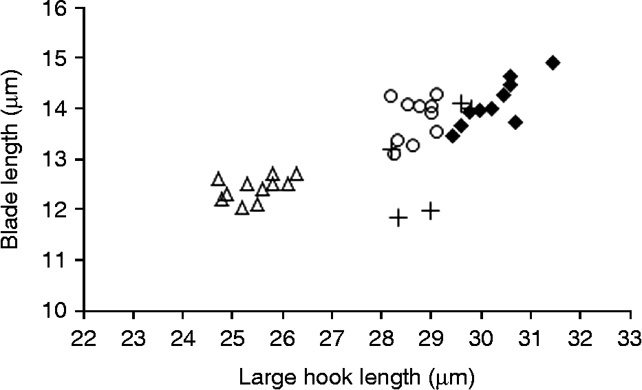
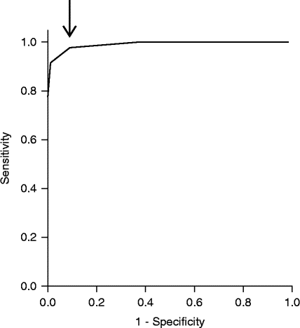
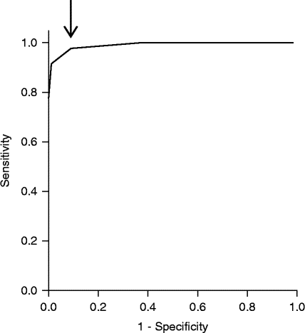
A scatterplot of LBL against LTL showed that Neuquén isolates of E. canadensis (G6 and G7) group together and separately from E. granulosus ss G1 isolates (fig. 2). On the basis of these results, the next step in this study was to determine a cut-off value based on LTL measurements to differentiate between E. granulosus ss G1 and E. canadensis larval isolates. ROC analysis was applied to all the individual LTL measurements of each E. granulosus sl species (E. canadensis, n= 450; E. granulosus ss, n= 440); the optimum cut-off value (better combination of SENS and SPEC) estimated by this method was LTL = 26.5 μm with an AUC = 99.0% (fig. 3). For this cut-off point in the ROC curve, the diagnostic parameters of the morphometric method were: SENS = 97.8%, SPEC = 91.1%, PPV = 96.5%, NPV = 94.3% and DE = 95.9%.
Scatterplot of large hook and blade lengths (μm, mean) of individual isolates from sheep, goats, cattle and pigs characterized in this study; Δ, G1 sheep; ♦, G6 goat; +, G6 cattle; ○, G7 pig.
Receiver operating characteristic (ROC) curve representing the plot of sensitivity versus 1-specificity for the larval rostellar hook morphometry method; cut-off (arrowed) corresponds to large hook total length = 26.5 μm (sensitivity 0.978; specificity 0.911).
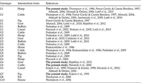
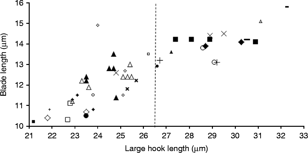
Due to local epidemiological characteristics, it was only possible to analyse isolates belonging to one species/ genotype of E. granulosus sl from each intermediate host species. A comprehensive review of the literature was carried out, and published morphometric data of larval isolates from different intermediate hosts and different species of E. granulosus sl were registered (table 2). The cut-off value for LTL determined in this study was then applied to those data in addition to ours, to validate that it can be used to distinguish between E. granulosus ss and other species of E. granulosus sl (fig. 4).
Genotypes, intermediate hosts and reference sources of morphometrics of Echinococcus granulosus sensu lato including the present study.
Scatterplot of large hook and blade lengths (μm, mean) from isolates of E. granulosus sl characterized in this study as well as previously published by other authors;  , proposed cut-off based on large hook total length (26.5 μm); see table 2 for references. Δ, G1 sheep; ♦, G1 goat; ▲, G1 cattle; ■, G1 buffalo; □, G1 pig; ◇, G1 camel; +, G2 cattle; ◇, G3 sheep; x, G3 goat; ●, G3 cattle; □, G3 buffalo;
 , G3 pig;
 , G4 horse; X, G5 cattle; ○, G5 buffalo; ●, G5 pig; ▲, G6 sheep; ♦, G6 goat; +, G6 cattle; ■, G6 camel; ○, G7 pig; Δ, G8 elk; −, G10 moose.
Discussion
Previous studies worldwide have included both molecular and morphological methods to characterize metacestode isolates from different intermediate hosts (Ponce Gordo & Cuesta Bandera, Reference Ponce Gordo and Cuesta Bandera1997; Harandi et al., Reference Harandi, Hobbs, Adams, Mobedi, Morgan-Ryan and Thompson2002, Reference Harandi, Hajialilo and Shokouhi2012; Tashani et al., Reference Tashani, Zhang, Boufana, Jegi and McManus2002; Ahmadi, Reference Ahmadi2004; Ahmadi & Dalimi, Reference Ahmadi and Dalimi2006; Thompson et al., Reference Thompson, Boxel, Ralston, Constantine, Hobbs, Shury and Olson2006; Almeida et al., Reference Almeida, Rodrigues-Silva, Neves, Romani and Machado-Silva2007; Andresiuk et al., Reference Andresiuk, Ponce Gordo, Cuesta Bandera, Elissondo, Dopchiz and Denegri2009; Pednekar et al., Reference Pednekar, Mukulesh, Gatne, Thompson and Traub2009; Latif et al., Reference Latif, Tanveer, Maqbool, Siddiqi, Kyaw-Tanner and Traub2010; Calderini et al., Reference Calderini, Gabrielli and Cancrini2012; Mowlavi et al., Reference Mowlavi, Salehi, Eshraghian, Rokni, Harandi, Mohajeran and Salahi-Moghaddam2012; Rajabloo et al., Reference Rajabloo, Hosseini and Jalousian2012). Most of those studies have shown acceptable correlations between the results obtained by each of the two approaches; however, validation of the larval rostellar hook morphometry method against molecular genotyping as gold standard is yet to be done.
Rostellar hook measurements of PSCs of E. granulosus ss have been frequently reported from sheep, cattle, camels, goats and pigs (Thompson et al., Reference Thompson, Kumaratilake and Eckert1984, Reference Thompson, Boxel, Ralston, Constantine, Hobbs, Shury and Olson2006; Ponce Gordo & Cuesta Bandera, Reference Ponce Gordo and Cuesta Bandera1997; Harandi et al., Reference Harandi, Hobbs, Adams, Mobedi, Morgan-Ryan and Thompson2002; Tashani et al., Reference Tashani, Zhang, Boufana, Jegi and McManus2002; Ahmadi, Reference Ahmadi2004; Ahmadi & Dalimi, Reference Ahmadi and Dalimi2006; Almeida et al., Reference Almeida, Rodrigues-Silva, Neves, Romani and Machado-Silva2007; Andresiuk et al., Reference Andresiuk, Ponce Gordo, Cuesta Bandera, Elissondo, Dopchiz and Denegri2009; Pednekar et al., Reference Pednekar, Mukulesh, Gatne, Thompson and Traub2009; Latif et al., Reference Latif, Tanveer, Maqbool, Siddiqi, Kyaw-Tanner and Traub2010; Rajabloo et al., Reference Rajabloo, Hosseini and Jalousian2012). However, there is less information concerning metacestode hook morphometry of other species of E. granulosus sl. Morphometric characteristics of E. canadensis G6 larval rostellar hooks have been described from camels in a few reports (Eckert et al., Reference Eckert, Thompson, Michael, Kumaratilake and El-Sawah1989; Harandi et al., Reference Harandi, Hobbs, Adams, Mobedi, Morgan-Ryan and Thompson2002; Ahmadi, Reference Ahmadi2004; Ahmadi & Dalimi, Reference Ahmadi and Dalimi2006; Mowlavi et al., Reference Mowlavi, Salehi, Eshraghian, Rokni, Harandi, Mohajeran and Salahi-Moghaddam2012) but only two previous studies from Iran have analysed a small number of G6 isolates from sheep (n= 3), cattle (n= 2) and recently from goats (n= 3) (Harandi et al., Reference Harandi, Hobbs, Adams, Mobedi, Morgan-Ryan and Thompson2002; Rajabloo et al., Reference Rajabloo, Hosseini and Jalousian2012). This research analysed a larger number of E. canadensis G6 isolates from non-camel intermediate hosts (goats and cattle), and the morphometric results are consistent with those previous reports. The available data about E. canadensis G7 larval rostellar hook dimensions is limited to the original description of this variant in pigs from Poland (Eckert et al., Reference Eckert, Thompson, Lymbery, Pawlowski, Gottstein and Morgan1993). The current study is the first morphological characterization of E. canadensis G7 hydatid cysts of pigs from South America, and the findings related to hook morphometry are in agreement with and support that earlier description of the G7 genotype morphometric pattern in pigs. As was expected, hook dimensions of E. granulosus ss G1 sheep isolates in this study were similar to reports from various other countries (Thompson et al., Reference Thompson, Kumaratilake and Eckert1984, Reference Thompson, Boxel, Ralston, Constantine, Hobbs, Shury and Olson2006; Ponce Gordo & Cuesta Bandera, Reference Ponce Gordo and Cuesta Bandera1997; Harandi et al., Reference Harandi, Hobbs, Adams, Mobedi, Morgan-Ryan and Thompson2002; Tashani et al., Reference Tashani, Zhang, Boufana, Jegi and McManus2002; Ahmadi, Reference Ahmadi2004; Ahmadi & Dalimi, Reference Ahmadi and Dalimi2006; Almeida et al., Reference Almeida, Rodrigues-Silva, Neves, Romani and Machado-Silva2007; Andresiuk et al., Reference Andresiuk, Ponce Gordo, Cuesta Bandera, Elissondo, Dopchiz and Denegri2009; Pednekar et al., Reference Pednekar, Mukulesh, Gatne, Thompson and Traub2009; Latif et al., Reference Latif, Tanveer, Maqbool, Siddiqi, Kyaw-Tanner and Traub2010; Calderini et al., Reference Calderini, Gabrielli and Cancrini2012; Mowlavi et al., Reference Mowlavi, Salehi, Eshraghian, Rokni, Harandi, Mohajeran and Salahi-Moghaddam2012; Rajabloo et al., Reference Rajabloo, Hosseini and Jalousian2012).
In this study, four different hook parameters were evaluated: LTL, STL, LBL and SBL. Multivariate analysis of the data demonstrated that the measurement of LTL is the most useful criterion to distinguish between hosts and/or species/genotypes of E. granulosus sl, at least among Neuquén isolates. All the morphometric values of LTL were greater in E. canadensis G6 and G7 isolates than in E. granulosus ss G1 isolates, regardless of host origin. This result confirms previous findings from Spain and Iran, the authors of which have suggested that in some cases the single character of large hook total length would be sufficient to predict the E. granulosus sl strain (Ponce Gordo & Cuesta Bandera, Reference Ponce Gordo and Cuesta Bandera1997; Harandi et al., Reference Harandi, Hobbs, Adams, Mobedi, Morgan-Ryan and Thompson2002).
A validation of the larval hook morphometry method based on LTL measurements against molecular genotyping as gold standard was carried out for the first time. The optimum cut-off value to distinguish between E. granulosus ss G1 isolates and E. canadensis isolates was calculated as 26.5 μm, based on ROC curve analysis of the data. ROC curve analysis also demonstrated that this morphometric method can differentiate accurately between true positive (E. canadensis) and true negative (E. granulosus ss G1) samples, is highly sensitive (SENS = 97.8%) and specific (SPEC = 91.1%), and has an adequate global diagnostic performance (AUC = 99%).
The use of morphological criteria as the sole means for the differentiation of strains/genotypes has been questioned because of the problems of host-induced variation (Hobbs et al., Reference Hobbs, Lymbery and Thompson1990). Regarding this issue, the results of this research indicate that hook parameters are influenced not only by the host but also by the species of E. granulosus sl. Moreover, the analysis of a variety of published morphometric data, together with the results obtained in this study, showed that hook dimensions are mainly determined by the genetic identity of the isolates, while the influence of the host is of less relevance. All the isolates identified as E. granulosus ss group together and separately from most of those originated by other species of E. granulosus sl, regardless of host species (fig. 4). This comprehensive analysis also demonstrates that the cut-off value determined in this study could be useful not only to differentiate between E. granulosus ss G1 and E. canadensis isolates but also to distinguish between E. granulosus ss (G1, G2 and G3) and all the other species of E. granulosus sl. From a practical point of view, the proposed method of differentiation could be summarized as follows: the mean of LTL measurements of an individual isolate (a single hydatid cyst) must be compared with the cut-off value: values < 26.5 μm could be characterized as E. granulosus ss whereas values >26.5 μm should indicate that the isolate belongs to another species of the E. granulosus sl complex.
The fact that the larval rostellar hook morphometry method does not allow the precise characterization of each species of the E. granulosus sl complex is certainly a limitation of the procedure. However, considering that in most endemic regions in the world the molecular epidemiology of echinococcosis includes the coexistence of the widely distributed E. granulosus ss G1 strain and other species of the complex (Ponce Gordo & Cuesta Bandera, Reference Ponce Gordo and Cuesta Bandera1997; Zhang et al., Reference Zhang, Chai, Jiao, Osman and McManus1998; Harandi et al., Reference Harandi, Hobbs, Adams, Mobedi, Morgan-Ryan and Thompson2002; Tashani et al., Reference Tashani, Zhang, Boufana, Jegi and McManus2002; Ahmadi, Reference Ahmadi2004; Bart et al., Reference Bart, Bardonnet, Elfegoun, Dumon, Dia, Vuitton and Piarroux2004, Reference Bart, Morariu, Knapp, Hie, Pitulescu, Anghel, Cosoroaba and Piarroux2006; M'rad et al., Reference M'rad, Filisetti, Oudni, Mekki, Belguith, Nouri, Sayadi, Lahmar, Candolfi, Azaiez, Mezhoud and Babba2005; Ahmadi & Dalimi, Reference Ahmadi and Dalimi2006; De La Rue et al., Reference De La Rue, Dinkel, Mackenstedt and Romig2006; Maillard et al., Reference Maillard, Benchikh-Elfegoun, Knapp, Bart, Koskei, Gottstein and Piarroux2007; Varcasia et al., Reference Varcasia, Canu, Kogkos, Pipia, Scala, Garippa and Seimenis2007; Villalobos et al., Reference Villalobos, González, Morales, de Aluja, Jiménez, Blanco, Harrison, Parkhouse and Gárate2007; Manterola et al., Reference Manterola, Benavente, Melo, Vial and Roa2008; Moro et al., Reference Moro, Nakao, Ito, Schantz, Cavero and Cabrera2009; Soriano et al., Reference Soriano, Pierangeli, Pianciola, Mazzeo, Lazzarini, Saiz, Kossman, Bergagna, Chartier and Basualdo2010; Aaty et al., Reference Aaty, Abdel-Hameed, Alam-Eldin, El-Shennawy, Aminou, Makled and Darweesh2012; Konyaev et al., Reference Konyaev, Yanagida, Ingovatova, Shoikhet, Nakao, Sako, Bondarev and Ito2012; Mowlavi et al., Reference Mowlavi, Salehi, Eshraghian, Rokni, Harandi, Mohajeran and Salahi-Moghaddam2012; Rajabloo et al., Reference Rajabloo, Hosseini and Jalousian2012), this validated technique could be useful as a quick and economical tool for screening large numbers of E. granulosus sl isolates in epidemiological and surveillance field studies in endemic areas, when fertile cysts are present.
Acknowledgements
The authors are grateful to Romina Nuñez and Lourdes Vittori for their efficient technical assistance and to Professors Luciana Di Pascuale, Irene Bruno and Karin Ruckhaus for their language revision. We are also thankful to Karina Chartier and Ramona Gomez for collection of parasite samples from rural areas. This study was supported by grant 04/N013 from the Research Department of the Comahue National University.