Introduction
The communication between the cells of a multicellular organism is vital to maintain the body homeostasis. In addition to juxtacrine communication mediated by intimate junctions, such as the gap junctions, cells constantly secrete a variety of individual components of different nature into the extracellular environment, such as hormones, peptides, cytokines, growth factors, among others, to establish endocrine, paracrine and/or autocrine communication (Imura and Fukata, Reference Imura and Fukata1993). In the 1990s, it was confirmed for the first time that somatic cells can communicate through ‘information packages’ capable of carrying different types of molecules (Yáñez-Mó et al., Reference Yáñez-Mó, Siljander, Andreu, Zavec, Borràs, Buzas, Buzas, Casal, Cappello, Carvalho, Colás, Cordeiro-da Silva, Fais, Falcon-Perez, Ghobrial, Giebel, Gimona, Graner, Gursel and Gursel2015). These information packages are complexes of lipid nature, called extracellular vesicles (EVs), and are secreted by cells to initiate endocrine and/or paracrine communication (Tetta et al., Reference Tetta, Ghigo, Silengo, Deregibus and Camussi2013). For decades, the role of EVs in cell communication went unnoticed, until Raposo and colleagues in 1996 demonstrated for the first time that these vesicles were capable of transferring transmembrane proteins from one cell to another (Raposo et al., Reference Raposo, Nijman, Stoorvogel, Liejendekker, Harding, Melief and Geuze1996). In 2007 the presence of functional nucleic acids in the cargo of EVs from mast cells was reported (Valadi et al., Reference Valadi, Ekström, Bossios, Sjöstrand, Lee and Lötvall2007). This finding would imply that EVs, in addition to carrying loads of lipids, sugars, and proteins to other cells, are also capable of interfering at the level of gene transcription in target cells, an ability thought almost exclusive to viruses at that time (Subra et al., Reference Subra, Laulagnier, Perret and Record2007; Batagov et al., Reference Batagov, Kuznetsov and Kurochkin2011; Théry, Reference Théry2011). Another possible similarity between EVs and viruses is their specificity. However, the mechanisms by which EVs select their target cell are still poorly understood and it is yet to be established whether this process is random or deterministic (Chivet et al., Reference Chivet, Javalet, Laulagnier, Blot, Hemming and Sadoul2014; Costa Verdera et al., Reference Costa Verdera, Gitz-Francois, Schiffelers and Vader2017). For example, it has been found, experimentally, that various cell types, including HeLa cells, can accept EVs from a wide variety of other cell types (Costa Verdera et al., Reference Costa Verdera, Gitz-Francois, Schiffelers and Vader2017), while certain types of EVs from primary neurons have a high specificity only for other neurons (Chivet et al., Reference Chivet, Javalet, Laulagnier, Blot, Hemming and Sadoul2014). Therefore, the level of specificity that the different types of EVs may have is a field in constant research. Once the EVs locate their target cell, they can be internalized by different mechanisms, such as direct fusion with the membrane, receptor-mediated endocytosis, and phagocytosis, among others (Denzer et al., Reference Denzer, Kleijmeer, Heijnen, Stoorvogel and Geuze2000; Soria et al., Reference Soria, Pampliega, Bourdenx, Meissner, Bezard and Dehay2017). Some of the proteins responsible for regulating the adhesion and fusion processes of some types of EVs with their target cells are tetraspanins such as CD9, CD63, CD81 and CD82 (Farooqi et al., Reference Farooqi, Desai, Qureshi, Librelotto, Gasparri, Bishayee, Nabavi, Curti and Daglia2018). However, new actors that participate in the modulation of these processes are still being elucidated.
The different types of EVs have been classified by various criteria such as their biogenesis pathway, size and protein markers, among others (Théry et al., Reference Théry, Witwer, Aikawa, Alcaraz, Anderson, Andriantsitohaina, Antoniou, Arab, Archer, Atkin-Smith, Ayre, Bach, Bachurski, Baharvand, Balaj, Baldacchino, Bauer, Baxter, Bebawy and Zuba-Surma2018). Depending on their biogenesis pathway, they can be classified into two main groups: exosomes and ectosomes. Exosomes present an endosomal biogenesis that culminates in the fusion of a multivesicular body with the plasma membrane of the emitting cell, which causes the release of exosomes to the extracellular environment, whereas ectosomes are the result of an evagination of the plasma membrane of the emitting cell. As for the classification of EVs by size, the International Society for Extracellular Vesicles (ISEV) states that small extracellular vesicles (sEVs) are those with a diameter of less than 200 nm, whereas medium and large extracellular vesicles (m/lEVs) are all those with a diameter larger than 200 nm. It is worth mentioning that, as the terms sEVs and m/lEVs consider only the diameter of the EVs for their classification, they exclude other biologically more relevant parameters such as the route of biogenesis or the cell type from which they are secreted. This inhibits the appreciation of the full panorama of the function/role that exosomes or ectosomes secreted by different cells or tissues exert. The main proteins used as markers for specific EVs populations include TSG101, HSPA8, CD9, CD63 and CD82. However, validating a given set of specific markers to identify a particular type of EVs secreted by a particular cell type or even tissue is complicated and there is still little consensus (Théry et al., Reference Théry, Witwer, Aikawa, Alcaraz, Anderson, Andriantsitohaina, Antoniou, Arab, Archer, Atkin-Smith, Ayre, Bach, Bachurski, Baharvand, Balaj, Baldacchino, Bauer, Baxter, Bebawy and Zuba-Surma2018). Many published papers aiming to analyze the effect of a particular type of EV, usually exosomes, have used purification protocols and/or markers that are currently of questionable accuracy. It is therefore difficult to refer to their results as a consequence of a specific type of vesicle (Niu et al., Reference Niu, Pang, Liu, Li, Cheng and Yeung2017; Mathieu et al., Reference Mathieu, Martin-Jaular, Lavieu and Théry2019, Reference Mathieu, Névo, Jouve, Valenzuela, Maurin, Verweij, Palmulli, Lankar, Dingli, Loew, Rubinstein, Boncompain, Perez and Théry2021). Therefore, due to the uncertainty mentioned and the lack of consensus in the classification criteria, this review will refer to the total set of EVs secreted by a given organ, tissue or cell type, according to the name they receive in conformity with their source of origin. For example folliculosomes, a term that comprises a heterogeneous population of exosomes and ectosomes secreted by the cells that make up the ovarian follicle.
It has been reported that virtually all cell types of organisms from different kingdoms (Archaea, Bacteria, Fungi, Plant and Animal) are capable of secreting EVs, being considered a constitutive and ubiquitous form of universal cell-to-cell communication (Yáñez-Mó et al., Reference Yáñez-Mó, Siljander, Andreu, Zavec, Borràs, Buzas, Buzas, Casal, Cappello, Carvalho, Colás, Cordeiro-da Silva, Fais, Falcon-Perez, Ghobrial, Giebel, Gimona, Graner, Gursel and Gursel2015; Milasan et al., Reference Milasan, Tessandier, Tan, Brisson, Boilard and Martel2016; Zamith-Miranda et al., Reference Zamith-Miranda, Nimrichter, Rodrigues and Nosanchuk2018; Liu et al., Reference Liu, Cvirkaite-Krupovic, Commere, Yang, Zhou and Shen2021). Different viruses are also able to hijack the endosomal machinery of cells to reach new hosts (Brasset et al., Reference Brasset, Taddei, Arnaud, Faye, Fausto, Mazzini, Giorgi and Vaury2006; Burns et al., Reference Burns, Brooks, Wildung, Navakanitworakul, Christenson and Spencer2014; Urbanelli et al., Reference Urbanelli, Buratta, Tancini, Sagini, Delo, Porcellati and Emiliani2019). Similarly, during the last decade, the role of these vesicles in animal reproduction has become apparent, as they participate in processes such as spermatogenesis, oogenesis and embryogenesis. This has made it necessary to start considering EVs as an important factor in the improvement of assisted reproductive technologies (ARTs) in the laboratory, due to the varied nature of their content and the determinant role they play in different in vivo environments. Furthermore, reading the ‘messages’ within EVs allows interpretation of the origin of different pathologies, which makes them promising candidates as molecular markers (Beg et al., Reference Beg, Wang, Saeed, Devaraj, Kamalesh, Nakshatri and Roudebush2017; Hamlett et al., Reference Hamlett, Goetzl, Ledreux, Vasilevko, Boger, LaRosa, Clark, Carroll, Carmona-Iragui, Fortea, Mufson, Sabbagh, Mohammed, Hartley, Doran, Lott and Granholm2017). Conversely, because the molecules present in the cargo of EVs can influence at metabolic and gene expression levels in target cells, EVs have become a possible biotherapy to induce a specific response in a certain cell type, either in vitro or in vivo (Piehl et al., Reference Piehl, Fischman, Hellman, Cisale and Miranda2013; Sohel et al., Reference Sohel, Hoelker, Noferesti, Salilew-Wondim, Tholen, Looft, Rings, Uddin, Spencer, Schellander and Tesfaye2013a; Rodrigues et al., Reference Rodrigues, Alli, Paula-Lopes and Hansen2018). To enhance such effects, some studies have gone even further by modifying the content of the EVs, adding and/or removing the specific factors that could cause the expected response in the target cell (Villarroya-Beltri et al., Reference Villarroya-Beltri, Gutiérrez-Vázquez, Sánchez-Cabo, Pérez-Hernández, Vázquez, Martin-Cofreces, Martinez-Herrera, Pascual-Montano, Mittelbrunn and Sánchez-Madrid2013; Yim et al., Reference Yim, Ryu, Choi, Lee, Lee, Choi, Kim, Shaker, Sun, Park, Kim, Heo and Choi2016).
Thereby, the present review aims to analyze the role played by EVs in mammalian reproduction, explaining the mechanisms by which the different molecules present in these vesicles can modulate important cellular processes. The review will also point out the gaps that currently exist in the understanding of these molecular mechanisms and it will propose new perspectives to improve ARTs in the laboratory through the use of the different EVs.
2 Interaction of EVs with sperm
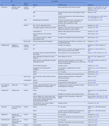
Studies of EVs in spermatozoa date back to the 1970s. These cells were one of the first models in which the mechanisms underlying EV-mediated cellular communication were studied. In 1978, Ronquist and colleagues described that there was a type of EVs in human seminal plasma that contained an Mg2+ and Ca2+-dependent ATPase system in their membranes (Ronquist et al., Reference Ronquist, Brody, Gottfries and Stegmayr1978). These EVs from the epithelial cells of the prostate would later be called prostasomes, which are secreted into the seminal fluid at the time of ejaculation. They have a diameter that varies between 30 and 500 nm and contain sphingomyelin, which is the main phospholipid in their membranes (Ronquist and Brody, Reference Ronquist and Brody1985). Moreover, when the spermatozoon leaves the testicle it must pass through the epididymis, which is a narrow and long convoluted tube whose length varies across species, reaching up to 80 m in Equus caballus. For this reason, the sperm that transits through this organ can take from 3 to 14 days depending on species (Sullivan et al., Reference Sullivan, Saez, Girouard and Frenette2005). As sperm make their way through this structure, the epididymal epithelial cells interact with them through different pathways, and which support the process of their maturation. Among these intercellular communication pathways, there is one mediated by epididymosomes, which was described for the first time in Chinese hamsters by Yanagimachi et al. (Reference Yanagimachi, Kamiguchi, Mikamo, Suzuki and Yanagimachi1985). Epididymosomes are a type of EVs with a diameter of 25–300 nm (Saez et al., Reference Saez, Frenette and Sullivan2003). They have two main subpopulations that seem to have different functions, which will be detailed in later sections (Yanagimachi et al., Reference Yanagimachi, Kamiguchi, Mikamo, Suzuki and Yanagimachi1985; Sullivan, Reference Sullivan2015). Another of the main sources of EVs that sperm receive comes from oviductal cells, and were termed oviductosomes by Al-Dossary et al. (Reference Al-Dossary, Strehler and Martin-DeLeon2013); they consist of vesicles with a diameter of 25–100 nm in murine and 50–250 nm in humans, and deliver essential biomolecule loads in the sperm while they travel through the female reproductive tract (Al-Dossary et al., Reference Al-Dossary, Strehler and Martin-DeLeon2013; Bathala et al., Reference Bathala, Fereshteh, Li, Al-Dossary, Galileo and Martin-DeLeon2018). These three types of EVs are the most studied among those that interact with sperm cells. Others EVs have also been reported, such as EVs from Sertoli cells (Mancuso et al., Reference Mancuso, Calvitti, Milardi, Grande, Falabella, Arato, Giovagnoli, Vincenzoni, Mancini, Nastruzzi, Bodo, Baroni, Castagnola, Marana, Pontecorvi, Calafiore and Luca2018), uterosomes from luminal fluid from the uterus (Martin-DeLeon, Reference Martin-DeLeon2016), vaginosomes from the epithelial and stromal cells of the vagina (Fereshteh et al., Reference Fereshteh, Bathala, Galileo and Martin-DeLeon2019), among others.
Accordingly, this section will detail the content of the main EVs that interact with sperm cells, i.e. prostasomes, epididymosomes and oviductosomes, and will also tackle how they modulate different processes in spermatozoa.
2.1 Prostasomes
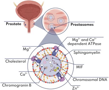
Different research groups have investigated the interactions of prostasomes with sperm cells. For example, it was discovered that the prostasomes found in the seminal plasma of mammals have a greater specificity for sperm than for other cells such as lymphocytes (Kumar et al., Reference Kumar, Pandita, Ganguly, Soren and Pagrut2018). In this sense, it should be mentioned that the sperm surface has a slightly lower net negative charge than the prostasome one (Ronquist et al., Reference Ronquist, Nilsson and Hjertën1990), which together with the probable existence of a binding protein mechanism, similar to that described by Al-Dossary et al., (Reference Al-Dossary, Bathala, Caplan and Martin-DeLeon2015), could explain their specificity and ease of merging with each other. However, this has not yet been elucidated (Al-Dossary et al., Reference Al-Dossary, Bathala, Caplan and Martin-DeLeon2015). Prostasomes contain divalent cations of calcium (Ca2+), zinc (Zn2+) and magnesium (Mg2+) and, as mentioned, their particular membrane is rich in sphingomyelin, also containing a 2:1 ratio of cholesterol/phospholipids (Kumar et al., Reference Kumar, Pandita, Ganguly, Soren and Pagrut2018). The Ca2+ cation participates together with bicarbonate in sperm capacitation, stimulating the soluble enzyme adenylate cyclase, which when activated generates an increase in the synthesis of cyclic AMP that activates protein kinase A, triggering the phosphorylation of tyrosines of various proteins sperm cells (Visconti et al., Reference Visconti, Moore, Bailey, Leclerc, Connors, Pan, Olds-Clarke and Kopf1995; Litvin et al., Reference Litvin, Kamenetsky, Zarifyan, Buck and Levin2003). Ca2+ is also the trigger for the acrosomal reaction, a process that allows sperm to penetrate the outer coatings of the oocyte (Visconti et al., Reference Visconti, Moore, Bailey, Leclerc, Connors, Pan, Olds-Clarke and Kopf1995; Wong et al., Reference Wong, Flik, Groenen, Swinkels, Thomas, Copius-Peereboom, Merkus and Steegers-Theunissen2001; Flesch et al., Reference Flesch, Wijnand, Van de Lest, Colenbrander, Van Golde and Gadella2001; Chávez et al., Reference Chávez, De Blas, De La Vega-Beltrán, Nishigaki, Chirinos, González-González, Larrea, Solís, Darszon and Treviño2011). The Mg2+ cation acts as a cofactor of many enzyme systems and has been related to the modulation of sperm motility (Wong et al., Reference Wong, Flik, Groenen, Swinkels, Thomas, Copius-Peereboom, Merkus and Steegers-Theunissen2001). Similarly, the Zn2+ cation acts as a cofactor for many metalloenzymes, and has been associated with different processes related to DNA, such as stabilization and decondensation of sperm chromatin (Kvist, Reference Kvist1980; Kvist et al., Reference Kvist, Kjellberg, Björndahl, Soufir and Arver1990), and expression of genes that encode for steroid receptors, whose transcription factors include those that contain zinc fingers (Mackeh et al., Reference Mackeh, Marr, Fadda and Kino2018). It is also involved in the activity of acrosin, an important hydrolytic enzyme present in the acrosome (Steven et al., Reference Steven, Griffin and Chantler1982). As far as sphingomyelin is concerned, it has been observed that it slows down the efflux of sterols in human spermatozoa, therefore preventing premature capacitation and acrosomal reaction (Cross, Reference Cross2000). In pigs, however, it inhibits the efflux of cholesterol and fluidity of the apical plasma membrane of the sperm head (manifestations of sperm capacitation), but not the acrosomal reaction (Piehl et al., Reference Piehl, Fischman, Hellman, Cisale and Miranda2013), suggesting that the effect of this sphingolipid varies between sperm of different species.
One of the first proteins described in prostasomes is the Mg2+ and Ca2+-dependent ATPase, which can increase the availability of ATP for sperm, which is necessary for motility (Ronquist et al., Reference Ronquist, Brody, Gottfries and Stegmayr1978). Prostasomes also contain the macrophage migration inhibitory factor (MIF), a pro-inflammatory cytokine classified as a ‘moonlighting’ protein, which was purported to be associated with the regulation of the innate immune response of the vagina to the presence of sperm proteins; nevertheless, this is yet to be experimentally confirmed (Frenette et al., Reference Frenette, Légaré, Saez and Sullivan2005).
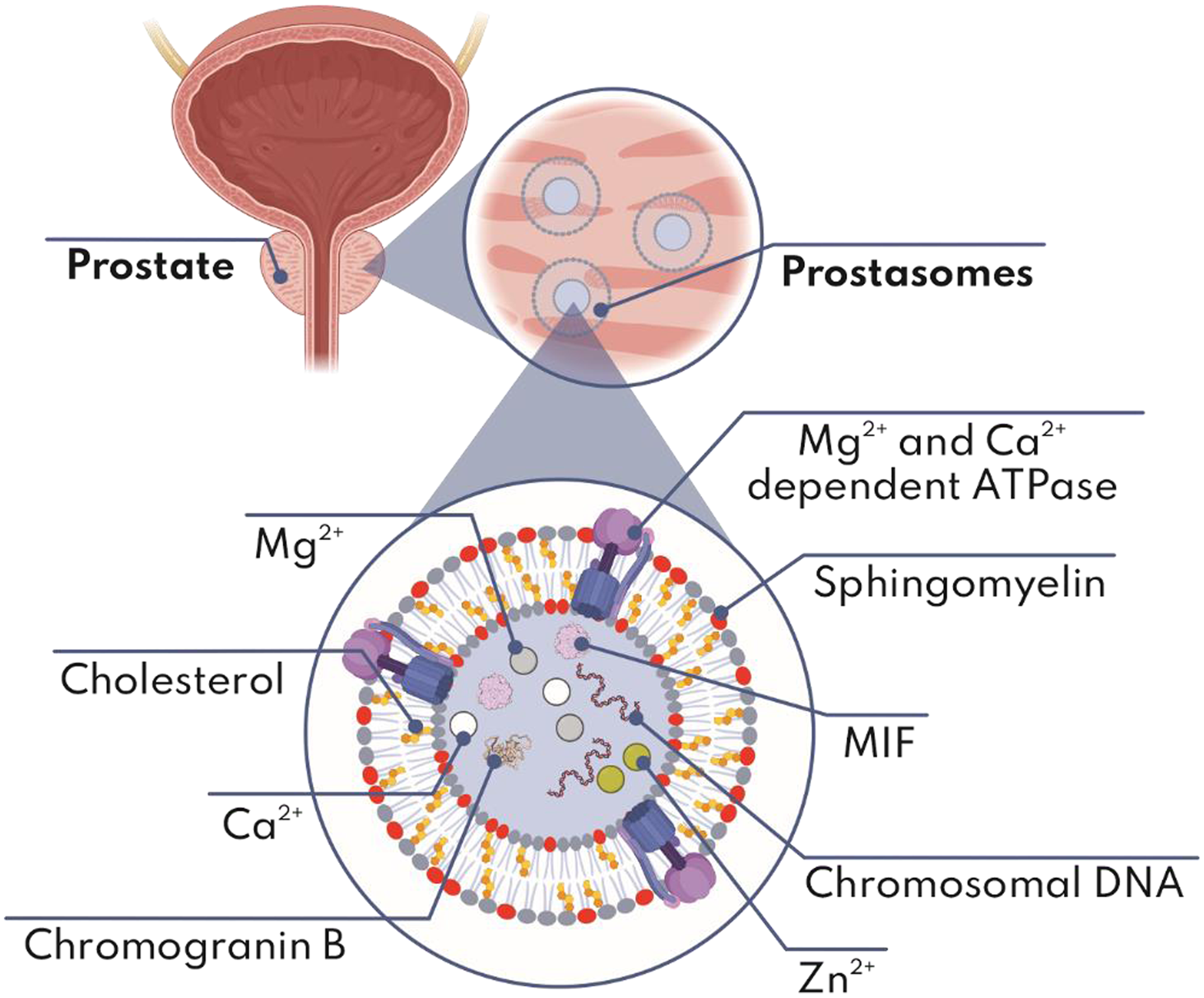
Additionally, prostasomes have been described as containing paternal chromosomal DNA fragments, which transfer to sperm (Ronquist et al., Reference Ronquist, Ronquist, Carlsson and Larsson2009, Reference Ronquist, Larsson, Ronquist, Isaksson, Hreinsson, Carlsson and Stavreus-Evers2011). It has been proposed that this process is performed to guide homology-directed repair (HDR) mechanisms in the embryo in case of sperm DNA damage caused, for example, by oxidative stress, which has not been confirmed experimentally (Ronquist et al., Reference Ronquist, Larsson, Ronquist, Isaksson, Hreinsson, Carlsson and Stavreus-Evers2011). Conversely, human prostasomes were reported to exert an antibiotic effect against Bacillus megaterium, because these EVs contain large amounts of chromogranin B, a protein that permeabilizes bacterial walls and membranes, therefore protecting sperm from possible attack by some microorganisms (Carlsson et al., Reference Carlsson, Nilsson, Larsson, Stridsberg, Sahlén and Ronquist2003). Figure 1 and Table 1 show the contents and effects of prostasomes on sperm.
Prostasomes. The figure shows the environment of the prostasomes and possible subvesicular localization of their components (enlarged images in digital format show the molecules in detail). Created with BioRender.com
Sperm-related EVs

* Experimentally not proven or mechanism not known.
2.2 Epididymosomes
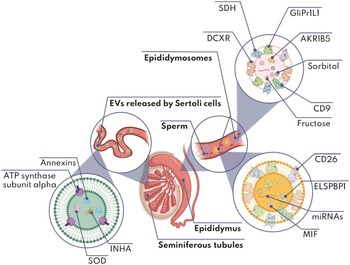
Sperm motility is a process with great energy expenditure. Zn2+ prevents sperm from wasting energy during their stay in the testes, by preventing the thiol groups of outer dense fibres (ODFs) from oxidizing and therefore forming disulphide bonds, necessary for proper motility. When sperm travel through the epididymis, epididymosomes enter the scene, delivering MIF to these gametes, which is responsible for removing Zn2+ from ODFs (Frenette et al., Reference Frenette, Légaré, Saez and Sullivan2005). However, as for Zn2+, MIF is a repressor of premature sperm motility, as it also reduces the thiol groups of ODFs, preventing the formation of disulphide bonds. Therefore it must also be eliminated at some point in order for sperm to become fully competent in the female environment, although the mechanisms involved in that removal are still unknown (Eickhoff et al., Reference Eickhoff, Baldauf, Koyro, Wennemuth, Suga, Seitz, Henkel and Meinhardt2004; Frenette et al., Reference Frenette, Légaré, Saez and Sullivan2005).
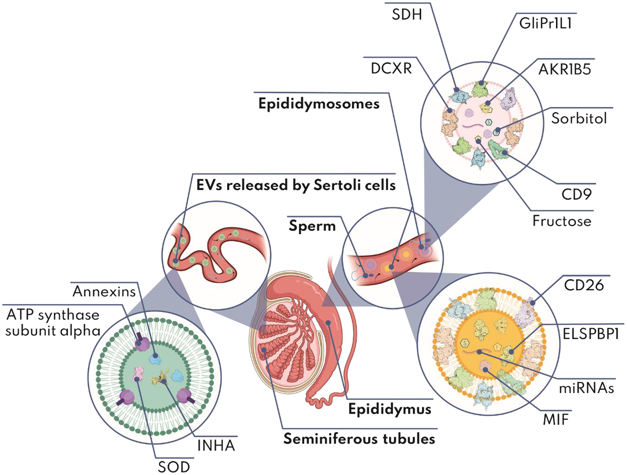
Conversely, while sperm travel through the epididymis during their maturation process, they receive dicarbonyl/l-xylulose reductase (DCXR) from epididymosomes, another moonlighting protein with various functions (Ebert et al., Reference Ebert, Kisiela and Maser2015). DCXR bound to the outer acrosomal membrane can modulate the primary adhesion of sperm to glycoproteins in the zona pellucida of oocytes during the fertilization process, presenting specificity at the species level (Boué et al., Reference Boué, Blais and Sullivan1996). It has been described under the name P34H in humans, P26h in hamsters and P25b in cattle, and it has also been found in mice and pigs (Tumova et al., Reference Tumova, Zigo, Sutovsky, Sedmikova and Postlerova2021). Glioma pathogenesis-related1-like protein 1 (GliPr1L1) is another protein that is delivered to sperm via epididymosomes. When blocked by anti-GliPr1L1 antibodies, the percentage of sperm that bind to the zona pellucida of oocytes is reduced, demonstrating its relevance in this process (Caballero et al., Reference Caballero, Frenette, D’Amours, Belleannée, Lacroix-Pepin, Robert and Sullivan2012). However, the molecular mechanisms through which this protein contributes to the sperm binding to the zona pellucida have not yet been elucidated (Yudin et al., Reference Yudin, Li, Robertson, Tollner, Cherr and Overstreet2002; Caballero et al., Reference Caballero, Frenette, D’Amours, Belleannée, Lacroix-Pepin, Robert and Sullivan2012).
In cattle (and humans; Frenette et al., Reference Frenette, Thabet and Sullivan2006), epididymosomes have been seen to contain two enzymes involved in the synthesis of fructose via the polyol pathway: aldose reductase 1 B5 (AKR1B5) and sorbitol dehydrogenase (SDH) (Frenette et al., Reference Frenette, Lessard and Sullivan2004). AKR1B5 reduces glucose generating sorbitol, a polyol that is not very permeable to the plasma membrane, through a reaction is optimal at pH 6.5, which can be found throughout the caput and corpus of the epididymis (Burg, Reference Burg1995; Frenette et al., Reference Frenette, Lessard and Sullivan2004). Then, sorbitol is oxidized by SDH into fructose, the main carbohydrate used by sperm to produce energy through glycolysis, which is also more permeable to the membrane than sorbitol (Cao et al., Reference Cao, Aghajanian, Haig-Ladewig and Gerton2009). SDH optimally generates fructose at pH 7.2, which can be found in the cauda of the epididymis and in semen (Frenette et al., Reference Frenette, Lessard and Sullivan2004, Reference Frenette, Thabet and Sullivan2006). However, it has not yet been determined whether epididymosomes can deliver both enzymes to sperm. In this sense, AKR1B5 is associated with this type of EVs in cattle and animals with a developed epididymis, it could modulate a mechanism to decrease the energy metabolism of sperm during their journey through the caput and corpus of the epididymis by transforming glucose into sorbitol to limit the entry of carbohydrates into sperm, in addition to generating a hyperosmotic milieu that can improve the survival and storage of these gametes (Crichton et al., Reference Crichton, Hinton, Pallone and Hammerstedt1994; Frenette et al., Reference Frenette, Lessard and Sullivan2004). Then, during the ejaculation process, SDH associated to epididymosomes transforms sorbitol into fructose, which is capable of crossing the sperm membrane to serve as an energy source that supplies the subsequent sperm processes that culminate in fertilization (Frenette et al., Reference Frenette, Thabet and Sullivan2006).
Moreover, miRNA transfer from epididymosomes to sperm has been described in mice, establishing that epididymosomes and sperm share 82% of the miRNA profile. It is worth mentioning miR-30, miR-465, miR-466, miR-467, and miR-669, which could be involved in the translational regulation in embryos during their early stages and in epigenetic inheritance processes (Reilly et al., Reference Reilly, McLaughlin, Stanger, Anderson, Hutcheon, Church, Mihalas, Tyagi, Holt, Eamens and Nixon2016).
As mentioned above, there are two subpopulations of epididymosomes, one being positive for CD9 and CD26 tetraspanins, and the other being positive for epididymal sperm binding protein 1 (ELSPBP1; Sullivan, Reference Sullivan2015). CD9+ epididymosomes are 10–100 nm in diameter and contain high levels of proteins such as MIF, DCXR, GliPr1L1 and AKR1B5 (Sullivan, Reference Sullivan2015). Tests with anti-CD9 and anti-CD26 antibodies indicated that the blockade of both tetraspanins prevents the fusion of CD9+ epididymosomes with sperm, which implicates both tetraspanins in the fusogenic process (Caballero et al., Reference Caballero, Frenette, Belleannée and Sullivan2013). Furthermore, CD9+ epididymosomes were described as having a preference for living and functional sperm (Caballero et al., Reference Caballero, Frenette, Belleannée and Sullivan2013). Conversely, in cattle, it has been observed that the ELSPBP1+-epididymosome subpopulation has greater affinity for dead spermatozoa during their journey through the caput and cauda of the epididymis, which is reinforced by the presence of the Zn2+ cation (D’Amours et al., Reference D’Amours, Frenette, Bordeleau, Allard, Leclerc, Blondin and Sullivan2012). It should also be noted that such EVs do not fuse with dead sperm in vitro (D’Amours et al., Reference D’Amours, Frenette, Bordeleau, Allard, Leclerc, Blondin and Sullivan2012). The function of ELSPBP1 has not yet been determined nor have the mechanisms underlying the specificity of ELSPBP1+ EVs for dead male gametes in vivo (D’Amours et al., Reference D’Amours, Frenette, Caron, Belleannée, Guillemette and Sullivan2016). See Figure 2 and Table 1 for a description of the contents and effects of epididymosomes on sperm.
Epididymosomes and EVs released by Sertoli cells. The figure shows the environment of the epididymosomes and EVs released by Sertoli cells, and possible subvesicular localization of their components (enlarged images in digital format show the molecules in detail). Created with BioRender.com
2.3 Oviductosomes, uterosomes, vaginosomes and EVs released by Sertoli cells
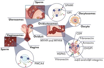
In mice and cattle, it has been seen that when sperm reach the female reproductive tract they interact with oviductosomes, which are capable of transferring proteins such as plasma membrane calcium ATPase 4 (PMCA4), which is the main Ca2+ flow pump in these gametes (Al-Dossary et al., Reference Al-Dossary, Strehler and Martin-DeLeon2013). Another study that delved into the interactions of oviductal fluid proteins with bovine spermatozoa (Lamy et al., Reference Lamy, Nogues, Combes-Soia, Tsikis, Labas, Mermillod, Druart and Saint-Dizier2018) identified 21 probable oviductosomal proteins that interact with spermatozoa. These include annexins A1 (ANXA1) and A2 (ANXA2), non-muscle myosin heavy chain IIa (MYH9) and IIc (MYH14), heat shock 70 kDa protein 5 (HSPA5), and oviduct-specific glycoprotein (OVGP1). These proteins are involved in the modulation of bovine sperm capacitation, motility and viability, as was observed in vitro and in silico (King and Killian, Reference King and Killian1994; Abe et al., Reference Abe, Sendai, Satoh and Hoshi1995; Almiñana et al., Reference Almiñana, Corbin, Tsikis, Alcântara-Neto, Labas, Reynaud, Galio, Uzbekov, Garanina, Druart and Mermillod2017; Lamy et al., Reference Lamy, Nogues, Combes-Soia, Tsikis, Labas, Mermillod, Druart and Saint-Dizier2018). It was described that murine uterosomes, (as well as murine, human and bovine prostasomes, epididymosomes and oviductosomes) can deliver proteins such as PMCA4 and sperm adhesion molecule 1 (SPAM1) to spermatozoa, with the expression levels of both proteins being dependent on hormonal stimuli throughout the estrous or menstrual cycle (Martin-DeLeon, Reference Martin-DeLeon2016). SPAM1 is a hyaluronidase anchored to glycosylphosphatidylinositol (GPI) on the sperm surface and in the inner membrane of the acrosome, which participates in processes such as the acrosomal reaction, binding and degradation of hyaluronic acid from the matrix of cumulus cells and binding to the zona pellucida (Griffiths et al., Reference Griffiths, Galileo, Reese and Martin-DeLeon2008). Other less studied EVs of the female reproductive tract such as vaginosomes and EVs from the endometrium (endometriosomes) have been shown to modulate different processes in sperm in vitro, such as capacitation and acrosomal reaction, because they contain proteins such as PMCA4 and SPAM1 (Franchi et al., Reference Franchi, Cubilla, Guidobaldi, Bravo and Giojalas2016; Fereshteh et al., Reference Fereshteh, Bathala, Galileo and Martin-DeLeon2019).
Moreover, it is important to mention that the mechanism involved in the fusogenic process for the delivery of the molecular cargo to spermatozoa by oviductosomes has been proposed to be mediated by tetraspanin CD9, which serves as a scaffolding protein binding to adhesion proteins such as αvβ3 and α5β1 integrins, and whose ligands are vitronectin and fibronectin, respectively (Al-Dossary et al., Reference Al-Dossary, Bathala, Caplan and Martin-DeLeon2015). CD9, the two integrins and their respective ligands are present in oviductosomes, as well as throughout the head and mid-piece of sperm. These are places where the receptor–ligand interaction should take place and give way to the fusogenic process and the subsequent delivery of the different molecules from EVs to sperm. Although it was confirmed by confocal microscopy that epididymosomes and uterosomes also interact with the head and mid-piece of murine sperm, this has not been shown to occur through the same mechanism (Griffiths et al., Reference Griffiths, Galileo, Reese and Martin-DeLeon2008).
Finally, the EVs released by Sertoli cells were reported to contain proteins involved in the conditioning of the testicular environment to give way to spermatogenesis, highlighting that the protein profiles of these EVs respond in vitro to testosterone and FSH stimuli (Mancuso et al., Reference Mancuso, Calvitti, Milardi, Grande, Falabella, Arato, Giovagnoli, Vincenzoni, Mancini, Nastruzzi, Bodo, Baroni, Castagnola, Marana, Pontecorvi, Calafiore and Luca2018). Figure 2, Figure 3 and Table 1 show the contents and effects of oviductosomes, uterosomes, vaginosomes and EVs released by Sertoli cells on sperm.
Oviductosomes, vaginosomes, uterosomes and oocytosomes. The figure shows the environment of the oviductosomes, vaginosomes, uterosomes and oocytosomes, and possible subvesicular localization of their components (enlarged images in digital format show the molecules in detail). Created with BioRender.com

2.4 New perspectives
The properties exhibited by the EVs mentioned in section 2 can be exploited to enhance the efficacy of ARTs from the sperm factor, with the most reasonable being to use their vesicular content, such as cations, carbohydrates, lipids, proteins or nucleic acids, to intervene in important processes such as capacitation, acrosomal reaction and/or binding to the zona pellucida. However, slightly broader perspectives can be proposed for its application. For example, as mentioned above, prostasomes have affinity for sperm and show an intrinsic ability to transfer chromosomal DNA molecules to them. This can be of great interest to improve sperm-mediated gene transfer (SMGT), therefore allowing animal transgenesis in different species without the need for expensive equipment, and also to reduce the percentage of embryos with mosaicism (Smith and Spadafora, Reference Smith and Spadafora2005; Canovas et al., Reference Canovas, Gutierrez-Adan and Gadea2010).
Human prostasomes contain large amounts of chromogranin B, which, in addition to the specificity and low immunogenicity of EVs in general (Kim et al., Reference Kim, Yang, Oh, Hong, Seo and Jang2017), offer an interesting alternative as the delivery vehicle for the antibiotic compound to combat resistant infections. However, further clinical trials are needed to validate this approach. Conversely, the high sphingomyelin and cholesterol content of proteasomal membranes that is transferred to sperm membranes could make sperm cryopreservation techniques more efficient by stabilizing their membranes, therefore reducing cryocapacitation, risk of biological contaminants, as for using egg yolk, and cryodamage that compromises sperm viability (Singh et al., Reference Singh, Kumar and Atreja2014). This makes these EVs a relatively inexpensive and accessible alternative to increase the percentage of competent sperm post-thaw. As for the DCXR protein in the spermatozoa, transferred by the epididymosomes, this can be used as a marker of epididymal maturation, to select the most suitable spermatozoa for different ARTs. Similarly, the subpopulation of ELSPBP1+ epididymosomes, which have affinity for dead sperm in vivo, could help to improve the selection of sperm to be used in intracytoplasmic sperm injection (ICSI), especially when such gametes come from individuals with fertility problems due to low or no sperm motility. Finally, EVs from Sertoli cells could provide a range of different biomarkers of infertility related to hormones such as FSH and testosterone, as hormonal stimuli modify the protein profiles of these EVs (Mancuso et al., Reference Mancuso, Calvitti, Milardi, Grande, Falabella, Arato, Giovagnoli, Vincenzoni, Mancini, Nastruzzi, Bodo, Baroni, Castagnola, Marana, Pontecorvi, Calafiore and Luca2018).
3. Interaction of EVs with follicular cells and the oocyte
The research on EVs that can influence the oocyte or its environment is relatively recent. However, during the 1990s the oocyte was for the first time suggested to be capable of modifying its follicular microenvironment, controlling the differentiation of granulosa and cumulus cells through labile paracrine factors (Eppig et al., Reference Eppig, Chesnel, Hirao, O’Brien, Pendola, Watanabe and Wigglesworth1997). One of the first reports of the interaction of EVs with oocytes was made in Caenorhabditis elegans, where it was described how their spermatids and spermatozoa secrete a specific type of EVs (spermatosomes). These EVs were found to target the oocyte and deliver the major sperm protein (MSP), which acts as a hormone that modulates the meiotic maturation of oocytes (Kosinski et al., Reference Kosinski, McDonald, Schwartz, Yamamoto and Greenstein2005). In mammals, the existence of spermatosomes was not reported, but they have been synthesized in vitro from pieces of murine sperm membrane, to use them as vehicles that mediate the delivery of molecules (Atif et al., Reference Atif, Salam, Ahmad, Hasan, Jamal, Sudhanshu, Azevedo and Owais2008). So far, spermatosomes seem to be an interesting quirk of nature limited to invertebrates. For this reason, studies of EVs involved in the development of mammalian oocytes have focused on the maturation microenvironment, that is, the ovarian follicle, where the oocyte can interact with different cell types such as theca cells, granulosa cells (mural granulosa cells) and cumulus cells (cumulus granulosa cells). One of the first studies that described the existence of EVs released in the microenvironment of the ovarian follicle by somatic cells (folliculosomes) was carried out in Drosophila melanogaster, where it was observed how the ZAM retrotransposon uses folliculosomes to infect oocytes to be transmitted to the offspring (Brasset et al., Reference Brasset, Taddei, Arnaud, Faye, Fausto, Mazzini, Giorgi and Vaury2006). Mammalian folliculosomes were not observed until 2012, when da Silveira and colleagues described them as CD63+ EVs, with a fairly heterogeneous diameter from 50 nm to 1000 nm. The content of these EVs is influenced by different factors and seems to be essential in processes such as folliculogenesis and oogenesis (da Silveira et al., Reference da Silveira, Veeramachaneni, Winger, Carnevale and Bouma2012), as will be detailed in later paragraphs.
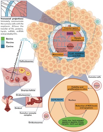
When the oocyte is ovulated, it is arrested at metaphase (MII) and leaves the follicular microenvironment to face a new milieu dominated by oviductal cells and their respective oviductosomes (detailed in the previous section). Although the oocyte reaches the MII stage within the ovarian follicle in most mammals, this is not the case in canines, as meiotic maturation is completed within the oviductal environment; therefore, oviductosomes could have a relevant role in this process (Lee et al., Reference Lee, Oh, Kim and Lee2020b). The fact that oocytes are ovulated at the MII stage in most mammals does not rule out the possible role of oviductosomes on these gametes; these potential interactions, however, have not been addressed to our knowledge. Conversely, it is well known that among the components of the follicular fluid, there is a portion that corresponds to constituents of blood plasma (Sohel et al., Reference Sohel, Hoelker, Noferesti, Salilew-Wondim, Tholen, Looft, Rings, Uddin, Spencer, Schellander and Tesfaye2013a), where it would be expected to find EVs from different organs of the body that could interact with the follicular cells and the oocyte. Although the latter was not described experimentally, it has been shown under in vitro conditions that EVs from other cellular environments have a positive effect on the correct competition of in vitro-matured oocytes (Ding et al., Reference Ding, Zhu, Shen, Lu, Zou, Huang, Li and Huang2020; Yang et al., Reference Yang, Zhang, Xu, He, Liu, Li, Zhang, Lin, Su, Wu and Li2020). It is important to note at this point that, while the oocyte has been described as a receptor cell for EVs in invertebrates (Kosinski et al., Reference Kosinski, McDonald, Schwartz, Yamamoto and Greenstein2005; Brasset et al., Reference Brasset, Taddei, Arnaud, Faye, Fausto, Mazzini, Giorgi and Vaury2006), this is still in question in mammals. In effect, an experimental confirmation (with the exception of canines, which have physiologically different oogenesis; Lee et al., Reference Lee, Oh, Kim and Lee2020b) about whether the EVs of the cumulus cells or any subtype of folliculosome are capable of crossing and/or interacting with the zona pellucida and discharge its content to the ooplasm is not available. Therefore, the effects observed in vitro when EVs are added to the oocyte maturation medium could be an indirect consequence orchestrated by the cumulus cells (Russell et al., Reference Russell, Gilchrist, Brown and Thompson2016). However, it is important to highlight that, in cattle, communication between cumulus cells and the oocyte was confirmed by intracellular vesicles (IVs) loaded with mRNA and non-coding RNA that contributed to the reserve of maternal RNAs in the oocyte (Macaulay et al., Reference Macaulay, Gilbert, Caballero, Barreto, Fournier, Tossou, Sirard, Clarke, Khandjian, Richard, Hyttel and Robert2014). These IVs reached the ooplasm through transzonal projections from the cumulus cells, which are in close contact with the zona pellucida and the oolemma (Figure 5) (Macaulay et al., Reference Macaulay, Gilbert, Caballero, Barreto, Fournier, Tossou, Sirard, Clarke, Khandjian, Richard, Hyttel and Robert2014). Regarding the role of mammalian oocytes as emitters of EVs, this has been observed experimentally in murine, whose oocytosomes can interact with sperm and most likely with follicular cells, such as cumulus cells (Figure 3; Eppig et al., Reference Eppig, Chesnel, Hirao, O’Brien, Pendola, Watanabe and Wigglesworth1997; Miyado et al., Reference Miyado, Yoshida, Yamagata, Sakakibara, Okabe, Wang, Miyamoto, Akutsu, Kondo, Takahashi, Ban, Ito, Toshimori, Nakamura, Ito, Miyado, Mekada and Umezawa2008; Gupta et al., Reference Gupta, Primakoff and Myles2009). Therefore, as folliculosomes are by far the most studied EVs in the mammalian oocyte microenvironment, this section will mainly focus on detailing the different effects that folliculosomes and their content can have upon follicular cells and the oocyte.
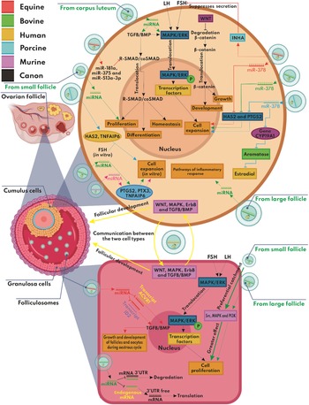
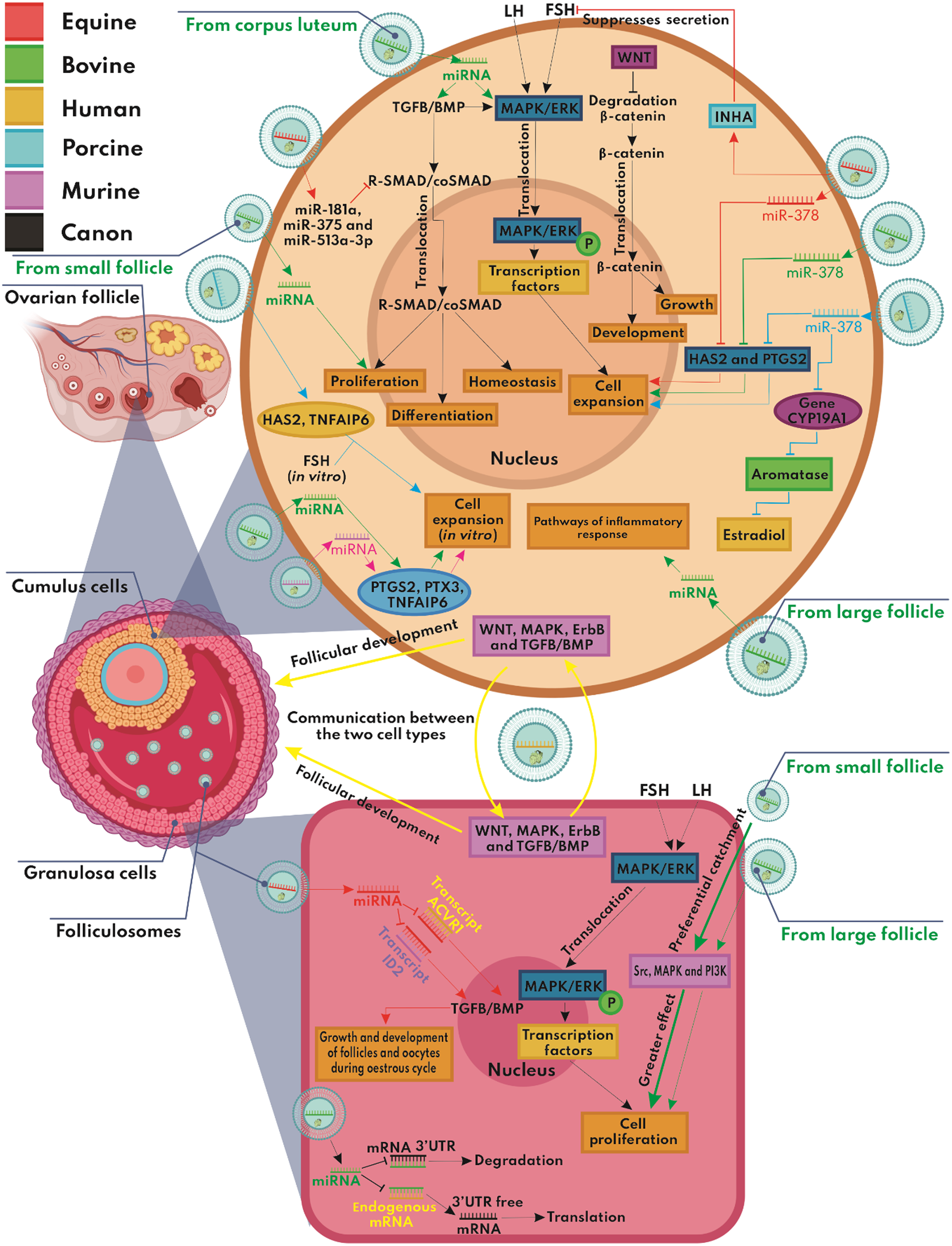
Folliculosomes. Effects of folliculosomes on cumulus cells (upper region) and granulosa cells (lower region). Canonical signalling pathways are represented in black. Signalling pathways involving folliculosomal content are represented in different colours depending on the species in which they have been studied, as indicated in the legend at the top left. Arrows (→) represent activation, stimulation or effects. Truncated lines (—|) represent inhibition or suppression. Different coloured rectangles symbolize proteins or signalling pathways, whereas orange rectangles symbolize effects. Ovals symbolize genes (enlarged images in digital format show the molecules in detail). P, phosphorylation. Created with BioRender.com
3.1 Folliculosomes
In folliculosomes, the main biomolecules that have been studied are miRNAs. These small ‘pieces’ of ribonucleic acid have attracted the interest of different research groups due to their ability to intervene in the expression process of different genes. Either by complementing themselves with the 3′-untranslated region (3′-UTR) of an mRNA for its subsequent degradation (decreasing its expression), or by complementing itself with an endogenous miRNA from the target cell, which prevents the translation of an mRNA of interest, avoiding the degradation of the latter (increasing its expression) (Figure 4). In silico analyses predicted that folliculosomal miRNAs can modulate mitogen-activated protein kinases/extracellular signal-regulated kinases (MAPK), transforming growth factor beta (TGFβ) and canonical WNT signalling pathways (da Silveira et al., Reference da Silveira, Veeramachaneni, Winger, Carnevale and Bouma2012; Sohel et al., Reference Sohel, Salilew-Wondim, Hölker, Rings, Schellander and Tesfaye2013b). In follicular cells, the activation of the MAPK/extracellular signal-regulated kinases (MAPK/ERK) signalling pathway depends on hormones such as FSH and LH, which trigger a molecular mechanism that culminates in the activation of the ERK protein. This serine/threonine kinase is capable of translocating to the nucleus and from there regulate the transcriptional processes of various genes through phosphorylating transcription factors, which ultimately stimulates the proliferation of granulosa cells and the expansion of cumulus cells (Orton et al., Reference Orton, Sturm, Vyshemirsky, Calder, Gilbert and Kolch2005; Santonocito et al., Reference Santonocito, Vento, Guglielmino, Battaglia, Wahlgren, Ragusa, Barbagallo, Borzì, Rizzari, Maugeri, Scollo, Tatone, Valadi, Purrello and Di Pietro2014). However, the activation of MAPK in cumulus cells also requires factors of the TGFβ family, such as bone morphogenetic protein (BMP), which are produced by the oocyte and are involved in the formation of complexes R-SMAD/coSMAD that are translocated to the cell nucleus to act as transcription factors of multiple genes, therefore modulating the proliferation, differentiation and homeostasis of follicular cells (Lyons and Prasad, Reference Lyons and Prasad2012; Santonocito et al., Reference Santonocito, Vento, Guglielmino, Battaglia, Wahlgren, Ragusa, Barbagallo, Borzì, Rizzari, Maugeri, Scollo, Tatone, Valadi, Purrello and Di Pietro2014). In addition, the canonical WNT pathway culminates in blocking the protein complex involved in the degradation of β-catenin, which translocates to the cell nucleus and acts as a transcription factor for multiple genes involved in the growth and development of follicular cells (Figure 4) (Santonocito et al., Reference Santonocito, Vento, Guglielmino, Battaglia, Wahlgren, Ragusa, Barbagallo, Borzì, Rizzari, Maugeri, Scollo, Tatone, Valadi, Purrello and Di Pietro2014; Nusse and Clevers, Reference Nusse and Clevers2017).
Folliculosomes and oviductosomes. Effects of folliculosomes (light blue) on oocytes (upper region) and oviductosomes (pink) on cumulus cells (lower region). Signalling pathways involving folliculosomal or oviductosomal content are represented as different colours depending on the species in which they have been studied, as indicated in the legend at the top left. Arrows (→) represent activation, stimulation or effects. Truncated lines (—|) represent inhibition or suppression. Different coloured rectangles symbolize proteins or signalling pathways, while orange rectangles symbolize effects. Ovals symbolize genes (enlarged images in digital format show the molecules in detail). Ac, acetylation. Created with BioRender.com

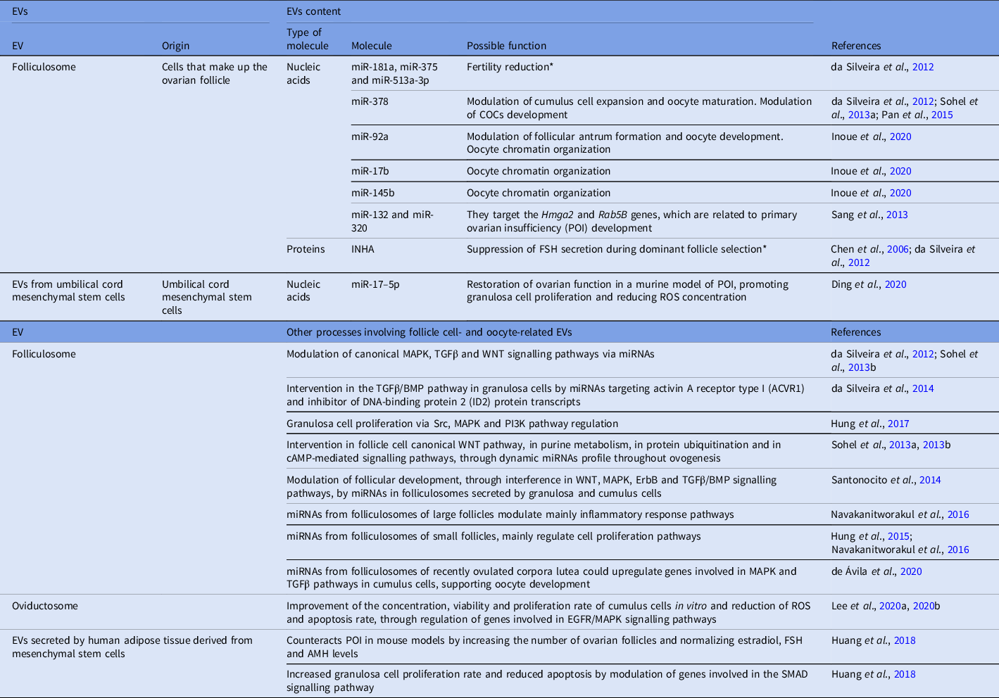
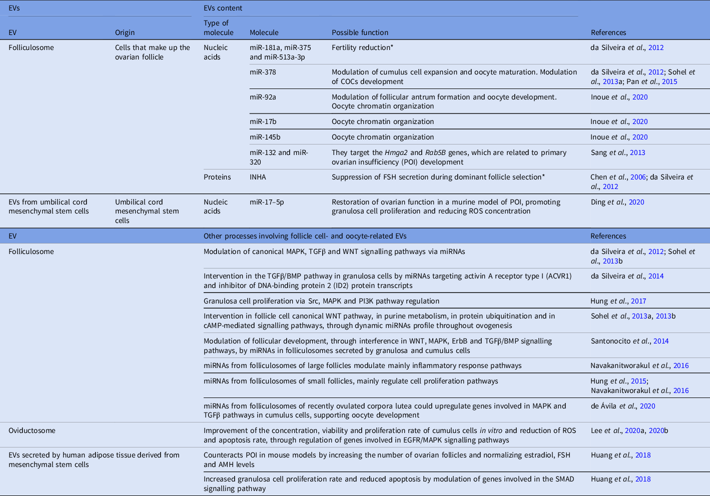
In 2012, da Silveira and colleagues reported the profiles of miRNAs and folliculosomal proteins in young and old mares. Specifically, the transcriptomic analysis of these vesicles revealed that 120 miRNAs were differentially expressed according to the age of the animals, highlighting that miR-181a, miR-375 and miR-513a-3p were more abundant in old mares. In silico analyses predicted that these miRNAs target TGFβ-pathway genes, coinciding with the reduction in fertility at older ages, probably due to abnormal folliculogenesis and oogenesis (da Silveira et al., Reference da Silveira, Veeramachaneni, Winger, Carnevale and Bouma2012). Regarding the proteomic analysis in the folliculosomes, the presence of the alpha chain of inhibin (INHA) was detected. This hormone suppresses FSH secretion during the selection of the dominant follicle through a molecular mechanism that has not yet been clarified (Chen et al., Reference Chen, Wang, Lin, Chang, Chuang and Ying2006; da Silveira et al., Reference da Silveira, Veeramachaneni, Winger, Carnevale and Bouma2012). In the horse, follicular sEVs were described to be able to modulate the growth and development of the follicle and the oocyte throughout the oestrous cycle. These follicular sEVs intervene in the TGFβ/BMP pathway of granulosa cells through miRNAs that target the transcripts of activin A receptor, type I (ACVR1) and DNA-binding protein inhibitor 2 (ID2) proteins (da Silveira et al., Reference da Silveira, Carnevale, Winger and Bouma2014). Furthermore, in cattle, it has been described that folliculosomes support the proliferation of granulosa cells by regulating Src, MAPK and PI3K pathways, highlighting that EVs from small follicles have a more marked effect than those from the large ones and that granulosa cells, regardless of the size of their follicle of origin, have a preferential uptake of small follicle folliculosomes (Hung et al., Reference Hung, Navakanitworakul, Khan, Zhang, Davis, McGinnis and Christenson2017). Also in cattle, it has been reported that folliculosomes contain a dynamic miRNA profile throughout oogenesis, some of which may intervene in both the canonical WNT pathway of follicular cells and purine metabolism, protein ubiquitination and cAMP-mediated signalling pathways (Sohel et al., Reference Sohel, Hoelker, Noferesti, Salilew-Wondim, Tholen, Looft, Rings, Uddin, Spencer, Schellander and Tesfaye2013a, Reference Sohel, Salilew-Wondim, Hölker, Rings, Schellander and Tesfaye2013b). In humans, it has been reported that the miRNAs present in folliculosomes and secreted by granulosa and cumulus cells can interfere with the WTN, MAPK, ErbB and TGFβ/BMP signalling pathways, therefore modulating follicular development (Santonocito et al., Reference Santonocito, Vento, Guglielmino, Battaglia, Wahlgren, Ragusa, Barbagallo, Borzì, Rizzari, Maugeri, Scollo, Tatone, Valadi, Purrello and Di Pietro2014). In porcine (Pan et al., Reference Pan, Toms, Shen and Li2015), bovine (Sohel et al., Reference Sohel, Hoelker, Noferesti, Salilew-Wondim, Tholen, Looft, Rings, Uddin, Spencer, Schellander and Tesfaye2013a) and equine (da Silveira et al., Reference da Silveira, Veeramachaneni, Winger, Carnevale and Bouma2012), the levels of folliculosomal miR-378 were found to decrease as the oocyte matured. This miRNA was proposed to modulate cumulus cell expansion and oocyte maturation through the translational disruption of proteins involved in these processes, such as HAS2 and PTGS2, and CX43, ADAMTS1 and PGR, respectively (Pan et al., Reference Pan, Toms, Shen and Li2015). In addition, miR-378 targets the Cyp19A1 gene that codes for aromatase, a key enzyme in steroidogenesis. Such translational disruption inhibits oestradiol synthesis in cumulus cells, which suggests that this miRNA modulates the development of cumulus–oocyte complexes (COCs) (Pan et al., Reference Pan, Toms, Shen and Li2015). In cattle and mice, another way in which folliculosomes, especially those from small antral follicles, support the expansion of cumulus cells is via the upward expression of genes involved in this process, such as prostaglandin-endoperoxide synthase 2 (Ptgs2), pentraxin-related protein 3 (Ptx3) and tumour necrosis factor alpha-induced protein 6 (Tnfaip6) (Hung et al., Reference Hung, Hong, Christenson and McGinnis2015). The explanation about why folliculosomes from smaller follicles have greater effects on the expansion of cumulus cells has come from the work of Navakanitworakul et al. (Reference Navakanitworakul, Hung, Gunewardena, Davis, Chotigeat and Christenson2016). These authors determined that while large follicles contain fewer folliculosomes than their small counterparts and that their borne miRNAs mainly modulate inflammatory response pathways, the miRNAs of small follicle folliculosomes mostly regulate the pathways of cell proliferation (Navakanitworakul et al., Reference Navakanitworakul, Hung, Gunewardena, Davis, Chotigeat and Christenson2016). In contrast, pig folliculosomes from 2 to 5 mm (small) follicles do not have a significant effect on the expansion of cumulus cells in vitro, despite upregulating the expression of Has2 and Tnfaip6, both are involved in that process when added together with FSH to the culture medium (Matsuno et al., Reference Matsuno, Kanke, Maruyama, Fujii, Naito and Sugiura2019). A recent study, also in pigs, found that miR-92a modulated the formation and diameter of the follicular antrum and the acetylation levels of the H4K12 protein in the oocyte, which is associated with correct oocyte development (Inoue et al., Reference Inoue, Munakata, Shinozawa, Kawahara-Miki, Shirasuna and Iwata2020). According to this, it is worth mentioning that miR-92a together with miR-17b and miR-145b contribute to the organization of oocyte chromatin (Inoue et al., Reference Inoue, Munakata, Shinozawa, Kawahara-Miki, Shirasuna and Iwata2020). Regarding the folliculosomes from recently ovulated corpora lutea, which contain low levels of progesterone, they present a high content of miRNAs that could upwardly modulate genes involved in the MAPK and TGFβ pathways in cumulus cells, therefore supporting oocyte development (de Ávila et al., Reference de Ávila, Bridi, Andrade, del Collado, Sangalli, Nociti, da Silva Junior, Bastien, Robert, Meirelles, Perecin and da Silveira2020).
The aforementioned studies showed the dynamics of the content of folliculosomes throughout the oestrous cycle and how they can modulate different pathways in granulosa and cumulus cells through their borne miRNAs. However, there are other factors that can modify the characteristics and content of folliculosomes. It has been reported, for example, that mastitis reduces the diameter of folliculosomes in bovine (Santos et al., Reference Santos, Bottino, Santos, Simões, Souza, Ferreira, da Silveira, Ávila, Bride and Sales2018), and that the metabolic state influences the miRNA profile of these EVs in cattle and humans (Hailay et al., Reference Hailay, Hoelker, Poirier, Gebremedhn, Rings, Saeed-Zidane, Salilew-Wondim, Dauben, Tholen, Neuhoff, Schellander and Tesfaye2019; Martinez et al., Reference Martinez, Baccarelli, Liang, Dioni, Mansur, Adir, Bollati, Racowsky, Hauser and Machtinger2019). Exposure to oxidative stress conditions such as hyperthermia has also been shown to modulate the folliculosomal content, to protect the oocyte from possible damage caused by non-physiological conditions (Saeed-Zidane et al., Reference Saeed-Zidane, Linden, Salilew-Wondim, Held, Neuhoff, Tholen, Hoelker, Schellander and Tesfaye2017; Morales Dalanezi et al., Reference Morales Dalanezi, Mogollon Garcia, de Andrade Ferrazza, Fagali Franchi, Kubo Fontes, de Souza Castilho, Gouveia Nogueira, Dos Santos Schmidt, Sartori and Pinheiro Ferreira2019). However, it has been observed that folliculosomes produced under non-stressful conditions have the intrinsic ability to protect oocytes from the stress caused in extreme circumstances, such as cryopreservation (Ferraz et al., Reference Ferraz, Fujihara, Nagashima, Noonan, Inoue-Murayama and Songsasen2020) or hyperthermia (Rodrigues et al., Reference Rodrigues, Tuna, Alli, Tribulo, Hansen, Koh and Paula-Lopes2019). Figure 4, Figure 5 and Table 2 summarize the contents and effects of folliculosomes on the oocyte and follicular cells.
Follicle cell- and oocyte-related EVs
* Experimentally not proven or mechanism not known.
3.2 Oviductosomes and other EVs
As mentioned above, the dog oocyte completes its meiotic maturation in the oviduct, that is why the role of oviductosomes in canine oogenesis has been studied. It was reported that, in vitro, oviductosomes improve the concentration, viability and proliferation rate of cumulus cells and also reduces the accumulation of reactive oxygen species (ROS) and the rate of apoptosis, through regulating genes involved in the EGFR/MAPK signalling pathways (Egfr, Pka, Tace/Adam17, Mapk1/3, Mapk14, Ptgs2, Tnfaip6, Gdf9 and Bmp15; Lee et al., Reference Lee, Oh, Kim and Lee2020a, Reference Lee, Oh, Kim and Lee2020b). It is important to note that, at least in dogs, it was confirmed that cumulus cells and oocytes absorbed oviductosomes (Lee et al., Reference Lee, Oh, Kim and Lee2020b).
Similarly, the effect of EVs from other cell types on the follicular environment was investigated. EVs secreted by human adipose tissue derived from mesenchymal stem cells has been showed to counteract primary ovarian insufficiency (POI) in mouse models. Increasing the number of ovarian follicles and normalizing the levels of oestradiol, FSH and AMH (Huang et al., Reference Huang, Lu, Ding, Zou, Wang and Li2018). In addition, such EVs showed the increment of the proliferation rate of granulosa cells, also reducing apoptosis through the modulation of genes involved in the SMAD signalling pathway (Huang et al., Reference Huang, Lu, Ding, Zou, Wang and Li2018). Comparable effects were observed with EVs from human umbilical cord mesenchymal stem cells, as they restored ovarian function in a POI murine model, promoting the proliferation of granulosa cells and reducing the concentration of ROS via inhibiting the expression of Sirt7 through the delivery of miR-17-5p (Ding et al., Reference Ding, Zhu, Shen, Lu, Zou, Huang, Li and Huang2020). See Figure 5 and Table 2 for a description of the contents and effects of oviductosomes on cumulus cells.
3.3 New perspectives
Although spermatosomes do not appear to be present in mammals, it has been shown that they can be synthesized in vitro (Atif et al., Reference Atif, Salam, Ahmad, Hasan, Jamal, Sudhanshu, Azevedo and Owais2008). However, due to the nature of their membrane proteins (Tumova et al., Reference Tumova, Zigo, Sutovsky, Sedmikova and Postlerova2021) and the fact that oocytes are physiologically adapted to the presence of spermatozoa, they become an attractive vehicle with high affinity and low cytotoxicity to guide different molecules such as miRNAs, mRNAs or proteins towards COCs to enhance their maturation in vitro. For example, synthetic miRNAs that decrease Fatty Acid Binding Protein 3 (FABP3) synthesis in cumulus cells of COCs in vitro, could be delivered through spermatosomes to reduce excessive lipid accumulation in the oocyte, which is detrimental to these gametes (del Collado et al., Reference del Collado, da Silveira, Sangalli, Andrade, da Silva Sousa and Silva2017). Conversely, many of the folliculosomal miRNAs can be used as biomarkers (Yang et al., Reference Yang, Liu and Huang2017) or therapeutic targets for different conditions. In particular, miR-132 and miR-320 levels could be used to diagnose polycystic ovary syndrome and therapeutic targets for that condition (and its follow-up), as indicated by Sang et al. (Reference Sang, Yao, Wang, Feng, Wang, Zhao, Xing, Jin, He, Wu and Wang2013). Another interesting target is the folliculosomal miR-378, which can be blocked in COCs to significantly improve the rate of MII oocytes in vitro, as it is likely that many aspirated oocytes are in an immature state in which the concentration of this miRNA is high (Pan et al., Reference Pan, Toms, Shen and Li2015). While there are not studies about the effect of oviductosomes on COCs in other domestic mammals, the possibility exists that these EVs could have a positive effect on in vitro oocyte maturation. This possibility should not be dismissed due to the existing differences in canine oogenesis relative to other mammals. Finally, just as applications to improve ARTs from the oocyte point of view may arise using oviductosomes, it is likely that the in-depth study of the other biomolecules contained in the folliculosomes, such as lipids and proteins, may open new possibilities. In this context, not discarding of course the content of EVs from other cell types, for example, from mesenchymal stem cells derived from adipose tissue or umbilical cord.
4. Interaction of EVs with preimplantation embryos and their environment
After fertilization, a series of molecular processes is triggered to culminate in the alignment of the pronuclei of both gametes, initiating the first divisions of the zygote until the blastocyst stage. In parallel, the new embryo begins its journey from the oviduct to the uterus, where implants to give rise to the conceptus. In this journey, the embryo interacts with at least two types of EVs that support its correct development: oviductosomes, secreted by the different cells of the oviduct, which modulate different processes in the embryo, mainly until its morula stage, and the second type called uterosomes, secreted by the different cells that make up the uterine tissue. A specific type of uterosomes are endometriosomes, which are secreted by endometrial cells (epithelial and stromal) and contain different molecules that support the embryo during its last preimplantation stage and during the rest of the gestation process. Moreover, it has been described that the embryo can also secrete EVs both in vivo and in vitro, modulating different processes in the cells of its environment and in other embryos with which it could be sharing its microenvironment (Wehman et al., Reference Wehman, Poggioli, Schweinsberg, Grant and Nance2011; Mapes et al., Reference Mapes, Chen, Kim, Mitani, Kang and Xue2012; Saadeldin et al., Reference Saadeldin, Kim, Choi and Lee2014; Qu et al., Reference Qu, Zhao, Wang, Zhang, Li, Fan and Liu2019).
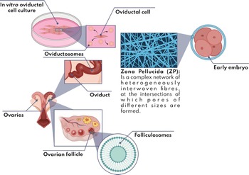
The study of the different types of EVs involved in embryogenesis is no more than a decade old. The early research was carried out in C. elegans and described EVs as modulators of important processes during early embryogenesis (Wehman et al., Reference Wehman, Poggioli, Schweinsberg, Grant and Nance2011; Mapes et al., Reference Mapes, Chen, Kim, Mitani, Kang and Xue2012). It was not until 2013 that the first studies about the effect of EVs on early embryogenesis were conducted in mammals. This research revealed that human epithelial endometrial cells secrete endometriosomes that have an average diameter of 50 to 150 nm, and are positive for CD9 and CD63 tetraspanins. Also it contains molecules that modulate the development and implantation of embryos (Ng et al., Reference Ng, Rome, Jalabert, Forterre, Singh, Hincks and Salamonsen2013). A year later, another study confirmed that in vitro cultured porcine embryos secreted EVs and, what is even more relevant, that neighbouring embryos in the same microdroplet can capture them. This evidence indicated that such EVs can bypass the zona pellucida in some way by generating a microenvironment of mutual support between the different embryos cultured in a group (Saadeldin et al., Reference Saadeldin, Kim, Choi and Lee2014). As mentioned in the previous section, the absorption of EVs by the oocytes of mammals such as bovine, equine or human, is in question due to the physical barrier that the zona pellucida represents (Lee et al., Reference Lee, Oh, Kim and Lee2020b). The zona pellucida is a complex network of heterogeneously intertwined fibres that form pores of different sizes between the intersections (Figure 6) (Van Soom et al., Reference Van Soom, Wrathall, Herrler and Nauwynck2010). However, it has been observed that the porosity of the zona pellucida differs between embryos produced in vivo and in vitro, the latter presenting larger pores and therefore being more receptive to larger diameter nanostructures (such as EVs), probably because of their in vitro manipulation (Kim et al., Reference Kim, Funahashi, Abeydeera, Moon, Prather and Day1996; Funahashi et al., Reference Funahashi, Ekwall, Kikuchi and Rodriguez-Martinez2001; Saadeldin et al., Reference Saadeldin, Kim, Choi and Lee2014). Conversely, it is known that during the fertilization process the different enzymes are released following the acrosomal reaction of sperm which modify the density of the zona pellucida. This makes the zona more permeable to nanostructures of different sizes such as EVs (Álvarez et al., Reference Álvarez, García, Corzo, Gutiérrez, de Bustamante, Barcia, Álvarez, Matorras and Hernández2007). Due to these reasons, the zona pellucida of embryos appears to be more permeable to EVs than that of oocytes, which suggests that adding EVs to embryo culture medium may support in vitro embryogenesis. In this way, Saadeldin et al. (Reference Saadeldin, Kim, Choi and Lee2014) described that the zona pellucida of porcine embryos generated by somatic cell nuclear transfer (SCNT) can allow the passage of EVs with a diameter up to 200 nm. According to the results that Saadeldin and colleagues observed, another important conclusion that can be drawn from is that communication mediated by EVs can explain to some extent the beneficial effect of group embryo culture and co-culture systems of embryos with different types of somatic cells, in which the interlocking communication could modulate both the content of the EVs released and the embryonic development. In this way, the current section will focus mainly on the EVs present in in vivo and in vitro environments of embryos, that is oviductosomes and uterosomes. The role of endometriosomes and EVs released by the embryos themselves will be reviewed, exploring their content in depth and how they can modulate different processes during embryogenesis.
Folliculosomes and oviductosomes. The figure shows the environment of the folliculosomes and oviductosomes, and possible subvesicular localization of their components (enlarged images in digital format show the molecules in detail). Created with BioRender.com

4.1 Folliculosomes and oviductosomes
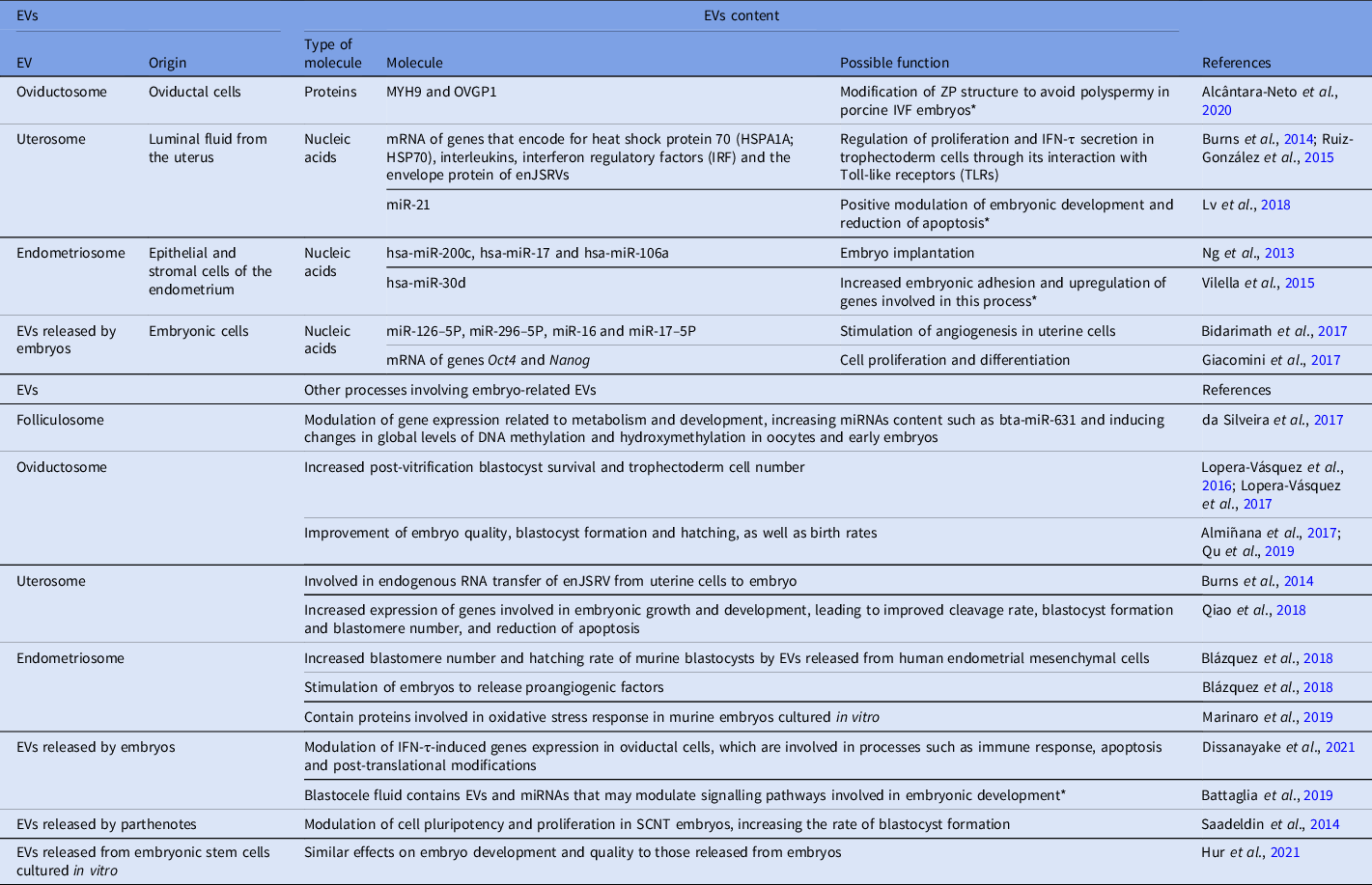
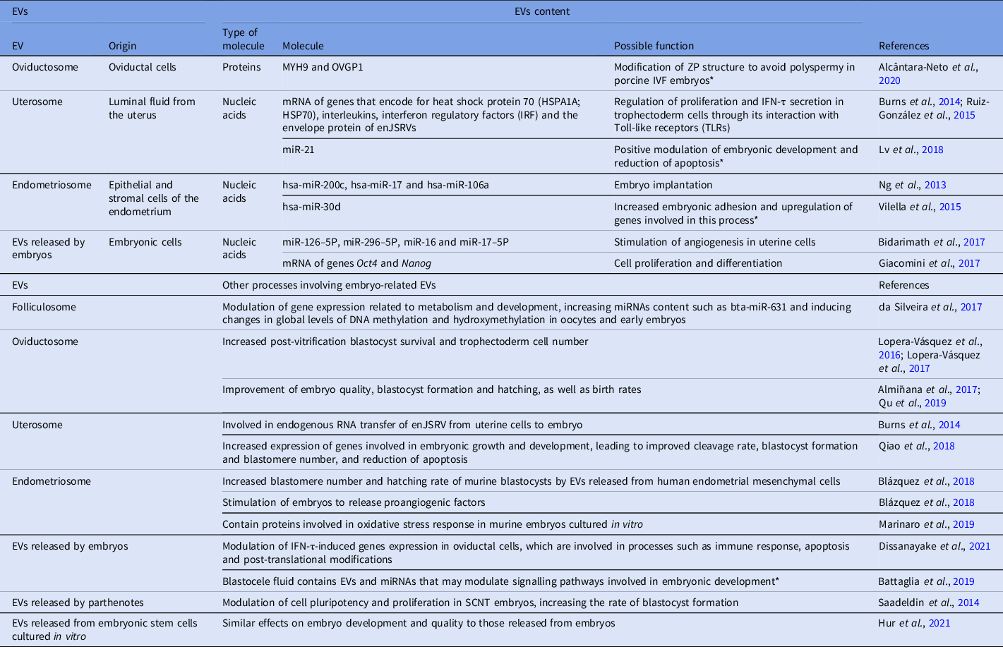
The previous section discussed many of the positive effects that folliculosomes can have on folliculogenesis and oogenesis. However, a study carried out by da Silveira et al. (Reference da Silveira, Andrade, Del Collado, Sampaio, Sangalli, Silva, Pinaffi, Jardim, Cesar, Nogueira, Cesar, Coutinho, Pereira, Perecin and Meirelles2017) showed that the benefits of such EVs can even go beyond early embryonic development. Indeed, these authors reported that the addition of folliculosomes to the in vitro maturation medium of oocytes and bovine embryo culture with a maximum of four cells modulates the expression of genes related to metabolism and development by increasing the content of different miRNAs, such as bta-miR-631, and inducing changes in the global levels of DNA methylation and hydroxymethylation (da Silveira et al., Reference da Silveira, Andrade, Del Collado, Sampaio, Sangalli, Silva, Pinaffi, Jardim, Cesar, Nogueira, Cesar, Coutinho, Pereira, Perecin and Meirelles2017). It is likely that the positive effect of these EVs on early embryos is a reflection that folliculosomes are present in the oviduct after ovulation and during fertilization. Nevertheless, the stellar EVs in this scene are undoubtedly the oviductosomes. As mentioned previously, oviductosomes are an important component during the early days of embryonic development after conception. It was reported that culturing bovine embryos in medium conditioned by bovine oviduct epithelial cells (BOEC) and in medium supplemented with fresh and thawed oviductosomes (isolated from the conditioned medium) increased the survival of post-vitrification blastocysts and the number of trophectoderm cells, supporting the idea that oviductosomes are the main cause of the beneficial effect of conditioned medium (Lopera-Vásquez et al., Reference Lopera-Vásquez, Hamdi, Fernandez-Fuertes, Maillo, Beltrán-Breña, Calle, Redruello, López-Martín, Gutierrez-Adán, Yañez-Mó, Ramirez and Rizos2016). These results are consistent with those obtained in another study carried out by the same group, in which supplementing the culture medium of preimplantation bovine embryos with oviductosomes secreted in vivo by the oviductal isthmus during the early luteal phase was found to improve embryo survival after vitrification (Lopera-Vásquez et al., Reference Lopera-Vasquez, Hamdi, Maillo, Gutierrez-Adan, Bermejo-Alvarez, Ramírez, Yáñez-Mó and Rizos2017). The beneficial effects on embryo survival in both works could be orchestrated to some degree by genes that are upregulated in embryos exposed to oviductosomes. Among them are the Aqp3 gene, which encodes for the Aquaporin 3 channel involved in the rapid transport of water and cryoprotectants across the plasma membrane, something that could be vital during vitrification/warming procedures. Other genes are Dnmt3A, which encodes for DNA (cytosine-5)-methyltransferase, essential during the epigenetic modifications necessary for correct embryonic development, such as the methylation of the second X chromosome; and Snrpn, which encodes for Small Nuclear Ribonucleoprotein-associated Protein N, a protein that has an important role in mRNA maturation, specifically during alternative splicing (Lopera-Vásquez et al., Reference Lopera-Vásquez, Hamdi, Fernandez-Fuertes, Maillo, Beltrán-Breña, Calle, Redruello, López-Martín, Gutierrez-Adán, Yañez-Mó, Ramirez and Rizos2016; Lopera-Vásquez et al., Reference Lopera-Vasquez, Hamdi, Maillo, Gutierrez-Adan, Bermejo-Alvarez, Ramírez, Yáñez-Mó and Rizos2017). In another study that compared the effects of fresh and post-thawed oviductosomes on the development and quality of bovine embryos, EVs that underwent storage at −80°C were found to increase the rate of blastocyst formation, their hatching and the number of total cells in in vitro produced bovine embryos, with respect to the presence of fresh EVs or the control without EVs (Almiñana et al., Reference Almiñana, Corbin, Tsikis, Alcântara-Neto, Labas, Reynaud, Galio, Uzbekov, Garanina, Druart and Mermillod2017). In the same work, differences were reported at the proteomic level in oviductosomes secreted by cells grown in vitro compared with those secreted in vivo, observing that the latter have a higher content of proteins that modulate the sperm–oocyte interaction and fertilization, and the development, quality, and implantation of bovine embryos generated in vitro (Almiñana et al., Reference Almiñana, Corbin, Tsikis, Alcântara-Neto, Labas, Reynaud, Galio, Uzbekov, Garanina, Druart and Mermillod2017). Such differences at the proteomic level could be related to the lack of cross-communication between oviductal cells cultured in vitro and other cell types. This contrasts with the in vivo scenario, in which cells are exposed to different hormonal concentrations throughout the oestrous or menstrual cycle. The latter was confirmed by another study carried out by the same group, in which the content dynamics of oviductosomes were analyzed throughout the bovine oestrous cycle. Significant changes were found in the composition of proteins, mRNA and non-coding RNA (such as miRNAs) (Almiñana et al., Reference Almiñana, Tsikis, Labas, Uzbekov, da Silveira, Bauersachs and Mermillod2018), evidencing the need of considering hormonal stimuli in oviductal cells cultured in vitro as an important factor that modulates the content of their EVs and, therefore, the effects that these may have on embryos. The latter cannot be ignored when it comes to purifying oviductosomes from samples obtained in slaughterhouses, making it necessary to consider the moment of the oestrous cycle in which the oviductal cells were found. In effect, this may be required to obtain the desired and reproducible results with respect to the effect of such EVs on the embryo. Another factor that can modulate the content of oviductosomes is the presence of zygotes in the oviduct, as shown by the results obtained by Qu et al. (Reference Qu, Zhao, Wang, Zhang, Li, Fan and Liu2019). These authors described that the supplementation of murine embryo culture medium with oviductosomes from pregnant females upregulates the expression of the Bcl2 (anti-apoptotic) and Oct4 (pluripotency) genes, and downregulates that of Bax (pro-apoptotic). This leads to the improvement in embryonic quality, blastocyst formation and birth rates according to the addition of oviductosomes from non-pregnant females (Qu et al., Reference Qu, Zhao, Wang, Zhang, Li, Fan and Liu2019). Conversely, in pigs it was shown that oviductosomes can modulate the hardening of the zona pellucida and reduce polyspermia during in vitro fertilization (IVF) (Alcântara-Neto et al., Reference Alcântara-Neto, Fernandez-Rufete, Corbin, Tsikis, Uzbekov, Garanina, Coy, Almiñana and Mermillod2020). The molecules involved in this mechanism still remain unknown, but MYH9 and OVGP1 proteins, both present in oviductosomes, have been purported to modify the structure of the zona pellucida once the oocyte was fertilized to avoid polyspermia (Alcântara-Neto et al., Reference Alcântara-Neto, Fernandez-Rufete, Corbin, Tsikis, Uzbekov, Garanina, Coy, Almiñana and Mermillod2020). Figure 6 and Table 3 show the contents and effects of folliculosomes and oviductosomes on the embryos.
Embryo-related EVs
* Experimentally not proven or mechanism not known.
4.2 Uterosomes and endometriosomes
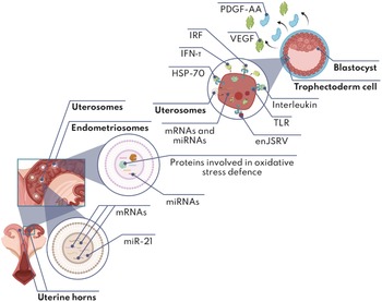
Uterosomes were demonstrated to be another important actor during in vivo and in vitro embryogenesis. In 2014, these EVs were described to be implicated in the transfer of endogenous beta retrovirus Jaagsiekte (enJSRVs) RNA from uterine cells to the conceptus (Burns et al., Reference Burns, Brooks, Wildung, Navakanitworakul, Christenson and Spencer2014). Later, another work showed that uterosomes secreted throughout the estrous cycle and during the first months of pregnancy stimulated embryo trophectoderm cells to proliferate and secrete interferon tau (IFN-τ). This protein is crucial during the first month of pregnancy in ruminants, as it coordinates the production of progesterone and regulates genes involved in the transport of nutrients to the embryo, among other functions (Bazer et al., Reference Bazer, Ying, Wang, Dunlap, Zhou, Johnson and Wu2015; Ruiz-González et al., Reference Ruiz-González, Xu, Wang, Burghardt, Dunlap and Bazer2015). The analysis of the content of uterosomes identified the presence of mRNA of genes that encode for heat shock protein 70 (Hspa1A; Hsp70), interleukins, interferon regulatory factors (IRF) and the envelope protein of the retrovirus in JSRVs (Burns et al., Reference Burns, Brooks, Wildung, Navakanitworakul, Christenson and Spencer2014; Ruiz-González et al., Reference Ruiz-González, Xu, Wang, Burghardt, Dunlap and Bazer2015). These proteins regulate the proliferation and secretion of IFN-τ via their interaction with the Toll-like receptors (TLR) of the trophectoderm cells, which demonstrates the importance of the communication of the uterus with the embryo mediated by uterosomes (Ruiz-González et al., Reference Ruiz-González, Xu, Wang, Burghardt, Dunlap and Bazer2015). Similar results were observed when adding early luteal phase uterosomes to the culture medium of bovine embryos, which increases the expression of genes involved in embryonic growth and development, such as acrogranin and Ifn-τ, and decreases that of Bax and Bip (rough endoplasmic reticulum stress marker). This leads to the improvement of cleavage rate, blastocyst formation and number of blastomeres and reduces the apoptosis rate, with respect to embryos grown in the absence of these EVs (Qiao et al., Reference Qiao, Ge, Ma, Zhang, Zuo, Wang, Zhang and Wang2018). In mice, uterosomes also modulate embryonic development positively and reduce apoptosis, via increasing the expression of genes such as Bcl2 and Oct4, and reducing that of Bax (Lv et al., Reference Lv, Yu, Wang, Yi, Zeng and Xiao2018). These effects could be regulated to a certain degree by miR-21 present in uterosomes, which increases its expression as the embryo develops, and has also been implicated in the modulation of anti-apoptotic processes (Lv et al., Reference Lv, Yu, Wang, Yi, Zeng and Xiao2018). Although more studies are needed to understand the mechanisms through which this particular miRNA acts.
Conversely, endometriosomes, which can be considered a specific type of uterosomes, were also demonstrated to exert interesting effects on embryonic development. It was observed that the system of co-culture of murine embryos with endometrial cells results in a higher rate of blastocyst formation than the co-culture with oviductal cells (Desai et al., Reference Desai, Kennard, Kniss and Friedman1994), which could be orchestrated mainly by the EVs released into the culture medium (Lopera-Vásquez et al., Reference Lopera-Vásquez, Hamdi, Fernandez-Fuertes, Maillo, Beltrán-Breña, Calle, Redruello, López-Martín, Gutierrez-Adán, Yañez-Mó, Ramirez and Rizos2016). The factors secreted by oviductal cells could be beneficial for the production of embryos in vitro, being an important component of the natural environment of the embryo during its first days of development (Lopera-Vásquez et al., Reference Lopera-Vásquez, Hamdi, Fernandez-Fuertes, Maillo, Beltrán-Breña, Calle, Redruello, López-Martín, Gutierrez-Adán, Yañez-Mó, Ramirez and Rizos2016). Some studies have demonstrated that they could also release anti-implantation and anti-hatching molecules, as during the embryo journey along the oviduct, implantation or premature hatching must be avoided to prevent ectopic gestation or abnormal development (Bongso et al., Reference Bongso, Ng and Ratnam1990; Pauerstein et al., Reference Pauerstein, Eddy, Koong and Moore1990). The confirmation of the presence of anti-implantation and anti-hatching molecules in oviductosomes would explain to some extent the observations of Lopera-Vásquez et al. (Reference Lopera-Vásquez, Hamdi, Fernandez-Fuertes, Maillo, Beltrán-Breña, Calle, Redruello, López-Martín, Gutierrez-Adán, Yañez-Mó, Ramirez and Rizos2016), who reported a reduction in the blastocyst formation rate on day 9 when oviductosomes were added to the embryo culture medium. For its part, the positive effect of endometriosomes were reported in multiple studies. In humans, these EVs contain miRNAs relevant to embryo implantation, including hsa-miR-200c, hsa-miR-17 and hsa-miR-106a (Ng et al., Reference Ng, Rome, Jalabert, Forterre, Singh, Hincks and Salamonsen2013). In another study, in which the effects of the presence of human endometriosomes on murine cultured embryos were analyzed, an increase in embryonic adhesion and an upregulation of genes involved in this process, such as Itgb3, Itga7 and Cdh5, were observed, suggesting hsa-miR-30d (present in these EVs) to be the main component responsible for such effects (Vilella et al., Reference Vilella, Moreno-Moya, Balaguer, Grasso, Herrero, Martínez, Marcilla and Simón2015). Similarly, murine embryonic development has been favoured by the EVs released by human mesenchymal endometrial cells, increasing the number of blastomeres and the hatching rate of blastocysts (Blázquez et al., Reference Blázquez, Sánchez-Margallo, Álvarez, Matilla, Hernández, Marinaro, Gómez-Serrano, Jorge, Casado and Macías-García2018). In addition, endometriosomes were seen to stimulate embryos to release pro-angiogenic factors such as VEGF and PDGF-AA, which could modulate endometrial angiogenesis, vascularization, differentiation and tissue remodelling (Blázquez et al., Reference Blázquez, Sánchez-Margallo, Álvarez, Matilla, Hernández, Marinaro, Gómez-Serrano, Jorge, Casado and Macías-García2018). Endometriosomes are also involved in the response to oxidative stress in murine embryos grown in vitro, as they contain at least 23 proteins involved in this process (Marinaro et al., Reference Marinaro, Macías-García, Sánchez-Margallo, Blázquez, Álvarez, Matilla, Hernández, Gómez-Serrano, Jorge, Vázquez, González-Fernández, Pericuesta, Gutiérrez-Adán and Casado2019). Moreover, as in oviductal cells, endometrial cells respond to different stimuli that affect their secretion rate and content. In sheep, for example, progesterone was reported to regulate 1611 genes and nine miRNAs in endometrial cells and increase the rate of secretion of endometriosomes (Burns et al., Reference Burns, Brooks, O’Neil, Hagen, Behura and Spencer2018). Another factor that was shown to modulate the secretion rate of endometriosomes was the concentration of O2. In effect, under hypoxia conditions (O2 at 1%), bovine endometrial cells release more EVs and their content in proteins involved in embryo development is higher (Koh et al., Reference Koh, Peiris, Vaswani, Reed, Rice, Salomon and Mitchell2016). See Figure 7 and Table 3 for a description of the contents and effects of uterosomes and endometriosomes on embryos.
Uterosomes and endometriosomes. The figure shows the environment of the uterosomes and endometriosomes, and possible subvesicular localization of their components (enlarged images in digital format show the molecules in detail). Created with BioRender.com

4.3 EVs released by embryos
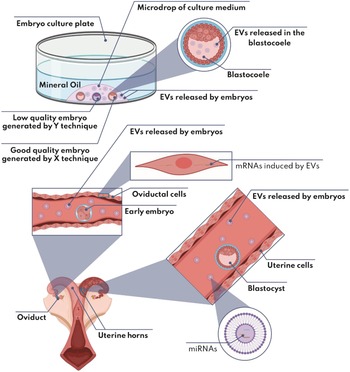
Regarding the EVs released by embryos, it has been reported that they can modulate their environment in vivo by delivering factors that promote angiogenesis and tissue remodelling to the different uterine cells. In this sense, it was described that, in pigs, EVs released by trophectoderm cells contain miRNAs such as miR-126–5P, miR-296–5P, miR-16 and miR-17–5P, which can stimulate angiogenesis in uterine cells (Bidarimath et al., Reference Bidarimath, Khalaj, Kridli, Kan, Koti and Tayade2017). EVs from good quality bovine embryos had also been shown to modify gene expression in oviductal cells, acting specifically on the expression of genes induced by IFN-τ such as Oas1y, Mx1 and Isg15 (Dissanayake et al., Reference Dissanayake, Nõmm, Lättekivi, Ord, Ressaissi, Godakumara, Reshi, Viil, Jääger, Velthut-Meikas, Salumets, Jaakma and Fazeli2021).
Saadeldin et al. (Reference Saadeldin, Kim, Choi and Lee2014) described that EVs released by parthenotes co-cultured with SCNT-generated porcine embryos contained mRNAs of genes involved in cell pluripotency and proliferation, such as Oct4, Sox2, Klf4, Nanog and c-Myc, which are captured by SCNT embryos and improve the rate of blastocyst formation (Saadeldin et al., Reference Saadeldin, Kim, Choi and Lee2014). Another study that investigated the EVs released by human embryos also reported that they contained transcripts of Oct4 and Nanog genes (Giacomini et al., Reference Giacomini, Vago, Sanchez, Podini, Zarovni, Murdica, Rizzo, Bortolotti, Candiani and Viganò2017). Similarly, the blastocoel fluid was reported to contain EVs and different miRNAs that could modulate critical signalling pathways involved in embryo development, such as pluripotency, cell reprogramming, epigenetic modifications, intercellular communication, cell adhesion and cell fate. This reveals the dynamic communication mediated by EVs between the different cells of the same embryo (Battaglia et al., Reference Battaglia, Palini, Vento, La Ferlita, Lo Faro, Caroppo, Borzì, Falzone, Barbagallo, Ragusa, Scalia, D’Amato, Scollo, Musumeci, Purrello, Gravotta and Di Pietro2019). Conversely, it has been observed that the quality of the embryo influences the content of the EVs that they release (Dissanayake et al., Reference Dissanayake, Nõmm, Lättekivi, Ord, Ressaissi, Godakumara, Reshi, Viil, Jääger, Velthut-Meikas, Salumets, Jaakma and Fazeli2021). In addition, it is likely that the vesicles secreted by embryos generated by different techniques, e.g. IVF, ICSI, SCNT or parthenogenetic activation (PA), have also differences in their content. According to this, it is worth mentioning that EVs secreted by IVF-generated bovine embryos differ morphologically from those released by bovine parthenotes, and that the rate of EV secretion from the embryos is higher than that of the parthenotes (Mellisho et al., Reference Mellisho, Velásquez, Nuñez, Cabezas, Cueto, Fader, Castro and Rodríguez-Álvarez2017). Figure 8 and Table 3 show the contents and effects of the EVs released by embryos on their environment and also other embryos.
EVs released by embryos. The figure shows the environment of the EVs released by embryos and possible subvesicular localization of their components (enlarged images in digital format show the molecules in detail). Created with BioRender.com
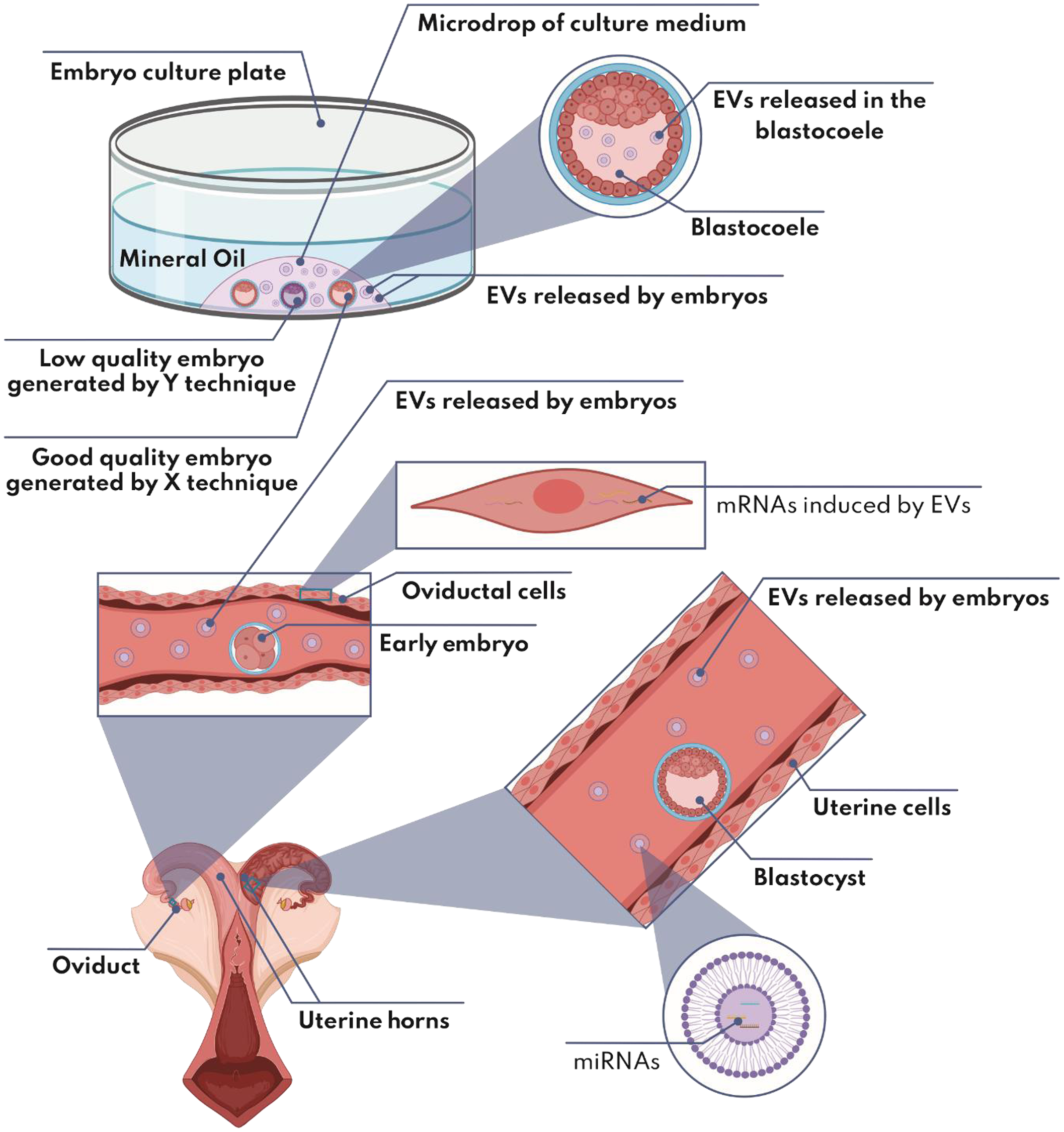
4.4 New perspectives
miR-21 is considered anti-apoptotic, and is present in conditions of high cell proliferation such as carcinogenesis (Shen et al., Reference Shen, Xu, Lv, Dai, Chen and Chen2020) and embryogenesis (Lv et al., Reference Lv, Yu, Wang, Yi, Zeng and Xiao2018). Supplementing the embryo culture medium with EVs rich in this miRNA appears to improve embryonic development, although an excess could be harmful as the overexpression of miR-21 has a pro-apoptotic effect (Yan et al., Reference Yan, Rao, Yuan, Gao, Huang, Zhao and Ren2017). Conversely, and as discussed previously, oviductosomes, uterosomes and endometriosomes can play an important role during the different embryonic stages, as they interact with the embryo sequentially during its journey to the uterus. Therefore, mimicking the maternal physiological environment in the best possible way makes the sequential culture necessary with each of these types of EVs. This could significantly improve embryonic quality and development in vitro. Another way to support the development of embryos is through their co-culture with other embryos generated through simpler techniques, to increase the amount in the same well, such as embryos generated by SCNT co-cultured with parthenotes (Saadeldin et al., Reference Saadeldin, Kim, Choi and Lee2014), where the EVs secreted by the latter improve the quality and development of the former. However, to take full advantage of this methodology, it is imperative to outline the content and determine the effects of EVs of embryos generated by different techniques on early embryogenesis. Being very likely that the results obtained will be different between the species, due to the efficiency rates achieved with each in vitro production technique. For example, in bovine, IVF-derived embryos could secrete EVs with greater beneficial effects on embryo quality and development than ICSI-derived embryos, as they have higher quality (Salamone et al., Reference Salamone, Canel and Rodríguez2017). Such EVs could also support the remodelling of the maternal environment during embryo transfer (Bidarimath et al., Reference Bidarimath, Khalaj, Kridli, Kan, Koti and Tayade2017). So co-transfer with EVs secreted by good quality embryos would be another interesting perspective. To have a more accessible stock of EVs without having to generate embryos, an interesting alternative would be the derivation of embryonic stem cells from embryos generated through ARTs that work better for each species. Although EVs released from embryonic stem cells had been demonstrated to have similar effects to those released from embryos (Hur et al., Reference Hur, Feng, Wilson, Cerione and Antonyak2021), it was not reported whether there were differences in the profile of EVs secreted by stem cells derived from embryos of different quality, so further studies are needed.
5. Final considerations
Many studies have demonstrated the biological functionality of EVs to enhance ARTs (Saadeldin et al., Reference Saadeldin, Kim, Choi and Lee2014; da Silveira et al., Reference da Silveira, Andrade, Del Collado, Sampaio, Sangalli, Silva, Pinaffi, Jardim, Cesar, Nogueira, Cesar, Coutinho, Pereira, Perecin and Meirelles2017; Almiñana et al., Reference Almiñana, Tsikis, Labas, Uzbekov, da Silveira, Bauersachs and Mermillod2018). In this sense, the amount of vesicles that must be applied to generate significant effects depends on factors mentioned throughout this review as the cell of origin, the target cell, the type of molecule to be interfered with, the species in which the changes will be made, among others. As a point of reference, for spermatozoa, oocytes and embryos, it was shown that to generate a significant change in their transcriptome or proteome, the amount of EVs to be added, expressed in μg of protein of EVs/ μl (final concentration), it is in the range 0.02–0.4 μg/ μl (Caballero et al., Reference Caballero, Frenette, Belleannée and Sullivan2013; Almiñana et al., Reference Almiñana, Tsikis, Labas, Uzbekov, da Silveira, Bauersachs and Mermillod2018; Blázquez et al., Reference Blázquez, Sánchez-Margallo, Álvarez, Matilla, Hernández, Marinaro, Gómez-Serrano, Jorge, Casado and Macías-García2018; Lee et al., Reference Lee, Oh, Kim and Lee2020b).
Conversely, it is worth mentioning that it is generally easier to isolate large amounts of such vesicles from in vivo samples such as tissues or biological fluids than from conditioned medium from in vitro, probably due to the obvious difference in the amount of cells secreting EVs in both cases (Ortega et al., Reference Ortega, Roefs, de Miguel Perez, Kooijmans, de Jong, Sluijter, Schiffelers and Vader2019). However, although fewer EVs per ml can be isolated from conditioned medium, they have the advantage that they can be purified relatively easier compared with EVs from biological fluids such as serum or blood plasma, due to the difference in the complexity of the components between the two cases. As a point of reference, it was observed that 5 ml of conditioned medium from confluent cells, cultured in 100 mm plates for 48 h, and using different isolation and purification methodologies, were sufficient to isolate the EVs necessary to generate a significant change in the transcriptome and/or proteome of the target cell (Patel et al., Reference Patel, Khan, Zubair, Srivastava, Khushman, Singh and Singh2019). While it is important to note that the secretion rate of EVs from different cells depends on the aforementioned factors, it has been shown that disruption of the Ndrg1 and Rab7A genes involved in endolysosomal trafficking significantly increased EV production in cancer epithelial cells cultured in vitro (Ortega et al., Reference Ortega, Roefs, de Miguel Perez, Kooijmans, de Jong, Sluijter, Schiffelers and Vader2019). This represents an interesting strategy to replicate in other cell types if a larger amount of EVs per ml of culture medium needed to be isolated, especially because the biological functionality of the EVs and their content are not affected.
Conversely, the ideal methodology to isolate EVs from tissues, biological fluids or in vitro conditioned medium, depends on how purified a EV population is required and/or the source of origin. The most common methodology to isolate EVs is differential ultracentrifugation, although it was shown that this methodology appears to be too aggressive to preserve the biological functionality of the vesicles and has also the disadvantages of being cumbersome and requiring expensive equipment (Niu et al., Reference Niu, Pang, Liu, Li, Cheng and Yeung2017). Therefore, different commercial alternatives have emerged to facilitate the isolation of EVs, such as polymer-based precipitation, affinity columns or selection with specific antibodies targeting certain vesicle subpopulations, among others, with polymer-based precipitation being one of the best alternatives due to its simplicity, low cost, high efficiency and preservation of the biological functionality of EVs (Niu et al., Reference Niu, Pang, Liu, Li, Cheng and Yeung2017; Patel et al., Reference Patel, Khan, Zubair, Srivastava, Khushman, Singh and Singh2019). However, this method has been shown to isolate all types of EVs with a diameter of 30–150 nm, not differentiating between ectosomes and exosomes (Niu et al., Reference Niu, Pang, Liu, Li, Cheng and Yeung2017). Other isolation and purification alternatives present difficulties in separating EVs from cellular debris, proteins or even vesicles of larger diameter, so we encourage interested readers to investigate the literature and scientific forums on the real efficiency of the different commercial kits to isolate and purify the different EVs, according to their needs, especially if they wish to maintain good manufacturing practices (GMP) and avoid biological contaminants.
After isolating and purifying a certain type of EV, the question arises whether the different batches of isolated vesicles will have the same quality to generate the desired effect. Even controlling the isolation methodology and the amount of EVs applied, this is not a guarantee that the same desired effect will be obtained in 100% of the cases, especially for EVs isolated from tissues, biological fluids and primary cultures, from slaughterhouse samples. This is due to factors mentioned above such as age, health condition of the animals, stage of the estrous cycle, genetics, epigenetics, environmental factors, among others, which can considerably modify the content of EVs, while altering their effect (da Silveira et al., Reference da Silveira, Veeramachaneni, Winger, Carnevale and Bouma2012; Eaton et al., Reference Eaton, Jayasooriah, Buckland, Martin, Cropley and Suter2015; Almiñana et al., Reference Almiñana, Corbin, Tsikis, Alcântara-Neto, Labas, Reynaud, Galio, Uzbekov, Garanina, Druart and Mermillod2017; Saeed-Zidane et al., Reference Saeed-Zidane, Linden, Salilew-Wondim, Held, Neuhoff, Tholen, Hoelker, Schellander and Tesfaye2017; Santos et al., Reference Santos, Bottino, Santos, Simões, Souza, Ferreira, da Silveira, Ávila, Bride and Sales2018; Hailay et al., Reference Hailay, Hoelker, Poirier, Gebremedhn, Rings, Saeed-Zidane, Salilew-Wondim, Dauben, Tholen, Neuhoff, Schellander and Tesfaye2019). Therefore, to maintain GMP, we urge that to isolate EVs from established cell lines under standardized culture conditions or for vesicles that must necessarily be obtained from living organisms, an ideal scenario would be to maintain standardized environmental conditions and use specific animal strains or breeds to preserve the maximum genetic and epigenetic heritage. It may even be necessary to use SCNT-generated animals, all to maintain the same quality in the effect of EVs to avoid ‘unwanted vesicular content’.
It is also important to mention that non-biological contaminants that may remain after isolation and purification of EVs should not be taken lightly, especially in clinical applications, as it was shown that even polymer-based precipitation can leave some traces of the patented polymer in the final purification result (Patel et al., Reference Patel, Khan, Zubair, Srivastava, Khushman, Singh and Singh2019). Therefore, depending on the methodology used, it is also necessary to consider extra steps to remove such contaminants or reduce them to percentages harmless to human or animal health, to maintain GMP.
Undoubtedly, EVs are a powerful ally to improve ARTs that may applied as modulators of a specific desired effect in different therapies. Such a premise is more than plausible due to the different characteristics of its content that depend on its cellular origin. Furthermore, it should be noted that these lipid nanostructures are essentially a physiological component of cells and their environment, moulded by evolution to express a certain degree of specificity for different cell types, with low cytotoxicity and immunogenicity. Moreover, and compared with their synthetic counterparts, they are biodegradable, a necessary characteristic to avoid generating long-term damage by bioaccumulation (Barkalina et al., Reference Barkalina, Jones, Wood and Coward2015; Kim et al., Reference Kim, Yang, Oh, Hong, Seo and Jang2017). Conversely, although the EVs produced by the different animal cells are beginning to be considered as important modulators in reproduction, it is important to consider that the different populations of bacteria and other microorganisms of the male and female microbiome also secrete EVs (Yáñez-Mó et al., Reference Yáñez-Mó, Siljander, Andreu, Zavec, Borràs, Buzas, Buzas, Casal, Cappello, Carvalho, Colás, Cordeiro-da Silva, Fais, Falcon-Perez, Ghobrial, Giebel, Gimona, Graner, Gursel and Gursel2015). These EVs could positively and/or negatively interfere with the different processes and pathways mentioned throughout this review, making the evaluations of the effect of this type of vesicles on mammalian reproduction necessary.
One factor to consider regarding the use of EVs, especially those obtained from samples collected in slaughterhouses, is that they could also be involved in the mechanism that underlies the epigenetic inheritance of accumulated environmental experiences in life. Therefore, the effects of exposure to a certain stimulus, such as hunger (Heijmans et al., Reference Heijmans, Tobi, Stein, Putter, Blauw, Susser, Slagboom and Lumey2008), obesity (Ng et al., Reference Ng, Lin, Laybutt, Barres, Owens and Morris2010), smoking (Pembrey et al., Reference Pembrey, Bygren, Kaati, Edvinsson, Northstone, Sjöström and Golding2006), psychological trauma (Matthews and Phillips, Reference Matthews and Phillips2010) or simply fear of something specific (Dias and Ressler, Reference Dias and Ressler2014), could be transmitted to the germ line by the different EVs (Eaton et al., Reference Eaton, Jayasooriah, Buckland, Martin, Cropley and Suter2015). This could be used to obtain animals with a particular behaviour, such as a more docile horse, without the need for genetic editing. Physiological (Almiñana et al., Reference Almiñana, Corbin, Tsikis, Alcântara-Neto, Labas, Reynaud, Galio, Uzbekov, Garanina, Druart and Mermillod2017; Saeed-Zidane et al., Reference Saeed-Zidane, Linden, Salilew-Wondim, Held, Neuhoff, Tholen, Hoelker, Schellander and Tesfaye2017; Martinez et al., Reference Martinez, Baccarelli, Liang, Dioni, Mansur, Adir, Bollati, Racowsky, Hauser and Machtinger2019) and pathological aspects (Santos et al., Reference Santos, Bottino, Santos, Simões, Souza, Ferreira, da Silveira, Ávila, Bride and Sales2018) must also be considered when using EVs from slaughterhouse samples, as mentioned earlier in this review, there are various factors that influence the content of EVs and, therefore, their effect. Alternatively, there could be reproducibility problems in the results. This also requires, especially at the clinical level (human and veterinary), the EVs used in different therapies to come from standardized sources, for example cell lines. Moreover, it is also recommended that the FBS used in the embryo culture medium to be depleted of EVs, as its content is variable and is subject to each particular production batch. Also reporting that such EVs are harmful to the embryo culture (Lopera-Vásquez et al., Reference Lopera-Vásquez, Hamdi, Fernandez-Fuertes, Maillo, Beltrán-Breña, Calle, Redruello, López-Martín, Gutierrez-Adán, Yañez-Mó, Ramirez and Rizos2016). Almiñana et al. (Reference Almiñana, Corbin, Tsikis, Alcântara-Neto, Labas, Reynaud, Galio, Uzbekov, Garanina, Druart and Mermillod2017) demonstrated that the content of EVs from cell lines is poorer than that of EVs secreted in vivo. This problem could be corrected with hormonal supplementation to the culture medium or even by site-specific genetic engineering techniques, such as CRISPR-Cas9. This would open up the possibility of a new approach by engineering extracellular vesicles from cell lines specifically designed to secrete EVs with certain characteristics (Villarroya-Beltri et al., Reference Villarroya-Beltri, Gutiérrez-Vázquez, Sánchez-Cabo, Pérez-Hernández, Vázquez, Martin-Cofreces, Martinez-Herrera, Pascual-Montano, Mittelbrunn and Sánchez-Madrid2013; Yim et al., Reference Yim, Ryu, Choi, Lee, Lee, Choi, Kim, Shaker, Sun, Park, Kim, Heo and Choi2016; Ortega et al., Reference Ortega, Roefs, de Miguel Perez, Kooijmans, de Jong, Sluijter, Schiffelers and Vader2019).
The study of the different types of EVs is expanding the frontiers of our understanding about cellular communication and many other molecular mechanisms hidden so far, giving way to possible neoteric applications in areas such as medicine, ectogenesis or genetic engineering, which are just beginning to be unveiled.
Acknowledgements
The authors gratefully acknowledge funding support from project DI21–0040, Universidad de La Frontera and Doctoral scholarship CONICYT – ANID 21191434, Chile.
Financial support
This work was supported by the Universidad de La Frontera, through project DI21-0040 and Doctoral scholarship CONICYT – ANID 21191434, Chile.
Conflict of interest
None of the author have any conflict of interest to declare.
Ethical standards
With reference to this type of article (review), no animals were involved.