Introduction
Drooling (sialorrhoea) is the spillage of oral secretions out of the mouth. It is a normal phenomenon in infancy, particularly during teething, but usually subsides by two years of age. Persistent drooling after the age of four years is considered abnormal, and can be distressing to both child and carer. Consequences include social isolation and embarrassment, skin excoriation, an increased burden of patient care due to frequent changes of clothing or bibs and cleaning of furniture, and soiling of school books.
Drooling occurs due to: impaired swallowing; impaired oral performance (caused by loss of tongue movement, malocclusion, oral anaesthesia or a constantly open mouth); and occasionally due to over-production of saliva. Children with neurological disability have functional impairment at many levels, resulting in significant drooling (seen in 10–20 per cent of children with cerebral palsy).Reference Crysdale, Raveh, McCann, Roske and Kotler1 It is usually this group of patients for whom treatment is sought, and who go on to surgical management.
Many approaches have been documented in the management of drooling. Behaviour modification, oral motor training, correction of aggravating factors and pharmacotherapy have been used with varying degrees of success. Surgery is indicated when conservative measures have failed or cognitive impairment renders them impractical, or when drooling is constant and profuse. Surgical treatment may comprise salivary gland excision, duct rerouting, duct ligation or tympanic neurectomy. Each of these may be used as a solitary procedure or in combination with others, and all have variable results in the control of sialorrhoea. Patients often have inadequate control of symptoms following the first surgical treatment and require further procedures. Many surgical options are relatively risky; submandibular gland excision, for example, is prone to significant morbidity due to factors such as prolonged anaesthesia, external incisions, pain and elimination of a large proportion of salivary secretion.
On the contrary, parotid duct ligation is a quick and simple procedure conducted via an intra-oral approach. It is frequently performed as an adjunct to other operations. However, few authors have studied its applicability as a separate procedure.Reference Crysdale, Raveh, McCann, Roske and Kotler1 In this study, we aimed to evaluate bilateral parotid duct ligation as a discrete procedure in the clinician's armamentarium for the treatment of drooling.
Patients and methods
Patients
All children who had undergone bilateral parotid duct ligation at Great Ormond Street Hospital for Children between February 2003 and September 2006 were identified from a prospective ENT surgical database. Those who had undergone the procedure as the solitary surgical intervention to treat drooling at that time, either as first-line surgical management or subsequent to other operations, were studied. Retrospective data were obtained from the case notes by a member of the clinical team. This included patient demographics, prior surgical and non-surgical management, length of hospital stay, post-operative complications, length of follow up, outcome, and subsequent management. Assessment of outcome was based on the qualitative classification system previously used by Bailey and Wadsworth.Reference Bailey and Wadsworth2 Patients were stratified into the categories ‘much better’, ‘better’, ‘no change’ or ‘worse’, based on subjective parental reports at the post-operative follow-up appointments.
Surgical procedure
The procedure was performed under general anaesthesia. The parotid duct was cannulated using a lacrimal probe. An island of mucosa was isolated around the duct papilla then retracted with Allis forceps. The distal 1 cm of the duct was dissected circumferentially and ligated with a silk 4–0 suture. The papilla was excised. The duct was then tucked into the buccal fat pad and the mucosal opening closed over it with a Vicryl 4–0 suture. The procedure was repeated on the contralateral parotid duct. Oral co-amoxiclav (or erythromycin in penicillin-sensitive patients) was administered for one week post-operatively. Paracetamol and ibuprofen were given as required.
Results
Ten patients were identified, seven boys and three girls. Their age range at operation was two to 13 years (mean, nine years). All patients had significant neurological disability (nine cases of cerebral palsy and one of chromosomal abnormality). Five patients had associated symptoms of aspiration; the remaining five did not aspirate.
The youngest child to be operated on was only two years three months of age, well below the six year age limit at which one would usually consider surgical intervention for drooling. This child had laryngomalacia, vocal fold weakness and severe gastroesophageal reflux. She had suffered a cardiorespiratory arrest, thought to be secondary to aspiration, at the age of eight months, with severe neurological consequences. Her drooling worsened following a revision fundoplication, and saliva-reducing surgical intervention was thus indicated. This patient's drooling was reported to have ‘improved dramatically’ following the operation. However, unfortunately she died three months later of causes unrelated to the surgical procedure.
There were no specified indications for surgical intervention. Each case was assessed on its merits. Seven patients had had prior pharmacotherapy, but either had required more effective treatment or had suffered intolerable side effects. Eight patients had undergone previous surgical treatment, including bilateral submandibular gland excision, bilateral submandibular duct transposition, unilateral sublingual gland excision and tympanic neurectomy; however, they continued to drool unacceptably. The number of bib changes per day had been recorded for six children, and ranged from four to 15. Five children had significant skin maceration around the mouth and chin. One nine-year-old child was particularly distressed by social problems and stigmatisation at school due to being constantly wet with saliva.
None of the children experienced complications at the time of surgery. The children's post-operative stay ranged from zero to eight nights (mode, one night). All patients experienced mild post-operative swelling in the parotid region, which quickly subsided. Post-operative discomfort was controlled adequately by oral administration of paracetamol and/or ibuprofen as required. One patient (10 per cent) developed a localised wound infection that was successfully treated with oral antibiotic therapy. Two patients had post-operative medical complications (respiratory distress post-extubation and pulmonary hypertension), which prolonged their hospital stay (two and eight nights, respectively). These events were felt to be unrelated to the surgical procedure itself. There were no cases of xerostomia, based on parental report.
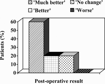
The follow-up period ranged from three to 54 months (mean, 27 months). The post-operative outcomes are depicted in Figure 1. Overall, 80 per cent of patients experienced an improvement in their drooling. Two patients (20 per cent) went on to have further surgical intervention to manage their drooling.
Effect of parotid duct ligation on drooling.

Discussion
Saliva is of vital importance for dental health, antibacterial function, lubrication of the mouth, bolus formation and buffering of the contents of the oral cavity. The management of drooling can therefore only be considered successful when drooling is reduced enough to prevent sequelae, but salivary flow remains adequate to maintain a moist, healthy oral cavity. Patients presenting to the otolaryngologist for treatment of drooling are usually children with neurological disability.
Drooling in neurologically impaired children is usually of multifactorial aetiology. The oral phase of swallowing is typically dysfunctional, resulting in pooling of saliva in the mouth with over-spill. Impaired head control, lip and jaw closure, and tongue movement compound the problem. Patients who aspirate also have incoordination of the pharyngeal phase of swallowing. The combination of these factors and significant cognitive impairment means that conservative management options are at best of limited value, and generally precluded altogether in these children. Surgery is thus indicated for definitive management. Surgical options should be considered in order, from least to most invasive.
Surgical procedures for the management of drooling fall into two main groups. Saliva can be redirected to the oropharynx to be swallowed more readily, which is the aim of submandibular and parotid duct transposition. Mankarious et al. Reference Mankarious, Bottrill, Huchzermeyer and Bailey3 reported a long-term success rate of 80 per cent following submandibular duct rerouting, and others have had similar results.Reference Shirley, Hill, Woolley and Wiatrak4 Unfortunately, these procedures are contraindicated in children who aspirate, in whom increased salivary flow to the oropharynx is likely to cause salivary contamination of the lower respiratory tract.
Alternatively, the salivary output can be decreased by submandibular gland excision, submandibular and parotid duct ligation, or tympanic neurectomy. Shirley et al. Reference Shirley, Hill, Woolley and Wiatrak4 reviewed 21 cases of four duct ligation and found 81 per cent to be ‘much better’ or ‘better’ after surgery. Of note, five of the six complications quoted in this series were related to ligation of the submandibular ducts: ranula, submandibular gland sialoadenitis, and prolonged submandibular gland swelling. There were no such complications relating to the parotid glands. Bilateral submandibular gland excision with parotid duct ligation was reported by Stern et al. in 2002;Reference Stern, Feinmesser, Collins, Shott and Cotton5 62 of 72 families (87 per cent) reported either no further drooling or significant improvement. However, submandibular gland excision is associated with substantial morbidity due to the requirement for an external incision, with potential for wound infection and subsequent scarring, damage to the marginal mandibular nerve, and damage to lingual and hypoglossal nerves.Reference Klem and Mair6 Tympanic neurectomy has been used in the past, but drooling frequently returns to pre-operative levels and therefore this procedure is now performed infrequently.Reference Crysdale, Bluestone, Stool, Alper, Arjmand, Casselbrant, Dohar and Yellon7
Botulinum toxin has been introduced as an alternative therapeutic option in the management of drooling over the last decade. This treatment is gaining popularity; however, the most effective serotype, dose and injection site have yet to be determined, and further randomised controlled trials are required to evaluate these further.Reference Lim, Mace, Reza Nouraei and Sandhu8
Parotid duct ligation is a further option in the surgical management of drooling. The physiological basis for duct ligation is atrophy of the affected gland. Complete occlusion of the main duct of the rat parotid has been shown to result in an 85 per cent absolute reduction of secretory tissue after two weeks.Reference Scott, Liu and Smith9 Parotid duct ligation has been performed in combination with bilateral submandibular gland excision,Reference Stern, Feinmesser, Collins, Shott and Cotton5 bilateral submandibular duct transpositionReference Wilson and Henderson10 and bilateral submandibular duct ligation (termed four duct ligation).Reference Shirley, Hill, Woolley and Wiatrak4, Reference Ceriati, De Peppo, Ciprandi, Marchetti, Silveri and Rivosecchi11 Simple ligation of the duct has been advocated in preference to parotid duct rerouting, due to the simplicity, low complication rate and good long-term outcome of the procedure.Reference Chang and Wong12 Obliteration of the duct has also been achieved by laser photocoagulation. Chang and Wong reported ‘remarkable’ or ‘significant’ improvement in 47 of 48 patients (98%) in the severity of drooling, and in 100% of patients in the frequency of drooling following this procedure.Reference Chang and Wong12
Despite these apparent advantages, bilateral parotid duct ligation is not widely performed as a separate operation. It is quick and simple to perform, with none of the potential risks to nerves posed by submandibular gland procedures. There is no external scar as it can be performed through a small, intra-oral incision. It is well tolerated with minimal morbidity. It reduces salivary production, which is of benefit to patients who aspirate but could theoretically cause xerostomia.
We undertook our study of 10 neurologically impaired children in order to assess the effectiveness of bilateral parotid duct ligation as a discrete procedure in the surgical treatment for drooling. Data were collected by a member of the clinical team and may therefore have been subject to expectation bias; this bias may be reduced in future studies by employment of a blinded observer for data collection.
There were no intra-operative complications in this series. Our local complication rate of 10 per cent compares well with complication rates reported for other procedures used in the management of drooling.Reference Shirley, Hill, Woolley and Wiatrak4 The fact that two other patients had post-operative complications reflects the considerable comorbidity of our patient population.
All of our patients experienced transient, mild, post-operative swelling in the parotid region. It would seem logical that significant pain and swelling would occur following duct ligation, but this did not generally prove to be a problem in our experience or that of others.Reference Shirley, Hill, Woolley and Wiatrak4, Reference Klem and Mair6 Although there is no proven explanation for this, several factors may account for it. It has been postulated that submandibular duct ligation may predispose to calculus formation due to the higher alkalinity, viscosity, and calcium and phosphate salt concentration of submandibular saliva compared with parotid saliva.Reference Stern, Feinmesser, Collins, Shott and Cotton5 Inflammatory changes were demonstrated histologically in rats following parotid duct ligation, but were accompanied by cell apoptosis and acinar shrinkage.Reference Scott, Liu and Smith9 Parotid saliva is secreted when stimulated by eating and accounts for only 20–30 per cent of total saliva production. The combination of a smaller quantity of less viscous saliva, less tendency to stone formation and rapid glandular shrinkage may explain the relatively mild pain and swelling following parotid duct ligation. The post-operative discomfort which does occur may be attributed to obstructive glandular swelling and inflammation as well as to surgical trauma.
Post-operative pain was well controlled by simple analgesia in this series. Although all children had significant neurological impairment and therefore may not have complained of pain, parental feedback was also used to assess the need for analgesia.
Objective measurement of drooling has previously been carried out in order to quantify the relative success of surgery for drooling, including radioisotopic assessment,Reference Crysdale, Raveh, McCann, Roske and Kotler1 collection and measurement of stimulated saliva, and evaluation of salivary amylase levels.Reference Chang and Wong12 However, these are difficult to perform in children. Classification systems have also been developed with which to report the severity of drooling, such as the Teacher Drooling Scale,Reference Camp-Bruno, Winsberg, Green-Parsons and Abrams13 Drooling QuotientReference Jongerius, van den Hoogen, van Limbeek, Gabreëls, van Hulst and Rotteveel14 and visual analogue scale. However, all are of less value to the clinician than the outcome as reported by patients and their carers. As a result, we elected to assess post-operative outcome using the subjective classification already described above.Reference Bailey and Wadsworth2
Any intervention to reduce salivary output has the potential to cause xerostomia with a concomitant increase in dental disease. However, a resting salivary secretion rate of less than 4 per cent of the normal rate is usually required in order to cause xerostomia.Reference Bertram15 Even complete ablation of saliva production from all of the major salivary glands is therefore unlikely to result in xerostomia, since minor salivary gland secretion will persist.Reference Bailey16 Xerostomia was not evident in our study, as assessed by parental report, nor in the experience of others.Reference Stern, Feinmesser, Collins, Shott and Cotton5, Reference Klem and Mair6
• Most surgical treatment options for problematic drooling in neurologically impaired children have significant risks and variable results. Many patients require more than one operation
• Bilateral parotid duct ligation is frequently used as an adjunct to other operations, but there is little published data evaluating its relative success as a discrete procedure
• In this study, drooling improved in 80 per cent of children following parotid duct ligation
• Parotid duct ligation is quick, simple and effective and has a low morbidity rate. It can be considered in its own right as a primary procedure for droolers who aspirate, or subsequent to other interventions, as an option for the treatment of anterior and posterior drooling
Surgery was considered to be successful in 80 per cent of our cases, indicated by a ‘much better’ or ‘better’ outcome. This is very similar to the 81 per cent success rate reported for four duct ligation,Reference Shirley, Hill, Woolley and Wiatrak4 yet requires ligation of only two of the four ducts. It is slightly lower than the rates reported for other procedures performed in combination with parotid duct ligation for the management of drooling, for example, 98 per cent for bilateral submandibular duct transposition with bilateral parotid duct ligation,Reference Wilson and Henderson10 and 87 per cent for bilateral submandibular gland excision with bilateral parotid duct ligation.Reference Stern, Feinmesser, Collins, Shott and Cotton5 Simple bilateral parotid duct ligation was originally performed as a discrete procedure in our department, but only for troublesome drooling persisting after other surgical interventions (including submandibular gland excision and submandibular duct transposition). More recently, we feel that the success rate and less extensive nature of the operation warrant its use as a first-line surgical procedure in posterior droolers or aspirators. A less radical procedure can be performed initially, with the option to carry out more invasive surgery in an incremental fashion as required. It is also of benefit as part of a ‘ladder’ of intervention options in the management of anterior drooling (in our department, bilateral submandibular duct transposition remains the initial treatment of choice in such cases).
Conclusion
Several operative procedures are often required for adequate control of drooling. Bilateral parotid duct ligation is an effective yet conservative operation for drooling in children with neurological impairment. It requires minimal surgical dissection, has low morbidity and can be used in both anterior and posterior droolers. It should be considered as a potential first-line procedure in children who aspirate or have problems with copious secretions, and as a further treatment option in anterior droolers or those who continue to drool unacceptably following prior surgical intervention.