Introduction
Na+/H+ exchangers (NHE) are integral membrane proteins present in the plasma membrane of almost all vertebrate cells (Noel and Pouyssgur, Reference Noel and Pouyssgur1995; Orlowski and Grinstein, Reference Orlowski and Grinstein2004). Under normal physiologic conditions, they mediate an electroneutral exchange of one extracellular Na+ for an intracellular H+ that serves to maintain the intracellular pH near 7.1 and cell volume, provide H+ gradient to transport peptide (Collins et al., Reference Collins, Honda, Knobel, Bulus, Conary, DuBois and Ghishan1993). In mammals, molecular cloning studies have shown that NHE are encoded by a multigene family. cDNA encoding nine members of the family (NHE-1, NHE-2, NHE-3, NHE-4, NHE-5, NHE-6, NHE-7, NHE-8 and NHE-9) have been isolated and characterized in human and rodent (Wakabayashi et al., Reference Wakabayashi, Shigekawa and Pouyssegur1997; Zachos et al., Reference Zachos, Tse and Donowitz2005). The various NHE isoforms are known to be expressed in a tissue-specific manner, and presumably have different physiologic functions (Wakabayashi et al., Reference Wakabayashi, Shigekawa and Pouyssegur1997; Zachos et al., Reference Zachos, Tse and Donowitz2005). In the small intestine, three NHE isoforms (NHE-1, NHE-2 and NHE-3) have been identified. NHE-1 is the most ubiquitous isoform and is expressed on the basolateral membrane (BLM) of intestinal epithelial cells. NHE-1 is involved in cell volume regulation as well as intracellular pH regulation (Orlowski et al., Reference Orlowski, Kandasamy and Shull1992). NHE-2 and NHE-3 are expressed on the apical membrane of intestinal epithelial cells. Both NHE-2 and NHE-3 are involved in transepithelial Na+ absorption in the mammalian gastrointestinal tract (Tse et al., Reference Tse, Brant, Walker, Pouyssegur and Donowitz1992; Dudeja et al., Reference Dudeja, Rao, Syed, Joshi and Dahdal1996; Repishti et al., Reference Repishti, Hogan, Pratha, Davydova and Donowitz2001). However, most previous studies focused on the molecular characteristics of specific gene and their functions in human and rodent (Orlowski et al., Reference Orlowski, Kandasamy and Shull1992; Tse et al., Reference Tse, Brant, Walker, Pouyssegur and Donowitz1992; Wakabayashi et al., Reference Wakabayashi, Shigekawa and Pouyssegur1997; Repishti et al., Reference Repishti, Hogan, Pratha, Davydova and Donowitz2001; Zachos et al., Reference Zachos, Tse and Donowitz2005). Information is not available for pig NHE-2 sequence and the developmental regulation of this gene involved in small intestinal mucosa secretion and maintaining intracellular pH process is not studied.
Taken together, the objectives of this study were to clone the full sequence of NHE-2 and investigate the tissue distribution and the developmental regulation of NHE-2 mRNA abundance along the intestine, which will enrich our understanding of the relationship among age, NHE-2 gene expression, peptide absorption and mucosa secretion.
Material and methods
Animals and experimental procedure
A total of 35 littermate purebred Landrace gilts were divided into seven groups at the ages of day 1, 7, 26, 30 (2 days post-weaning), 60, 90 and 150, respectively, and provided with food and water ad libitum throughout the duration of the experiment. All procedures were approved by the Animal Care Committee at the South China Agricultural University. The animals used in this experiment were cared for in accordance with the guidelines established by University Council of Animal Care. Intestinal tissue samples were collected from a total of 35 pigs at different ages: suckling (1, 7 and 26 days), post-weaning (30 days) and growing (60, 90 and 150 days).
Tissue sample collection
Pigs were euthanized with an overdose injection of 10% sodium pentobarbital before sampling. The brain, lung, liver, kidney, muscle and heart were collected. The entire intestine was then rapidly removed and dissected free of mesenteric attachments and placed on a smooth, cold surface tray. The duodenum, jejunum, ileum and colon were separated. The duodenal tissue collected was the first 30 cm of the small intestine beginning at the pyloric sphincter. The ileal tissue was the 30 cm distal portion of the small intestine that ended at the ileocecocolic junction. Approximately 30 cm from the middle of the small intestine was taken as the jejunal tissue. The colon was taken starting from the ileocecocolic junction. The isolated intestinal segments were immediately opened lengthwise following the mesentery line and flushed with ice-cold saline (154 mM NaCl, 0.1 mM PMSF, pH 7.4) and divided into 15-cm segments. Each tube, which contained approximately 15 g of tissue, was tightly capped and frozen in liquid nitrogen immediately and stored in a freezer at −80°C before tissue pulverization under liquid nitrogen.
RNA extraction and cDNA synthesis
Total RNA was isolated from 100 mg of liquid nitrogen pulverized tissue using the TRIZOL reagent (Invitrogen, Shanghai, China) and treated with DNase I (Invitrogen) according to the manufacturer’s instructions. The RNA quality was checked by electrophoresis on 1% agarose gel, and stained with 10 μg/ml ethidium bromide. The RNA had an OD260 : OD280 ratio between 1.8 and 2.0. Synthesis of the first-strand cDNA was performed with oligo(dt)20 and Superscript II reverse transcriptase (Invitrogen).
5′ RACE and 3′ RACE
Porcine NHE-2 gene-specific primers (GSP) were designed with Primer 3 (http://primer3.sourceforge.net/) based on porcine NHE-2 partial sequence (AF184171) and synthesized. 3′/5′ RACE was carried out according to the manufacturer’s instructions (BD Biosciences Clontech, Franklin Lakes, NJ, USA). Briefly, for 3′ RACE, the amplification reaction was performed for 35 cycles (94°C for 30 s, 65°C for 40 s and 68°C for 7 min) using the forward primer (5′-GTCCCAGAAATCCTACACCACCATCA-3′) and the reverse primer (universal primer A mix (UPM); Clontech Laboratories Inc., Mountain View, CA, USA); for 5′ RACE, a similar amplification reaction but a 5-min elongation time was carried out using the forward primer (UPM) and reverse primer (5′-TGATGGTGGTGTAGGATTTCTGGGAC-3′).
cDNA cloning and sequence analysis
The RACE products were gel-purified and cloned into the pGEM-T Vector (Takara Biotechnology, Inc., Dalian, China). After transformation into Escherichia coli, plasmid purifications from the overnight-grown colonies were performed and the cloned cDNA was sequenced by dideoxy-mediated chain termination sequencing at Takara Biotechnology, Inc. Sequence multiple alignments and comparisons to other GenBank entries were performed using CLUSTAL W software (http://genome.cs.mtu.edu/map.html). The secondary structure prediction was performed using NTI Vector 7.1 (Lu and Moriyama, Reference Lu and Moriyama2004).
Quantification of mRNA by real-time RT-PCR analysis
Real-time PCR was performed using SYBR Green PCR Mix, containing MgCl2, dNTP and Hotstar Taq polymerase. Total RNA was prepared using the TRIZOL reagent (Invitrogen) and treated with DNase I (Invitrogen) according to the manufacturer’s instructions. Equal amounts (100 ng) of DNase I-treated RNA from all samples were used for synthesis of the first-strand cDNA with oligo(dt)20 and Superscript II reverse transcriptase (Invitrogen). The 2 μl cDNA template was added to a total volume of 25 μl containing 12.5 μl SYBR Green mix, and 1 μM each of forward and reverse primers. Primers for NHE-2 and 18S were designed with Primer 3 (http://primer3.sourceforge.net/) based on the porcine sequence to produce an amplification product that spanned at least two exons (Table 1). We used the following protocol: (i) denaturation program (15 min at 95°C); (ii) amplification and quantification program, repeated 45 cycles (15 s at 95°C, 15 s at 58°C, 15 s at 72°C); (iii) melting curve program (60–99°C with heating rate of 0.1°C/s and fluorescence measurement). We used an abundantly expressed gene, 18S, as the internal control to normalize the amount of starting RNA used for RT-PCR for all samples. Amplification and melt curve analysis were performed in ABI 7500 (Applied BioSystems, Foster City, CA, USA). Melt curve analysis was performed to confirm the specificity of each product, and the size of products was verified on ethidium bromide-stained 2% agarose gels in Tris–acetate–EDTA buffer. The identity of each product was confirmed by dideoxy-mediated chain termination sequencing at Takara Biotechnology, Inc. We calculated the relative expression ratio (R) of mRNA by R = 2(CT(18S)–CT(test)) (Livak and Schmittgen, Reference Livak and Schmittgen2001). Real-time PCR efficiencies were acquired by the amplification of dilution series of cDNA according to equation 10 (−1/slope) and were consistent between target mRNA and 18S. Negative controls were performed in which cDNA was substituted for water.
Primers used in real-time RT-PCR

Statistical analysis
To determine the change trend of NHE-2 mRNA expression, mRNA abundances among day 1, 7, 26, 30, 60, 90 and 150 were analyzed by one-way ANOVA, followed by a multiple comparison test (Turkey-test) and unequally spaced orthogonal polynomial contrast. Multiple comparisons of mRNA abundance among duodenum, jejunum, ileum and colon at day 60 were made using the GLM procedures and Tukey test by SAS (The SAS Institute, Cary, NC, USA). Data are presented as mean ± s.e. Significance was determined using the P < 0.05 levels.
Results
Growth performance
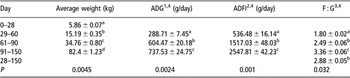
The growth performance and feed intake of pigs at different stages are shown in Table 2. Average weight, average day gain (ADG), average day feed intake (ADFI) and feed and gain ratio (F : G) increased with age.
Growth performances of pigs from 30 to 150 days

1ADG: average day gain (g/day).
2ADFI: average day feed intake (g/day).
3F : G: feed and gain ratio.
4Orthogonal polynomial contrast analyses of the data showed a linear change (P < 0.05).
a,b,c,dMeans in the same column with different superscript letters differ (P < 0.05).
Molecular cloning of porcine NHE-2
Two GSP were synthesized and 3′/5′ rapid amplifications of cDNA ends (RACE) were performed. 3′ RACE (1.2 kb) and 5′ RACE (1 kb) products were cloned into the pGEM-T vector and sequenced. The sequence of the 3′ RACE cDNA fragment is 1363 bp. Finally, 2872 bp of the cDNA was assembled from partial sequence (434 bp), 5′ RACE (1075 bp) fragments and 3′ RACE cDNA fragment (1363 bp) residues in the GenBank/EMBL data bank with accession number EF672046.
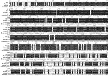
Sequence analysis of the porcine NHE-2 cDNA revealed (1) an ORF of 2445 bp that would encode a protein of 815 residues with a calculated molecular mass of 91.1 kDa, (2) 233 bp of the 5′ untranslated region (UTR) and (3) 194 bp of the 3′ UTR. BLASTn or BLASTp analysis demonstrated that the porcine sequence shares a high degree of sequence identity, both in the nucleotide sequences, especially in coding sequence (CDS) regions (80% and 70%), and in the deduced amino acid sequences (91% and 87%) with human (accession no. AF073299) and mouse (accession no. BC104737) NHE-2, respectively (Figure 1). Hydrophobicity prediction suggests 13 putative membrane-spanning domains within porcine NHE-2, similar to other mammalian NHE-2. Consistent with the results of homologous comparison, phylogenetic analysis shows that the divergence of porcine NHE-2 and human NHE-2 in evolution seems later than that of mouse NHE-2.
Deduced amino acid sequence comparison of porcine, human and mouse NHE. Deduced amino acids are indicated by their single-letter codes and are numbered to the right. The porcine-deduced amino acid sequence of NHE-2 (accession no. EF672046) shows 91% and 87% homology with the human (accession no. AF073299) and mouse (accession no. BC104737) NHE-2, respectively.

Tissue distribution of porcine NHE-2 mRNA
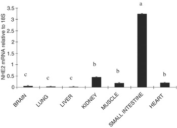
The relative NHE-2 mRNA expression in different tissues at day 60 is shown in Figure 2. The porcine NHE-2 mRNA was detected in the brain, liver, kidney, heart, lung, small intestine and muscle. The small intestine had the highest NHE-2 mRNA abundance and the brain, lung and liver had the lowest NHE-2 mRNA abundance (P < 0.05). However, there is no difference in NHE-2 mRNA abundance among the brain, lung and liver (P > 0.05). Compared with the small intestine, the kidney, muscle and heart had lower NHE-2 mRNA abundance (P < 0.05) and there is no difference among the kidney, muscle and heart (P > 0.05). Compared with the brain, lung and liver, the kidney, muscle and heart had higher NHE-2 mRNA abundance (P < 0.05).
Relative mRNA expression of porcine NHE-2 in different tissues of pig at day 150. All samples were normalized using 18S expression as an internal control in each real-time PCR. Relative level of NHE-2 mRNA was analyzed by the 2(−ΔCt) method. Bars that share a common superscript do not differ (P > 0.05). Data were presented as the mean ± s.e. (n = 5), in arbitrary units.

Expression of porcine NHE-2 mRNA along the longitudinal axis
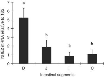
The intestinal distribution of NHE-2 mRNA at day 60 is shown in Figure 3. The NHE-2 mRNA level was decreased from the proximal to the distal part of the small intestine (P < 0.05) and stayed lower in the colon (P < 0.05). The duodenum had the highest NHE-2 mRNA abundance and the ileum and colon had the lowest NHE-2 mRNA abundance (P < 0.05). There was no difference between the ileum and the colon (P > 0.05). Compared to the ileum and the colon, the NHE-2 mRNA level was higher in the jejunum (P < 0.05).
Relative mRNA expression of porcine NHE-2 along the longitudinal axis of intestine at day 60. All samples were normalized using 18S expression as an internal control in each real-time PCR. Relative level of NHE-2 mRNA was analyzed by the 2(−ΔCt) method. Bars that share a common superscript do not differ (P > 0.05). Data were presented as the mean ± s.e. (n = 5), in arbitrary units.

Porcine NHE-2 mRNA relative abundance during ontogenesis
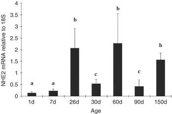
Developmental regulation of NHE-2 mRNA relative abundance in the duodenum is shown in Figure 4. The NHE-2 mRNA level was increased from day 1 to day 26 in the duodenum (P < 0.05) and dropped dramatically on day 30 (P < 0.05). There is no difference between day 1 and day 7 (P > 0.05). After day 30, the NHE-2 mRNA level remained the same except on day 90 (P > 0.05). There is no difference between day 30 and day 90 (P > 0.05).
Relative mRNA expression of porcine NHE-2 in pig duodenum during postnatal development. All samples were normalized using 18S expression as an internal control in each real-time PCR. Relative level of NHE-2 mRNA was analyzed by the 2(−ΔCt) method. Bars that share a common superscript do not differ (P > 0.05). Data were presented as the mean ± s.e. (n = 5), in arbitrary units. Orthogonal polynomial contrast analyses of the data showed a quadratic change (P < 0.05).

Discussion and conclusion
Members of the NHE gene family exhibit highly restricted temporal and spatial expression patterns. NHE-2 isoform is expressed at the apical membranes of the epithelial cells, where they play a major role in transepithelial Na+ absorption and proton efflux. To better understand the mechanisms underlying the regulation of the apical membrane NHE-2 isoform in pigs, we have focused our studies on molecular cloning, tissue distribution and ontogenetic regulation of NHE-2 mRNA expression in pig intestine. In this study, we have cloned the cDNA for the pig NHE-2 isoform. The pig NHE-2 cDNA encodes a polypeptide of 814 amino acid residues that exhibits an overall 90% amino acid identity to the human NHE-2 isoform and the calculated molecular mass of 91.1 kDa is also similar to human NHE-2 (90 kDa). The results indicated the molecular identity of cDNA to be NHE-2.
In the present study, we found that the porcine NHE-2 mRNA was detected in the brain, liver, kidney, heart, lung, small intestine and muscle. These results were in agreement with the previous study (Malakooti et al., Reference Malakooti, Dahdal, Schmidt, Layden, Dudeja and Ramaswamy1999). The abundance of the NHE-2 mRNA in these tissues may suggest that the expression of this isoform plays a role in transepithelial Na+ absorption in these tissues. However, the small intestine had the highest NHE-2 mRNA abundance compared with other tissues, which may suggest that it is particularly important in the intestine and may play a role other than transepithelial Na+ absorption in the intestine, for example, providing a proton gradient/drive force for peptide transporting (Repishti et al., Reference Repishti, Hogan, Pratha, Davydova and Donowitz2001).
In the present study, we found that mRNA levels for porcine NHE-2 demonstrated significant regional differences. The NHE-2 mRNA level was decreased from the proximal to the distal part of the small intestine (P < 0.05) and stayed lower in the colon (P < 0.05). The duodenum had the highest NHE-2 mRNA abundance and the ileum and colon had the lowest NHE-2 mRNA abundance (P < 0.05). These results were in agreement with the previous study in human and rat (Dudeja et al., Reference Dudeja, Rao, Syed, Joshi and Dahdal1996). Conversely, at the message level, NHE-2 is present in largest amount in the human distal colon > small intestine > proximal colon (Dudeja et al., Reference Dudeja, Rao, Syed, Joshi and Dahdal1996). This indicated that NHE gene regulation may be species specific. However, no careful quantitation of the amount of NHE-2 protein along the horizontal intestinal axis has been reported (Zachos et al., Reference Zachos, Tse and Donowitz2005). The different distribution of transporters along the intestinal axis from proximal to distal may be due to the unique morphological characteristics of the intestine and substrates, even though further research is needed to confirm this. Another concern of this study is that even though the intestine is physiologically divided into four segments, duodenum, jejunum, ileum and colon, there is still the possibility that the mRNA levels are different within each segment. More knowledge of the function of each gene is needed to further understand the precise pattern of distribution.
The intestine undergoes dramatic structural and functional changes after birth, such as increasing dry mass and absorptive surface area, and changing of membrane permeability and fluidity (Buddington et al., Reference Buddington, Elnif, Gardiner and Sangild2001). In addition to these non-specific changes, the absorptive capability per cell and the expression of transporters also alter with age. The ontogenic expression of NHE-2 has been reported in rat jejunum (Collins et al., Reference Collins, Xu, Kiela, Zeng and Ghishan1997 and Reference Collins, Kiela, Xu, Zeng and Ghishan1998). In rat jejunum there is a post-suckling (>2 weeks) increase in NHE-2 message, protein and contribution to BB NHE activity, which increases further at 6 weeks (post-weaning). However, at all times studied, BB NHE activity from NHE-2 was in the minority (maximum ∼25%). In the present study, the NHE-2 mRNA level in the duodenum was increased from day 1 to day 26 in the duodonum (P < 0.05) and dropped dramatically on day 30 (P < 0.05), which may have contributed to protein and BB NHE activity since previous studies indicated many of the responses occur via changes in gene transcription, although details generally are not available (Collins et al., Reference Collins, Xu, Kiela, Zeng and Ghishan1997 and Reference Collins, Kiela, Xu, Zeng and Ghishan1998). After weaning, the NHE-2 mRNA level dropped dramatically, which may be due to the disorder of intestinal function after weaning, for example, diarrheal disease (Moeser and Blikslager, Reference Moeser and Blikslager2007). However, the NHE-2 mRNA level is pretty stable during the growing period.
In previous studies, NHE-2 is regulated by several factors including growth factor (Xu et al., Reference Xu, Collins, Bai, Kiela, Lynch and Ghishan2001), protein kinase (Azarani et al., Reference Azarani, Goltzman and Orlowski1995), hormone (Ambuhl et al., Reference Ambuhl, Yang, Peng, Preisig, Moe and Alpern1999), cytokines (Rocha et al., Reference Rocha, Musch, Lishanskiy, Bookstein, Sugi, Xie and Chang2001) and other chronic extracellular stimuli. It is mainly at the transcriptional level regulation (Baum et al., Reference Baum, Moe, Gentry and Alpern1994). However, the mechanism of basal control of rat NHE-2 gene promoter activity is different in the renal and intestinal epithelium (Bai et al., Reference Bai, Collins, Xu and Ghishan2001). Sp3 is believed to be the major transcription of NHE-2 gene transcription in the intestinal epithelial cells. Xu indicated that rat NHE-2 activity can be stimulated by chronic epidermal growth factor treatment and that this response is at least partially mediated by gene transcription (Xu et al., Reference Xu, Collins, Bai, Kiela, Lynch and Ghishan2001). Infection-associated gut inflammation is very common in animal during postnatal development. Dramatic postnatal development of gut microbes also occurred in the gut. Diarrhea associated with infection by enteric pathogens could result from either increased chloride secretion, decreased NaCl absorption, or both. So the developmental changes of NHE-2 expression in postnatal pigs may be due to the combination of changes of substrate, cytokines, hormone, growth factors and extracellular stimuli during postnatal development.
The proton electrochemical gradient is considered as a driving force for solute transporter, including amino acid, peptides and fatty acids across the apical membrane and NHE is mainly responsible for maintaining the proton electrochemical gradient in mammalian small intestine (Thwaites and Anderson, 2007). Our previous study showed the mRNA expression of heterodimeric amino acid transporters (b0,+AT and y+LAT1) was not only differentially regulated by age but also differentially distributed along the intestine of piglets at early stages and growing stages of life (Feng et al., Reference Feng, Zhou, Zuo, Zhang, Yin, Wang and Wang2008). In the present study, we also found the same pattern with the NHE-2 transporter. So the developmental changes of NHE-2 expression in postnatal pigs may be related to the changes of the H+-coupled nutrient transporter during the postnatal development. However, how much contribution NHE-2 has in maintaining the proton electrochemical gradient in mammalian small intestine is still not known so far.
In conclusion, the mRNA expression of NHE-2 was not only differentially regulated by age but also differentially distributed along the small intestine of piglets at early stages and growing stages of life, which may be related to luminal substrate concentration (sodium loading) as well as disease and hormone. We still need further research on ontogenetic regulation of protein expression, and the total capacity of the whole intestine is important to fully understand ontogenetic changes.
Acknowledgements
This study was supported by the National Basic Research Program of China (Project no. 2004CB117501) and National Natural Science Foundation of China (Project no. 30671519).