Introduction
Necrotic enteritis (NE) is one of most important diseases of modern global poultry production. With an estimated cost per bird of at least US$0.05, NE is believed to cost the poultry industry around US$6 billion per annum in lost production and control strategies (Wade and Keyburn, Reference WADE and KEYBURN2015). For the individual producer, it has been calculated that more severe NE can erode profits by up to one third, with poorer feed conversion, reduced live weight gain and increased slaughter condemnations accounting for 44, 27 and 19% of this loss, respectively (Kaldhusdal et al., Reference KALDHUSDAL, BENESTAD and LOYLAND2016).
For many years, NE was not recognised as a major poultry disease (Williams, Reference WILLIAMS2005), but has grown in prominence, particularly since the European Union (EU) antibiotic growth promoter (AGP) ban in 2006 (Van Immerseel et al., Reference VAN IMMERSEEL, LYHS, PEDERSEN and PRESCOTT2016). In field studies, ‘drug-free’ broiler production results in a significant increase in NE occurrence (Smith, Reference SMITH2011; Gaucher et al., Reference GAUCHER, QUESSY, LETELLIER, ARSENAULT and BOULIANNE2015). This suggests that AGP (and anticoccidial) use plays a significant role in controlling the disease. However, many parts of the world have now either introduced legislation to reduce the use of antibiotics in animal production, or are in the process of reviewing antibiotic use, and so it seems inevitable that NE will continue to be both a major challenge and cost for the global poultry industry.
Necrotic enteritis
Necrotic enteritis in chickens is caused by pathogenic strains of the Gram-positive, spore-forming bacterium, Clostridium perfringens. C. perfringens are currently classified into five toxinotypes (A, B, C, D, E) according to the production of four different major toxins (alpha, beta, epsilon, iota). Toxinotype A strains of C. perfringens are generally associated with NE in broilers (Van Immerseel et al., Reference VAN IMMERSEEL, ROOD, MOORE and TITBALL2009), with strains expressing the more recently identified NetB toxin a definitive cause of NE in disease models (Keyburn et al., Reference KEYBURN, BOYCE, VAZ, BANNAM, FORD, PARKER, DI RUBBO, ROOD and MOORE2008). This pore forming toxin creates holes in cell membranes, which leads to the leakage of cell contents and cell death (Prescott et al., Reference PRESCOTT, PARREIRA, GOHARI, LEPP and GONG2016). Disease occurs when toxin production in the gastrointestinal tract (GIT) causes gross damage to the intestinal epithelium. Expression of a further toxin, tpeL, is thought to increase the virulence of NetB-expressing C. perfringens strains (Chalmers et al., Reference CHALMERS, BRUCE, HUNTER, PARREIRA, KULKARNI, JIANG, PRESCOTT and BOERLIN2008; Coursodon et al., Reference COURSODON, GLOCK, MOORE, COOPER and SONGER2012). C. perfringens are typically present in the GIT of healthy birds without NE but at numbers below 105 CFU per g of intestinal content (Caly et al., Reference CALY, D'INCA, AUCLAIR and DRIDER2015). However, these normal GIT inhabitants are (largely) considered to be non-pathogenic strains of C. perfringens, which do not cause NE (Lacey et al., Reference LACEY, JOHANESEN, LYRAS and MOORE2016). In the diseased situation, intestinal C. perfringens populations have been reported to increase to 107-109 CFU per g (Kondo, Reference KONDO1988). This increase is considered to be proliferation of low numbers of pathogenic C. perfringens following ingress into, or those already present in, the GIT. However, since a simple infection with pathogenic C. perfringens by itself is not sufficient to cause NE (Moore, Reference MOORE2016), other factors contribute to the proliferation of pathogenic strains, increased production of toxin(s) and the development of disease.
Necrotic enteritis typically affects broilers between two and six weeks of age, which may reflect diminishing maternal antibodies before the bird is immunologically mature, and presents as two forms, clinical and sub-clinical (Caly et al., Reference CALY, D'INCA, AUCLAIR and DRIDER2015). With the clinical form, the first sign can be the rapid, unexplained death of the affected bird. Other clinical signs can be reduced appetite, depression, diarrhoea, reluctance to move, ruffled feathers and closed eyes. With clinically affected flocks, mortality can be 1% per day with total mortality of up to 50% (Cooper and Songer, Reference COOPER and SONGER2009). The sub-clinical form is characterised by poor bird performance, but with no mortality. Both forms of NE can be definitively diagnosed by both identifying characteristic lesions in the intestine and confirming the presence of C. perfringens in those lesions (Smyth, Reference SMYTH2016). Reliable diagnosis of NE in live birds is, therefore, not currently possible. Subclinical NE lacks mortality that would assist with diagnosis, but is the form considered to have the greatest economic consequences through lost performance, slaughter condemnations and strategies (e.g. antibiotic use) to prevent disease (Shojadoost et al., Reference SHOJADOOST, VINCE and PRESCOTT2012).
There remain many gaps in our knowledge, particularly with regards to the pathogenesis of NE and immunity. While NetB seems key to the onset of NE, the target cell(s), cell receptor or how this toxin causes necrotic lesions with inflammation are currently unknown (Van Immerseel et al., Reference VAN IMMERSEEL, LYHS, PEDERSEN and PRESCOTT2016). This lack of knowledge currently undermines attempts to develop effective vaccines. Therefore, particularly with greater scrutiny on antibiotic use, the poultry industry's current focus is on addressing those key factors that are known to predispose to NE, which can often be influenced by diet.
Whilst this review considers C. perfringens in the context of an important poultry disease, it must be remembered that C. perfringens carriage by birds can also present a serious foodborne issue for humans (Van Immerseel et al., Reference VAN IMMERSEEL, DE BUCK, PASMANS, HUYGHEBAERT, HAESEBROUCK and DUCATELLE2004).
Predisposing factors
While many predisposing factors have been proposed to contribute to the development of NE, results can be contradictory and many conclusions are based on single experiments. Reproducible disease models probably provide the most reliable information on the initiators of NE. Various NE models use Eimeria challenge (coccidiosis) to induce disease and, under field conditions, coccidiosis may precede or co-occur with NE (Williams, Reference WILLIAMS2005). Although coccidiosis is commonly associated with NE, some experimental models successfully induce NE without coccidial challenge, confirming that a) Eimeria challenge is not essential and b) that other factors can initiate NE. For example, high protein, specifically fishmeal-based, diets are a common feature of reliable NE disease models.
It is apparent that compromised intestinal health can be considered the overriding trigger for pathogenic strains of C. perfringens to colonise, proliferate, express NetB toxin and cause NE. Clearly, numerous (dietary) factors can impact intestinal health, which underlines the complex, multifactorial nature of NE.
Coccidiosis
Coccidiosis is an enteric disease, which, in poultry, is caused by species of the Eimeria parasite. Eimeria invade the intestinal mucosa and damage the epithelium, which can lead to the leakage of serum into the gut and stimulation of mucus production, both of which can provide a rich source of nutrients for C. perfringens proliferation (Moore, Reference MOORE2016). Although not definitively proven, because NE is a disease of the small intestine, it seems logical that those species of Eimeria that specifically damage this part of the GIT (E. acervulina, E. maxima or E. necatrix) may be of particular relevance (Shojadoost et al., Reference SHOJADOOST, VINCE and PRESCOTT2012). Given the apparent role of Eimeria infection in the development of NE, mitigating Eimeria challenge is a key strategy in trying to prevent NE. Typically, coccidiosis control programmes have focused on the use of ionophores and chemicals and, more recently, vaccination. However, Eimeria drug resistance (Chapman and Jeffers, Reference CHAPMAN and JEFFERS2014) and reduced vaccine-related bird performance (Lee et al., Reference LEE, ECKERT, AMEISS, STEVENS, ANDERSON, ANDERSON, BARRI, MCELROY, DANFORTH and CALDWELL2011) encourages the review of coccidiosis control programmes and consideration of alternative options. Essential oils and, in particular, oregano (OEO) may be useful in this regard. Carvacrol is a major component of OEO and has been shown to inhibit E. tenella invasion of epithelial cells in-vitro (Burt et al., Reference BURT, TERSTEEG-ZIJDERVELD, JONGERIUS-GORTEMAKER, VERVELDE and VERNOOIJ2013). There have been a number of in-vivo studies demonstrating the benefit of OEO for coccidiosis management in broilers (Saini et al., Reference SAINI, DAVIS and DUDLEY-CASH2003a; Tsinas et al., Reference TSINAS, GIANNENAS, VOIDAROU, TZORA and SKOUFOS2011; Mohiti-Asli and Ghanaatparast-Rashti, Reference MOHITI-ASLI and GHANAATPARAST-RASHTI2015), including performance enhancement following Eimeria vaccination (Harrington et al., Reference HARRINGTON, KONSTANTI, MATHIS and BROOM2016). A further interesting feature of OEO is the ability to stimulate enterocyte proliferation (Levkut et al., Reference LEVKUT, MARCIN, REVAJOVA, LENHARDT, DANIELOVIC, HECL, BLANAR, LEVKUTOV and PISTL2011; Konstanti et al., Reference KONSTANTI, BROOM, OWUSU-ASIEDU, HARRINGTON and GIANNENAS2016). Enhanced enterocyte production would aid intestinal regeneration during either Eimeria infection and/or NE. In fact, OEO has been reported to be beneficial for NE (Saini et al., Reference SAINI, DAVIS and DUDLEY-CASH2003b).
Diet-related factors
protein
As mentioned above, high dietary protein is recognised as a significant predisposing factor for NE. Several NE disease models use fishmeal to increase dietary protein, while level of animal protein was shown to influence the relationship between feed and NE in a long-term, retrospective study from Norway (Kaldhusdal and Skjerve, Reference KALDHUSDAL and SKJERVE1996). Drew et al. (Reference DREW, SYED, GOLDADE, LAARVELD and VAN KESSEL2004) formulated broiler diets with 230, 315 or 400 g/kg crude protein (CP) using either soy protein concentrate (SPC) or fishmeal as the major protein sources. The authors showed that, in general, increasing dietary CP increased ileal and caecal C. perfringens numbers, regardless of protein source, while C. perfringens populations were greater for fishmeal vs. SPC for a given CP level. Amino acid (AA) analysis of the diets revealed higher glycine and methionine concentrations of the fishmeal than the SPC diets. Whilst the authors did not specifically enumerate pathogenic C. perfringens numbers, the work does confirm that higher protein levels and/or fishmeal promote proliferation of C. perfringens, with disparity in amino acid content the likely cause of differences between protein sources. While a specific role for methionine remains unclear (Widyaratne, Reference WIDYARATNE2012), the same group have shown a positive correlation between glycine content of both the diet and ileal contents, and ileal and caecal C. perfringens populations (Wilkie et al., Reference WILKIE, VAN KESSEL, WHITE, LAARVELD and DREW2005). Furthermore, when fat-encapsulated glycine was fed to broiler chickens to promote the slow release of the amino acid throughout the GIT, C. perfringens numbers were found to be higher in the ileum and/or caeca (Dahiya et al., Reference DAHIYA, HOEHLER, VAN KESSEL and DREW2007). Crystalline AAs are considered to be rapidly absorbed in the upper GIT and so use of these forms, particularly with regards to glycine, may contribute to preventing C. perfringens proliferation and NE. It is interesting that genome sequencing has identified that C. perfringens lack the ability to synthesis a number of AAs and so produce an array of hydrolytic enzymes to help them acquire substrates from their host/environment (Shimizu et al., Reference SHIMIZU, OHTANI, HIRAKAWA, OHSHIMA, YAMASHITA, SHIBA, OGASAWARA, HATTORI, KUHARA and HAYASHI2002). In addition, elevated protein and AAs have been associated with C. perfringens proliferation and toxin production (Titball et al., Reference TITBALL, NAYLOR and BASAK1999).
Clearly, protein source, level and dietary AA profile are important factors influencing intestinal health and NE development. With the need for more judicious use of antibiotics, it will be even more important that diet formulations more closely match the specific AA requirements of the bird to absolutely minimise protein and AA flow into the distal GIT. Part of this goal will include the provision of highly digestible proteins or AAs. A number of feed additives could be advantageous in this regard. Enzymes for various target substrates (e.g. non-starch polysaccharides and phytate) have been shown to increase ileal digestibility of CP and AAs, including glycine, in broilers (Selle et al., Reference SELLE, RAVINDRAN and PARTRIDGE2009). The need for better protein utilisation, particularly with the availability of cheaper, less digestible protein sources, could lead to greater use of proteases in poultry nutrition.
Organic acid-based (OAB) feed additives have been demonstrated to enhance ileal protein and AA digestibility in broilers (Khodambashi Emami et al., Reference KHODAMBASHI EMAMI, GRAYSTONE, DANESHMAND, AKBARI MOGHADDAM KAKHKI and BROOM2016). Moreover, NE and its predisposing factors are associated with reduced butyrate-producing bacteria in the intestine (Wu et al., Reference WU, STANLEY, RODGERS, SWICK and MOORE2014). Butyrate is recognised for having important beneficial functions in the intestine and the addition of OAB additives to broiler diets have been shown to increase intestinal butyrate (Graystone et al., Reference GRAYSTONE, KHODAMBASHI EMAMI and BROOM2016) and help reduce NE (Timbermont et al., Reference TIMBERMONT, LANCKRIET, DEWULF, NOLLET, SCHWARZER, HAESEBROUCK, DUCATELLE and VAN IMMERSEEL2010), potentially making such additives an integral part of a strategy to review dietary AA/CP content, enhance digestibility, improve gut health and prevent NE.
Whilst considering protein digestion, it should be noted that trypsin has been reported to degrade C. perfringens toxins in the small intestine (Songer, Reference SONGER1996). In support of this, work by Palliyeguru et al. (Reference PALLIYEGURU, ROSE and MACKENZIE2011), showed that trypsin inhibitors present in non-cooked soya bean-based diets increased the severity of NE lesions, with the relationship between severity and level of soya bean inclusion being directly proportional. This underlines the need to consider the wider implications of feed components and anti-nutritional factors for optimal protein digestion and absorption, and prevention of NE.
Non-starch polysaccharides
Non-starch polysaccharides (NSP) are key cell wall components of cereal grains, which are not hydrolysed by the endogenous enzymes of monogastric animals. In monogastric nutrition, NSP focus is generally on β-glucans and arabinoxylans, which invariably have a higher concentration in cereal grains such as barley, oats, rye and wheat (Bach Knudsen, Reference BACH KNUDSEN1997). Studies have shown that birds fed diets based on these cereal grains experience more severe NE than birds fed a corn-based diet (Riddell and Kong, Reference RIDDELL and KONG1992; Branton et al., Reference BRANTON, LOTT, DEATON, MASLIN, AUSTIN, POTE, KEIRS, LATOUR and DAY1997). This was supported by an observational study from Norway, which reported an increased incidence of NE for birds fed wheat and barley-based diets compared to those fed corn-based diets (Kaldhusdal and Skjerve, Reference KALDHUSDAL and SKJERVE1996). Cereal grains contain both soluble and insoluble NSPs. Soluble NSPs can increase digesta viscosity, which impedes both the diffusion of digestive enzymes and absorption of nutrients (Kumar et al., Reference KUMAR, SINHA, MAKKAR, DE BOECK and BECKER2012). This impairment of nutrient digestion and absorption will maintain more substrates in the intestine for the proliferation of the resident microbiota, including C. perfringens. The effect of ‘viscous’ grains on C. perfringens proliferation has been demonstrated in-vitro. Annett et al. (Reference ANNETT, VISTE, CHIRINO-TREJO, CLASSEN, MIDDLETON and SIMKO2002) reported that proliferation of C. perfringens type A was greater in both digested wheat and barley diets compared with a digested corn diet. In addition, NSP can stimulate greater mucin production in the intestine (Kleessen et al., Reference KLEESSEN, HARTMANN and BLAUTl2003). C. perfringens produce an array of hydrolases to degrade mucin for nutrient acquisition and to facilitate better access to the intestinal epithelium (Shimizu et al., Reference SHIMIZU, OHTANI, HIRAKAWA, OHSHIMA, YAMASHITA, SHIBA, OGASAWARA, HATTORI, KUHARA and HAYASHI2002).
Increased digesta viscosity is associated with reverse peristalsis and increased intestinal transit time (Sacranie et al., Reference SACRANIE, SVIHUS and IJI2012). Prolonging transit time, particularly if the digesta is sub-optimally digested, will provide more time for microbial utilisation of substrates and proliferation, while reverse peristalsis can move microbial communities from distal to more proximal GIT sites (Sacranie et al., Reference SACRANIE, IJI, MIKKELSEN and CHOCT2007). Both scenarios could be decisive factors in the development of NE.
Enzymes have become the accepted dietary strategy to combat the effects of NSP and digesta viscosity, particularly for ‘viscous’ grain-based diets. For example, xylanase and β-glucanase have been clearly demonstrated to reduce digesta viscosity (Bedford and Classen, Reference BEDFORD and CLASSEN1992), improve nutrient digestibility (Selle et al., Reference SELLE, RAVINDRAN and PARTRIDGE2009) and reduce the consequences of NE (Jia et al., Reference JIA, SLOMINSKI, BRUCE, BLANK, CROW and JONES2009; Liu et al., Reference LIU, GUO and GUO2012).
Wet litter
Numerous factors can contribute to wet litter, which is considered a predisposing factor for NE (Moore, Reference MOORE2016). Gaucher et al., Reference GAUCHER, QUESSY, LETELLIER, ARSENAULT and BOULIANNE2015, reported greater litter moisture, with increased incidence of NE, in a ‘drug-free’ programme. Litter is considered ‘wet’ when its water holding capacity is compromised, which is typically at a moisture content above 25% (Collett, Reference COLLETT2012). In effect, litter moisture reflects the balance between water addition and evaporation, which will be affected by housing, management, disease, as well as diet-related factors. Elevated digesta viscosity can result in birds increasing their intake and excretion of water (Van der Klis and De Lange, Reference VAN DER KLIS and DE LANGE2013). This can elevate litter moisture content, which facilitates C. perfringens sporulation (Moore, Reference MOORE2016) and makes the organism extremely tolerant of external challenges/stressors. Dietary electrolyte balance (DEB) can affect litter moisture. Ravindran et al. (Reference RAVINDRAN, COWIESON and SELLE2008) reported a positive linear relationship between DEB and both excreta moisture and score (with higher = poorer), demonstrating the importance of managing the electrolyte balance of poultry diets to minimise NE risk.
Phytases are commonly used in poultry diets to liberate phosphorus (P) from, and to counteract the anti-nutritional effects of, phytate (Selle and Ravindran, Reference SELLE and RAVINDRAN2007). Phytase use has been associated with increased excreta moisture (Debicki-Garnier and Hruby, Reference DEBICKI-GARNIER and HRUBY2003), which has been related to the release of bound minerals, for example calcium and sodium, from the phytate molecule during hydrolysis. These mineral ions have the potential to significantly influence DEB balance and negatively affect excreta characteristics (Dunlop et al., Reference DUNLOP, MOSS, GROVES, WILKINSON, STUETZ and SELLE2016). It is, therefore, imperative that the increased availability of these additional minerals (to P) is accounted for when formulating poultry diets with phytase to reduce potential issues with wet litter and NE.
Mycotoxins
Mycotoxins, which are secondary metabolites from fungi growing on feed materials, are known to disrupt intestinal health (Broom, Reference BROOM2015a). For example, Alassane-Kpembi et al. (Reference ALASSANE-KPEMBI, KOLF-CLAUW, GAUTHIER, ABRAMI, ABIOLA, OSWALD and PUEL2013) demonstrated that the Type B trichothecenes reduced intestinal cell viability, even at low concentrations, while Taranu et al. (Reference TARANU, BRAICU, MARIN, PISTOL, MOTIU, BALACESCU, BERIDAN NEAGOE and BURLACU2015) reported that low concentrations of zearalenone changed the expression of 1,954 intestinal cell genes. In the Taranu et al. work, the altered expression included genes involved in inflammatory and cellular repair pathways. Therefore, there is clear evidence of mycotoxins initiating intestinal damage that would promote C. perfringens proliferation and the development of NE. Antonissen et al. (Reference ANTONISSEN, CROUBELS, PASMANS, DUCATELLE, EECKHAUT, DEVREESE, VERLINDEN, HAESEBROUCK, EECKHOUT, DE SAEGER, ANTLINGER, NOVAK, MARTEL and VAN IMMERSEEL2015) showed that the addition of a modest concentration of fumonisins (B1 and B2) to the feed of broiler chickens reduced intestinal architectural parameters (e.g. villus height and crypt depth), while fumonisin feeding increased ileal (alpha toxin expressing) C. perfringens numbers prior to challenge with a NetB positive, type A strain. Following C. perfringens challenge, more birds fed the fumonisin contaminated feed subsequently developed subclinical NE (44.9 vs. 29.8%). The same authors had previously shown that deoxynivalenol (DON) caused intestinal epithelial damage and barrier disruption in broilers, and increased the incidence of sub-clinical NE following C. perfringens challenge (Antonissen et al., Reference ANTONISSEN, VAN IMMERSEEL, PASMANS, DUCATELLE, HAESEBROUCK, TIMBERMON, VERLINDEN, JANSSENS, EECKHAUT, EECKHOUT, DE SAEGER, HESSENBERGER, MARTEL and CROUBELS2014a).
Mycotoxins are able to affect the immune system and most of their effects would be regarded as negative (Girish and Smith, Reference GIRISH and SMITH2008). Deoxynivalenol is particularly well studied and numerous immune cells (e.g. macrophages, B and T lymphocytes and natural killer cells) are very sensitive to DON, while DON has been shown to alter cytokine secretion, increase cell apoptosis and suppress antibody responses to vaccination (Maresca, Reference MARESCA2013). In fact, vaccination is sometimes used in NE challenge models to compromise the immune system and thus reliably induce NE (Shojadoost et al., Reference SHOJADOOST, VINCE and PRESCOTT2012), which reaffirms the importance of the immune system in disease prevention.
Minimising mycotoxin contamination of poultry feed and controlling those mycotoxins that are still present prior to feeding can be important factors in preventing NE. With modern analytical techniques, it is now accepted that most raw materials and finished feeds are contaminated with multiple mycotoxins (Streit et al., Reference STREIT, SCHWAB, SULYOK, NAEHRER, KRSKA and SCHATZMAYR2013), which may have additive or even synergistic effects. As outlined, mycotoxins can damage the intestine and compromise the immune system. Good agronomy, harvesting and storage of grain all help prevent mycotoxin contamination but will not eliminate the risk. Therefore, nutritionists and producers should develop suitable mycotoxin management policies, which are likely to include effective in-feed mycotoxin control products, to help prevent NE. In addition, feed additives that have been demonstrated to suitably enhance immune responses, for example probiotics, prebiotics (Ajuwon, Reference AJUWON2016) and organic acids (Broom, Reference BROOM2015b), are useful candidates for inclusion in feed formulations for disease prevention. It should be remembered that mycotoxins can impair numerous factors (e.g. immunity, intestinal recovery, treatment, etc.) relating to coccidiosis alleviation (Antonissen et al., Reference ANTONISSEN, MARTEL, PASMANS, DUCATELLE, VERBRUGGHE, VANDENBROUCKE, LI, HAESEBROUCK, VAN IMMERSEEL and CROUBEL2014b), further underlining the necessity to manage mycotoxin contamination of feed.
Practical recommendations
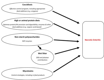
Several recommendations relating to the key NE predisposing factors and their dietary mitigation can be made, which are outlined in Figure 1 . Firstly, it is important to ensure an effective coccidiosis control programme is in place. Incorporation of alternative ingredients (e.g. oregano) can work in synergy to maximise the efficacy of control programmes and bird performance. Nutritionists need to ensure diets align as closely as possible to the nutrient requirements of the bird to maximise ileal AA digestibility. The use of fishmeal or other animal proteins, or those that are poorly digested, should be reviewed and alternatives considered to improve dietary AA balance and utilisation. Nutritionists should pay special attention to glycine content. Feeds should include specialist ingredients, including enzymes, or even organic acids, that are proven to increase AA/protein digestibility.
Major necrotic enteritis predisposing factors and diet-related mitigation.

It is essential to be mindful of viscous cereal grain inclusion in diets and thus NSP content, and use NSP enzymes as appropriate. Minerals from feed ingredients or those added to the feed should be carefully balanced to ensure that they do not adversely affect DEB, which can cause wet litter. When using phytase, consider mineral contributions to maintain DEB. All feed producers should implement an effective mycotoxin management strategy, and use appropriate, broad spectrum, mycotoxin control products as an ‘insurance’ against contamination which can damage the gut and impair the immune system, leaving poultry more vulnerable to NE. Finally, it is important to identify specific NE risk period(s) and minimise dietary changes at this time.
Conclusions
Necrotic enteritis is now a prominent and costly problem for the poultry industry. It is a complex disease with numerous factors that are considered to contribute to the development of NE. Many of these factors are related in some way to diet and this review has attempted to bring together, coherently, the latest information on those factors, along with practical recommendations. Duration and timing of predisposing factors are likely to affect the probability and severity of NE (Van Waeyenberghe et al., Reference VAN WAEYENBERGHE, DE GUSSEM, VERBEKE, DEWAELE and DE GUSSEM2016), and a better understanding of these aspects would be advantageous. Generally, NE arises when the intestinal environment has been compromised, which then allows pathogenic C. perfringens to become established and proliferate. NetB secretion, which is currently recognised as the key toxin, causes extensive damage to intestinal cells and thus the epithelium as a whole. Other diet-related factors, such as feed particle size, withholding feed, or timing of specification changes may be relevant factors for NE risk but are not part of this review. Moreover, other dietary components, such as correctly balanced vitamins and minerals, help to ensure optimum bird health and thus could reduce disease susceptibility, while other infections or stressors (e.g. stocking density) could mean that birds succumb to NE, but are not covered by this review. This review has focussed on those factors that are considered key to NE development and has outlined diet-related strategies that can play a crucial role in mitigating against disease. A coordinated approach to addressing the key NE predisposing factors, through appropriate dietary strategies, will help mitigate against NE, particularly in an era where more judicious use of antibiotics is a must.