




No CrossRef data available.
Article contents
Slow and wide QRS complex tachycardia as a unique complication following radiofrequency catheter ablation of a left-sided accessory pathway in a child
Published online by Cambridge University Press: 03 May 2005
Abstract
Radiofrequency lesions can, theoretically, be the substrate for new persistent arrhythmias. As far as we know, this has never previously been encountered after transcatheter ablation of accessory pathways. A child with Wolff–Parkinson–White syndrome was referred for radiofrequency catheter ablation of a left-sided accessory pathway. After successful ablation of the accessory pathway using a retrograde transaortic approach, the child developed an incessant wide QRS complex tachycardia at slow rate that was resistant to pharmacologic interventions. The focus of the tachycardia was identical to the ventricular site of insertion of the eliminated accessory pathway.
Information
- Type
- Brief Report
- Information
- Copyright
- © 2005 Cambridge University Press
In patients with the Wolff–Parkinson–White syndrome, left-sided accessory pathways can be ablated using a retrograde aortic approach, aiming at the ventricular insertion under the mural leaflet of the mitral valve. Complications after radiofrequency ablation of such pathways are rare. These are related mostly to the retrograde nature of the catheterization, such as damage to the aortic valve and the coronary arteries, endocarditis, dissection of the aorta, transient ischemic attacks, and those related to vascular access. The lesions induced by radiofrequency to ablate the pathway can also result in cardiac trauma, perforation, tamponade, myocardial infarction, and complete heart block.1, 2 Theoretically, the radiofrequency lesions can also produce an arrhythmogenic focus for new persistent arrhythmias, but as far as we are aware, this has not been seen clinically. We have now encountered such a complication.
Case report
A 12-year-old boy with Wolff–Parkinson–White syndrome and orthodromic atrioventricular reciprocating tachycardia was referred to our institution for transcatheter ablation. Analysis of his 12-lead electrocardiogram suggested that the accessory pathway was left-sided. Under general anesthesia, a decapolar diagnostic catheter was inserted into the coronary sinus via the right jugular vein. Quadripolar catheters, and a conventional bipolar pacing electrode, were positioned along the superoseptal region of tricuspid ring, the superior region of the right atrium, and the right ventricular apex. The antegrade activation sequence was consistent with a left lateral accessory pathway. This was confirmed by the eccentric retrograde atrial activation sequence, which also showed non-decremential properties of conduction. During orthodromic atrioventricular re-entrant tachycardia, the atrial activation sequence was identical to the observed sequence during ventricular pacing. The accessory pathway had a refractory period of 240 milliseconds.
Using a right femoral arterial approach, a Webster B curve thermocouple mapping/ablation catheter was advanced retrogradely through the aortic valve, and was used to map the mitral annulus and to deliver radiofrequency energy, supported by guidance using the LocaLisa navigation system (Medtronic Inc., Minneapolis, USA). During atrial pacing, a total of 13 radiofrequency energy pulses of 50 watts at 60 degrees Celsius were applied along the mitral annulus in the region with continuous atrial and ventricular signals. The total time used for ablation was 3 minutes. After the last application of radiofrequency, there was a sudden change in the antegrade activation sequence, and ventricular pacing revealed ventriculo-atrial decremental conduction, proving that antegrade and retrograde conduction across the accessory pathway had ceased. After the ablation, there were no signs of recurrence of conduction across the pathway. Intravenous administration of adenosine, at a does of 12 milligrams, resulted in a period of short-lasting complete atrioventricular block, confirming successful ablation of the accessory pathway. About 15 minutes after the last application, there emerged a sustained wide QRS complex tachycardia with a cycle length of 490 milliseconds. The patient was haemodynamically stable, and 2 boluses of lidocaine, at 1 milligram per kilogram of body weight, and 1 gram of magnesium sulphate, were administered without any effect. Atrial and ventricular pacing overdrove the arrhythmia, which recurred every time when the pacing was stopped after a short period of “warming-up”. The axis of the initial part of QRS during the tachycardia was identical to the axis of the delta wave before the ablation (Fig. 1). The ventricular signal recorded by the tip of the ablation catheter was entirely negative, suggesting that this was the site of origin of the tachycardia (Fig. 2). The patient was kept on strict monitoring in intensive care unit. The arrhythmia persisted for 48 hours, produced no haemodynamic impairment, and disappeared spontaneously.

Figure 1. 12-lead ECG of the patient in three different conditions: before ablation (a), during ventricular tachycardia (b), and after spontaneous resolution of the tachycardia (c). The axis of the delta wave is identical to the axis of the QRS during ventricular tachycardia.

Figure 2. Intracardiac recordings consistent with slow ventricular tachycardia after ablation. The earliest ventricular activation is at the tip of mapping/ablation catheter (black arrow). Earliest retrograde atrial activation is on the proximal His bundle electrode (white arrow). The cycle length of the tachycardia shows progressive shortening.
Discussion
Our case demonstrates that application of radiofrequency energy in the ventricle can induce new persistent arrhythmias.
Complications related to the use of radiofrequency energy. In adults,1, 2 as well as in children,3 treatment of the tachyarrhythmias produced by accessory pathways using radiofrequency ablation has a very high rate of success, at about 95 per cent, a rate of complications below 4 per cent, and a rate of mortality around 0.1 per cent. Persistent acute arrhythmic complications are far below 1 per cent,1–3 most often complete heart block. Incessant wide QRS complex tachycardia as an acute complication of ablation, as far as we know, has not previously been reported. Long term follow-up after successful ablation has shown paroxysmal or persistent atrial fibrillation to be the most common complicating arrhythmia, occurring almost always in older patients who have had paroxysmal atrial fibrillation before ablation. It is probably due to the electrical atrial remodeling, and not to the lesions induced by radiofrequency.4 On long-term follow-up, ventricular tachycardia after successful ablation has been reported only once, and was again not considered to be related to the lesion induced by the radiofrequency energy.5
Mechanism of the wide QRS complex tachycardia. Wide QRS complex tachycardias can occur spontaneously before ablation in patients with accessory pathways, due to their spontaneous automaticity, but they disappear after successful ablation.6 Isolated spontaneous automaticity in the pathway after ablation has also been reported,7 or as a cause of wide QRS complex tachycardia when associated with hypercatecholaminemia.8 The mechanism involved in our case is rather suggestive for enhanced automatism.9 The temporary overdrive suppression, and the “warming-up” phenomenon, are arguments in favour of this interpretation. Another argument in favour is the slow spontaneous disappearance, together with progressive prolongation of the cycle length, probably related to the recovery of the injured tissue. Presence of bidirectional block within the accessory pathway, however, together with overdrive suppression with either atrial or ventricular pacing, suggests a slow ventricular tachycardia rather than automaticity of the accessory pathway.
Alternative energy sources to reduce arrhythmogenicity. Although radiofrequency energy delivered through a catheter is highly successful for most ablations, safety has become a critically important issue in some types of arrhythmia, and in certain anatomical locations. The most advanced catheter-based system that can be used as an alternative to radiofrequency energy is cryothermy.
References
Hindricks G. The Multicentre European Radiofrequency Survey (MERFS): complication of radiofrequency catheter ablation of arrhythmia. The Multicentre European Radiofrequency Survey (MERFS) investigators of the Working Group on Arrhythmias of the European Society of Cardiology. Eur Heart J 1993; 14: 1644–1653.Google Scholar
Scheinman MM, Huang S. The 1998 NASPE prospective catheter ablation registry. Pacing Clin Electrophysiol 2000; 23: 1020–1028.Google Scholar
Kugler JD, Danford DA, Houston K, et al. Pediatric Radiofrequency Catheter Ablation Registry: Update of immediate results. In: Imai Y, Momma K (eds). Proceedings of the Second World Congress of Pediatric Cardiology and Cardiac Surgery. Futura, Armonk, NY, 1998, pp 604–605.
Aytemir K, Amasyali B, Kose S, et al. Maximum p-wave duration and p-wave dispersion predict recurrence of paroxysmal atrial fibrillation in patients with Wolff–Parkinson–White syndrome after successful radiofrequency catheter ablation. J Interv Card Electrophysiol 2004; 11: 21–27.Google Scholar
Oddsson H, Walfridsson H, Edvardsson N. Perception and documentation of arrhythmias after successful radiofrequency catheter ablation of accessory pathways. Ann Noninvasive Electrocardiol 2001; 6: 216–221.Google Scholar
Deam AG, Burton E, Walter P, Langberg JJ. Wide complex tachycardia due to automaticity in an accessory pathway. PACE 1995; 18: 2106–2108.Google Scholar
Tseng ZH, Yadav AV, Scheinman MM. Catecholamine dependent accessory pathway automaticity. PACE 2004; 27: 1005–1007.Google Scholar
Macle L, Shah DC, Jais P, Haissaguerre M. Accessory pathway automaticity after radiofrequency ablation. J Cardiovasc Electrophysiol 2002; 13: 285–287.Google Scholar
Josephson ME, Almendral JM, Buxton AE, Marchlinsky FE. Mechanisms of ventricular tachycardia. Circulation 1987; 75: III41–III47.Google Scholar

12-lead ECG of the patient in three different conditions: before ablation (a), during ventricular tachycardia (b), and after spontaneous resolution of the tachycardia (c). The axis of the delta wave is identical to the axis of the QRS during ventricular tachycardia.
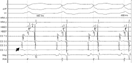
Intracardiac recordings consistent with slow ventricular tachycardia after ablation. The earliest ventricular activation is at the tip of mapping/ablation catheter (black arrow). Earliest retrograde atrial activation is on the proximal His bundle electrode (white arrow). The cycle length of the tachycardia shows progressive shortening.
 You have
Access
- 2
- Cited by