Introduction
Most of the species of Mesorhabditis Osche, Reference Osche1952 belonging to the Monhystera-group, are poorly described and illustrated. Males have not been reported in Mesorhabditis acuminata (Kreis, Reference Kreis1929) Dougherty, Reference Dougherty1955; Mesorhabditis capitata Loof, Reference Loof1964; Mesorhabditis cranganorensis (Khera, Reference Khera1968) Andrássy, Reference Andrássy1983; M. dunensis Khera, Reference Khera1971; Mesorhabditis sambharensis Khera, Reference Khera1971; and Mesorhabditis signifera (Baranovskaya, Reference Baranovskaya1959) Baranovskaya, 1962. Only a few species such as M. acuticauda (Shokoohi et al., Reference Shokoohi, Mehdizadeh, Amirzadi and Abolafia2014), Mesorhabditis minuta (Boström, Reference Boström1991; Abolafia & Peña-Santiago, Reference Abolafia and Peña-Santiago2009) belonging to the Spiculigera-group, and Mesorhabditis microbursaris (Mahboob and Jahan, Reference Mahboob and Jahan2021) belonging to the Monhystera-group have been described based on scanning electron microscopic observations. Likewise, Mesorhabditis acidophila (Borgonie et al., Reference Borgonie, Dierick, Houthoofd, Willems, Jacobs and Bert2010) and Mesorhabditis monhystera have been molecularly characterized. Lately, Launay et al. (Reference Launay, Félix, Dieng and Delattre2020) studied the relationship between the isolates of the Monhystera-group based on the D2/D3 domain of large subunit 28 rDNA.
The present study provides a detailed description of M. monhystera (Bütschli, Reference Bütschli1873) Dougherty, Reference Dougherty1955 based on morphometrics and morphological observations, molecular characterization and scanning electron microscopy (SEM). A phylogenetic analysis based on the D2/D3 segment of 28S rDNA, presents the precise status of the genus Mesorhabditis among closely related taxa. A pictorial key comparing the species of the Monhystera-group, is also provided. The phylogeography of haplogroups of Mesorhabditis using the median-joining network method (Bandelt et al., Reference Bandelt, Forster and Röhl1999) is used to infer the degree of mutation/divergence among the different isolates.
Materials and methods
Collection, extraction and culturing of nematodes
The sample containing M. monhystera was collected from soil contaminated with slaughter wastes in Vessu, Anantnag, Jammu and Kashmir, India. The samples were stored in plastic bags and brought to the laboratory. To extract nematodes, the samples were processed through Cobb's (Reference Cobb1918) sieving and decanting methods and the modified Baermann (Reference Baermann1917) funnel technique. Stock cultures of nematodes were maintained in 1.2% nematode growth medium.
Light microscopic observations
For light microscopy (LM), nematodes were fixed in 4% formaldehyde, dehydrated to pure glycerine (Seinhorst, Reference Seinhorst1959) and later mounted on slides using the wax ring technique (De Maeseneer & D'Herde, Reference De Maeseneer and D’ Herde1963). The nematodes were measured with an ocular micrometer and drawn using a drawing tube. LM photographs were taken with a Jenoptik digital camera, ‘ProgRes’ (Jena, Germany), mounted on an Olympus BX-51 DIC microscope.
Scanning electron microscopic observations
For SEM, live nematodes were picked from one-week-old culture. The nematodes (15 males and 15 females) were fixed in SEM fixative (1.6% paraformaldehyde and 2.5% glutaraldehyde) for 24 h at 4°C. The fixed nematodes were washed three times in phosphate buffer, dehydrated in ethanol series (30%–100%) and dried using hexamethyldisilazane. The dried nematodes were later mounted on stub and coated with 10 nm gold before being observed under 10–15 kV under a Hitachi TM4000 Plus scanning electron microscope (Hitachi, Singapore).
Molecular profiling
For DNA extraction, ten live individuals were transferred to an Eppendorf tube containing 20 μl lysis buffer (Williams et al., Reference Williams, Schrank, Huynh, Shownkeen and Waterston1992). The sample was kept at −20°C in a refrigerator for 24 h, and then incubated in a thermal cycler at 65°C for 45 min, followed by 15 min at 95°C. The samples were cooled at 4°C and stored at −20°C. For DNA amplification, 5 μl lysate was used in a 20 μl polymerase chain reaction (PCR) reaction mix following the manufacturer's protocol (GeNei, Bengaluru, India). The sequence of the D2/D3 expansion region of large subunit 28 s rDNA was amplified using the forward primer D2A 5′–ACAAG TACCGTGAGGGAAAGTTG–3′ and the reverse primer D3B 5′–TCCTC GGAAGGAACCAGCTACTA–3′. For amplification, 5 ml DNA lysate was used in a 20 ml PCR mix. The PCR parameters included: initial denaturation at 95°C for 5 min; followed by 35 cycles of 95°C for 30 s, 55°C for 30 s, 72°C for 1 min; and final extension for 10 min at 72°C. Aliquots of 5 ml of the PCR products were sized with a low DNA mass ladder and separated by electrophoresis in 1% agarose gel stained with ethidium bromide and observed under the Ultraviolet Transilluminator Dolphin View Gel Documentation system. Sequencing was done in both directions.
Evaluation of the phylogenetic framework
The obtained sequences were edited in Chromas version 2.6.6. (Technelysium Pty Ltd, www.technelysium.com.au), aligned and a consensus sequence generated in BioEdit (Hall, Reference Hall1999). The consensus sequence of 579 base pairs was submitted to GenBank with accession number ON693986. The sequence of M. monhystera (Bütschli, Reference Bütschli1873) Dougherty, Reference Dougherty1955 was aligned with GenBank sequences of 37 closely related taxa, in MEGA X (Kumar et al., Reference Kumar, Stecher, Li, Knyaz and Tamura2018) using the CLUSTAL_x alignment tool (Thompson et al., Reference Thompson, Gibson, Plewniak, Jeanmougin and Higgins1997). The ambiguously aligned sequences were removed using the online version of Gblocks 0.91b (Castresana, Reference Castresana2000). The phylogenetic tree with 481 characters in the final dataset was inferred by the Bayesian inference method, MrBayes version 3.1.2 (Huelsenbeck & Ronquist, Reference Huelsenbeck and Ronquist2001). For the analyses, the best model under the Akaike information criterion was determined to be (GTR + G + I) using jModelTest version 2.1.3 (Darriba et al., Reference Darriba, Taboada, Doallo and Posada2012). The Akaike-supported model, log-likelihood, state frequency of nucleotides, substitution rate across the sites, proportions of invariable sites, the shape parameter of the gamma distribution and rate of variation were examined during analysis. The obtained values of the above parameters were as follows: −InL = 2567.7772; freqA = 0.2457, freqC = 0.2052, freqG = 0.3231, freqT = 0.2260; R (AC) = 0.7687, R(AG) = 2.2213, R(AT) = 0.9403, R(CG) = 1.4047, R(CT) = 7.1119, R(GT) = 1.0000; p-inv = 0.1870; and gamma shape = 0.6110. The analysis was run with the Markov chain Monte Carlo for 4 × 10⁶ generations. ‘Burn-in’ samples were discarded every 2000 generations, and a consensus tree with a minimum 50% majority rule was used for analysis. The tree was visualized, edited and saved with FigTree 1.4.0 (Rambaut, Reference Rambaut2014).
Percentage similarity and genetic divergence
The percentage similarity and genetic divergence (base differences) among the sequences of selected species of Mesorhabditis were estimated as per Mahboob et al. (Reference Mahboob, Bashir, Asif, Nazir, Jahan and Tahseen2022). The number of base differences per sequence was computed in MEGAX with 262 positions including parts of both loci in the final dataset.
Systematics
Class: Chromadorea Inglis, Reference Inglis1983
Order: Rhabditida Chitwood, Reference Chitwood1933
Suborder: Rhabditina Chitwood, Reference Chitwood1933
Infraorder: Rhabditomorpha De Ley & Blaxter, Reference De Ley, Blaxter and Lee2002
Superfamily: Rhabditoidea Örley, Reference Örley1880
Family: Rhabditidae Örley, Reference Örley1880
Genus: Mesorhabditis Osche, Reference Osche1952
Diagnosis. Rhabditidae. Gonochoristic or hermaphroditic individuals with small to medium-sized, 400–944 μm long body. Cuticle finely transversely annulated. Lip region usually offset from adjoining body, rarely continuous. Lips well separated, rounded to globular, each with raised setose outer labial sensilla. Amphidial aperture small, on lateral lips. Stoma tubular, long. Cheilostomal walls usually not cuticularized. Gymnostom cuticularized constituting long tubular part of stoma. Stegostom with distinct metastegostomal swellings, each armed with two small denticles. Pharynx rhabditoid type with cylindrical corpus, swollen metacorpus, usually with zipper-like lumen and basal bulb with double-chambered haustrulum. Female reproductive system monodelphic, prodelphic. Ovary reflexed, oviduct continuing into a distinct spermatheca. Vagina obliquely oriented with post-equatorial vulval opening. Male reproductive system monorchic; vas deferens with paired ejaculatory glands. Spicules separated or distally fused, short to long and slender. Bursa well-developed or rudimentary, peloderan or leptoderan, anteriorly open. Genital papillae 5–9 pairs. Tail short conical to elongate conoid, moderately long. Phasmids at the level of or posterior to the anus.
Type species: Mesorhabditis spiculigera (Steiner, Reference Steiner1936) Dougherty, Reference Dougherty1955
Other species
-
Mesorhabditis acidophila Borgonie, Dierick, Houthoofd, Willems, Jacobs and Bert, Reference Borgonie, Dierick, Houthoofd, Willems, Jacobs and Bert2010
-
Mesorhabditis acuminata (Kreis, Reference Kreis1929) Dougherty, Reference Dougherty1955
-
Mesorhabditis acuticauda Ahmad, Shah and Mahamood, Reference Ahmad, Shah and Mahamood2010
-
Mesorhabditis africana Andrássy, Reference Andrássy1982
-
Mesorhabditis anisomorpha (Sudhaus, Reference Sudhaus1978) Andrássy, Reference Andrássy1983
-
Mesorhabditis belari (Nigon, Reference Nigon1949) Dougherty, Reference Dougherty, Dayal and Singh1953
-
Mesorhabditis capitata Loof, Reference Loof1964
-
Mesorhabditis carmenae Abolafia and Peña-Santiago, Reference Abolafia and Peña-Santiago2009
-
Mesorhabditis cranganorensis (Khera, Reference Khera1968) Andrássy, Reference Andrássy1983
-
Mesorhabditis denticulatus Mahboob and Jahan, Reference Mahboob and Jahan2021
-
Mesorhabditis dunensis Khera, Reference Khera1971
-
Mesorhabditis franseni Fuchs, Reference Fuchs1933
-
Mesorhabditis inarimensis (Meyl, Reference Meyl1953) Dougherty, Reference Dougherty1955
-
Mesorhabditis irregularis (Körner in Osche, Reference Osche1952) Dougherty, Reference Dougherty1955
-
Mesorhabditis kherai (Sudhaus, Reference Sudhaus1976) Sudhaus, Reference Sudhaus2011
-
Mesorhabditis kinchegensis Nicholas, Reference Nicholas1998
-
Mesorhabditis labiata (Völk, Reference Völk1950) Dougherty, Reference Dougherty1955
-
Mesorhabditis littoralis Yeates, Reference Yeates1969
-
Mesorhabditis longespiculosa (Schuurmans Stekhoven, Reference Schuurmans Stekhoven1951) Dougherty, Reference Dougherty1955
-
Mesorhabditis longistomis Massey, Reference Massey1974
-
Mesorhabditis megachilis (Sudhaus, Reference Sudhaus1978) Andrássy, Reference Andrássy1983
-
Mesorhabditis microbursaris (Steiner, Reference Steiner1926)
-
Mesorhabditis minuta Boström, Reference Boström1991
-
Mesorhabditis miotki (Sudhaus, Reference Sudhaus1978) Andrássy, Reference Andrássy1983
-
Mesorhabditis monhystera (Bütschli, Reference Bütschli1873) Dougherty, Reference Dougherty1955
-
Mesorhabditis oschei (Körner in Osche, Reference Osche1952) Dougherty, Reference Dougherty1955
-
Mesorhabditis paucipapillata (Paetzold, Reference Paetzold1955) Paetzold, Reference Paetzold1958
-
Mesorhabditis riparia (Brzeski, Reference Brzeski1985) Sudhaus, Reference Sudhaus2011
-
Mesorhabditis sambharensis Khera, Reference Khera1971
-
Mesorhabditis scanica (Allgén, Reference Allgén1949) Sudhaus, Reference Sudhaus2011
-
Mesorhabditis signifera (Baranovskaya, Reference Baranovskaya1959) Baranovskaya, 1962
-
Mesorhabditis simplex (Cobb, Reference Cobb1893) Sudhaus, Reference Sudhaus2011
-
Mesorhabditis spiculigera (Steiner, Reference Steiner1936) Dougherty, Reference Dougherty, Dayal and Singh1953
-
Mesorhabditis striatica Dassonville and Heyns, Reference Dassonville and Heyns1984
-
Mesorhabditis sudhausi Andrássy, Reference Andrássy1982
-
Mesorhabditis szunyoghyi Andrássy, Reference Andrássy1961
-
Mesorhabditis vernalis (Andrássy, Reference Andrássy1982)
Material examined
The voucher material representing nine females and nine males in good condition, was examined (figs 1–4).
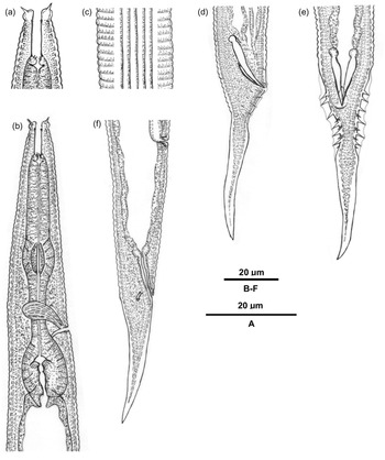
Line drawing of Mesorhabditis monhystera (Bütschli, Reference Bütschli1873) Dougherty, Reference Dougherty1955. (a–c, f) female and (d, e) male: (a) anterior end; (b) pharyngeal region; (c) lateral field with four prominent bands; (d) tail region (lateral view); (e) tail region (ventral view); and (f) tail region (lateral view). Scale bar = 20 μm.

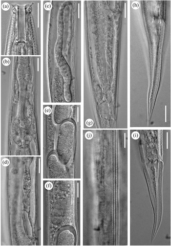
Light micrograph of Mesorhabditis monhystera (Bütschli, Reference Bütschli1873) Dougherty, Reference Dougherty1955 (female): (a) anterior end; (b) pharyngeal region; (c) distal part of the reproductive system showing ovarian flexure; (d) proximal part of the reproductive system showing seminal vesicle filled with sperms, and uterus; (e, f) part of reproductive system showing seminal vesicle, columella and uterus containing egg; (g) vulva–anus region; (h, i) tail region; and (j) lateral field with four bands. Scale bars = 10 μm.

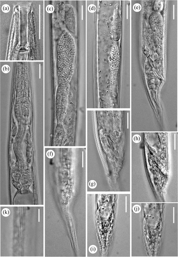
Light micrograph of Mesorhabditis monhystera (Bütschli, Reference Bütschli1873) Dougherty, Reference Dougherty1955 (male): (a) anterior end; (b) pharyngeal region; (c, d) genital branch with dorsally and laterally reflexed testis, respectively; (e) tail region showing spicule and gubernaculum; (f–h) tail region showing arrangement of genital papillae (lateral view); (i, j) tail region (ventral view); and (k) lateral field with four bands. Scale bars = 10 μm.

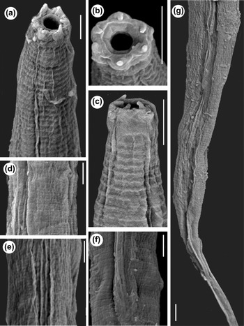
Scanning electron micrograph of Mesorhabditis monhystera (Bütschli, Reference Bütschli1873) Dougherty, Reference Dougherty1955: (a, c) anterior region; (b) en face view; (d) body region showing excretory pore; (e) body region showing lateral fields; (f) mid-body showing vulval lips; and (g) posterior region from the vulva to tail. Scale bars: 5 μm.

Measurements
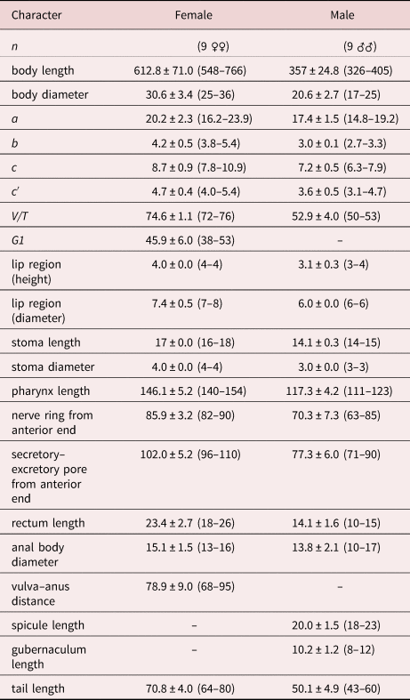
For measurements, see table 1.
Morphometric data of Mesorhabditis monhystera (Bütschli, Reference Bütschli1873) Dougherty, Reference Dougherty1955.

Measurements are in μm and in the form: mean ± standard deviation (range).
Note: a, total body length/body diameter; b, total body length/pharynx length; c, total body length/tail length; c’, tail length/anal body diameter; V/T, vulva percentage with respect to total body length/male gonad percentage with respect to total body length; and G1, female genital branch percentage with respect to total body length.
Description
Adult. Medium-sized nematodes, almost straight after fixation, tapering at both extremities, more in the posterior region. Cuticle 1–2 μm thick, annulated with transverse striations and punctations all over the body except tail region. Punctations conspicuous up to two stoma length in the anterior region. Lateral fields with four prominent bands/ridges. Lip region offset, about twice of its length. Lips six, globular, well separated, each with raised setose labial sensilla. Amphidial apertures slit-like, labial, minute, and indistinguishable under LM. Stoma long, 4–5 times longer than wide, constitute 11–12% of total pharyngeal length. Cheilostom a short tube with cuticularized walls. Gymnostom a long tube with parallel walls covering larger part of the stoma. Stegostom having two setose denticles at each metastegostomal swelling. Pharynx well-developed, highly muscular, covering about 20–26% of total body length; procorpus long, highly muscular with convoluted lumens (in some specimens), corpus lumen without conspicuous striation or zipper-like pattern, expanded posteriorly into a swollen metacorpus of about 14–18 × 10–15 μm in dimension; isthmus a narrow tube of 15–28 μm long, expanding posteriorly to form a well-developed pyriform basal bulb of about 17–20 μm × 12–15 μm dimension containing highly cuticularized grinder and double-chambered haustrulum. Nerve ring encircling the mid of isthmus at about 57–58% of total pharyngeal length from anterior end. Secretory–excretory duct opening at posterior level of nerve ring at 68–71% of pharyngeal length from anterior end. Cardia conoid, 3–5 μm long. Intestinal cells large with prominent nuclei. Rectum 1.3–1.6 times longer than anal body diameter. Tail conoid, shorter than vulva–anus distance or about 10–11% of the total body length. Phasmids open at the level of anus.
Female. Reproductive system monodelphic, prodelphic; ovary dorsally reflexed often reaching up to spermatheca. Oocytes arranged in three rows at the distal end of the ovary followed by two tiers and a single tier proximally. Oviduct indistinguishable. Spermatheca ovoid to oblong, axial with many spermatozoa followed by the uterus. Vagina thick-walled, 4–6 μm long or 1/4 of the corresponding body diameter obliquely oriented. Vulva posterior at about 72%–76% of body length from the anterior end with lips not protruded.
Male: Similar to female in general morphology except more arcuate ventrally in the posterior region. Testis monorchic, dorsally reflexed (lateral in few specimens). Seminal vesicle well differentiated, swollen containing numerous rounded minute sperms. Vas deferens a muscular narrow tube extending proximally into ejaculatory glands. Spicules small with prominent knob-like capitulum, indistinguishable neck, and slender calamus with a fused distal end. Gubernaculum trough-shaped, covering about 50% of spicule length. Bursa leptoderan, rudimentary, anteriorly open. Genital sensilla papilliform, nine pairs; two pairs precloacal and seven postcloacal pairs out of which three postcloacal pairs inside and four pairs outside bursal flaps, oriented dorsally; Tail conoid, constituting 13–15% of the total body length, usually shorter than vulva–anus distance.
Habitat and locality
The present population of M. monhystera (Bütschli, Reference Bütschli1873) Dougherty, Reference Dougherty1955 was collected from the soil sample contaminated with slaughter wastes in Vessu, Anantnag, Jammu and Kashmir, India at coordinates 33°40′17″ N 75°07′45″ E.
Voucher materials
Nine females and nine males on slides of M. monhystera (kmr/dist/Meso/1–10) were deposited in the Nematode Collection, Department of Zoology, Aligarh Muslim University, Aligarh, Uttar Pradesh, India.
Remarks
Mesorhabditis monhystera has been originally reported from soil around the roots of Plantago, Germany (Bütschli, Reference Bütschli1873) and subsequently from multiple terrestrial and also aquatic habitats of France (Launay et al., Reference Launay, Félix, Dieng and Delattre2020) and the United States (Chitwood & Chitwood, Reference Chitwood and Chitwood1934, Reference Chitwood and Chitwood1937; Sudhaus, Reference Sudhaus2018). The present population of M. monhystera (Bütschli, Reference Bütschli1873) Dougherty, Reference Dougherty1955 resembles those described by earlier workers (Bütschli, Reference Bütschli1873; Sudhaus & Fitch, Reference Sudhaus and Fitch2001; Andrássy, Reference Andrássy2005) including the original population, in most of the characteristics. However, the only population showing dissimilarity with the present population in the morphological characteristics is M. monhystera apud Abolafia & Peña-Santiago (Reference Abolafia and Peña-Santiago2009) that shows differences in the type of lip region (weakly vs. distinctly offset); length of labial sensilla (smaller vs. larger); and number (three vs. four) of ridges in the lateral fields, male tail and spicule shapes (with distinguishable vs. indistinguishable) calamus and the configuration of genital papillae (three vs. two) precloacal pairs. Coincidentally, M. monhystera apud Abolafia & Peña-Santiago (Reference Abolafia and Peña-Santiago2009) resembles M. vernalis Andrássy, Reference Andrássy1982 in all the above characteristics although the number of genital papillae is greater (nine vs. six) pairs.
Emended diagnosis
Mesorhabditis monhystera is characterized by small to medium-sized individuals with cuticle annulated with transverse striations and punctations; lateral fields with four prominent bands; lip region offset with six well-separated globular lips, each with a tentaculate labial sensilla; amphidial apertures labial, small, elliptical slit-like; corpus lumen occasionally striated, metacorpus swollen, basal bulb having a grinder with double-chambered haustrulum; reproductive system mono-prodelphic, vagina obliquely oriented, vulva posteriorly located at about 65%–76% from the anterior end without protruded lips; rectum usually 1.3–1.6 times longer than the anal body diameter; phasmids opening at the level of the anus; tail conoid, usually shorter than vulva–anus distance; male with spicules fused distally, each comprising of distinct knobbed manubrium, indistinguishable calamus, slender lamina; gubernaculum covering about 50% of the spicule length; bursa leptoderan largely rudimentary; genital papillae nine pairs; and two pairs precloacal and seven postcloacal pairs out of which three postcloacal pairs inside and four pairs outside bursal flap.
Discussion
Status of the genus Mesorhabditis among closely related genera
The Basic Local Alignment Search Tool (BLAST) results revealed similarities of the present population of M. monhystera with the monodelphic taxa viz., Crustorhabditis Sudhaus, Reference Sudhaus1974; Distolabrellus Anderson, Reference Anderson1983; Mesorhabditis Osche, Reference Osche1952 and Parasitorhabditis Fuchs, Reference Fuchs1937. These groups resembled in homologous traits such as offset lip region (except Parasitorhabditis obtusa possessing continuous lip region), vulva situated far posterior (except Rhabpanus ossiculum with a slightly posterior vulva) and tail hemispheroid to short conoid. However, the DNA sequence of the present population did not show similarity with monodelphic species of Cruznema Artigas, Reference Artigas1927 in the BLAST results. Nevertheless, the members of the genus Cruznema were included in the phylogenetic analysis due to being representatives of the family Rhabditidae and sharing some degree of homology. Panagrolaimus sp. (LT908055) was selected as an out-group with few common traits such as monodelphic female gonad with vulva situated far posterior and conoid tail. Besides these traits, Panagrolaimus spp. also showed similarity in having continuous lip region, fused lips and metastegostom without conspicuous denticles as observed in members of Matthesonema Osche, Reference Osche1955 and Parasitorhabditis, although warts were observed in some species of Parasitorhabditis.
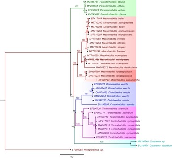
The tree topology of the selected genera revealed two major clades: one clade comprised the taxa of Mesorhabditis belonging to both Monhystera-groups and Spiculigera-groups suggesting monophyly; and the other clade represented the taxa of the genera Cruznema, Distolabrellus and Teratorhabditis, although Cruznema diverged earlier than the latter two genera. Moreover, the genus Parasitorhabditis formed a separate clade and diverged earlier than the rest of the groups.
Our analysis agreed well with the results of Launay et al. (Reference Launay, Félix, Dieng and Delattre2020) based on 28S rDNA and 1nternal transcribed spacer 2 region in the placement of the different isolates of the genus Mesorhabditis (Monhystera-group). However, the present analysis did not totally conform to that of Shokoohi et al. (Reference Shokoohi, Mehdizadeh, Amirzadi and Abolafia2014) based on small subunit (SSU) 28S rDNA, where Cruznema formed a clade with Buetschlinema Sudhaus, Reference Sudhaus2011 and diverged earlier than the genera Teratorhabditis and Distolabrellus. Also, the placement of the genus Parasitorhabditis and the species M. anisomorpha showed conflict where Parasitorhabditis diverged first and both M. anisomorpha and M. longispiculosa clustered together. The present analysis also differs from the phylogenetic inference made by Valizadeh et al. (Reference Valizadeh, Goldasteh, Rafiei-Karahroodi and Pedram2017) in the placement of Parasitorhabditis close to Mesorhabditis, although it showed agreement in the placement of M. longispiculosa and M. anisomorpha. However, the placement of Cruznema also differed as it clustered with Pellioditis Dougherty, Reference Dougherty, Dayal and Singh1953 and Rhabditella Cobb, Reference Cobb1929 (fig. 5).
Bayesian phylogenetic tree based on the D2/D3 domain of large subunit 28S rDNA inferred in MrBayes version 3.1.2 (Huelsenbeck & Ronquist, Reference Huelsenbeck and Ronquist2001). The evolutionary history was evaluated using the GTR + I + G model. The tree topology indicated the status of the present population of Mesorhabditis monhystera (Bütschli, Reference Bütschli1873) Dougherty, Reference Dougherty1955 among the congeners. The consensus tree with a minimum 50% majority rule was used for analysis. The posterior probability values are reflected at appropriate clades. The scale bar shows the number of substitutions per site.

Molecular status of the congeners of Mesorhabditis (Monhystera-group)
The phylogenetic tree demonstrated monophyletic status of the genus Mesorhabditis that formed a major clade of the subordinate taxa. The members were further divided into two subclades representing Monhystera-group and Spiculigera-group with 100% posterior probability values. The members of Monhystera-group further diverged into two groups with good branch support values: the isolates of M. monhystera with accession numbers (MT710269; ON693986; MT710271), clustered together showing similarity but differed with nearest M. denticulatus (MW763072) with 100% posterior probability values, whereas, most species – viz., M. belari (EF417149; MT710238), M. paucipapillata (MT710240), M. cranganorensis (MT710263), M. microbursaris (MT710259); M. vernalis (MT710258), M. littoralis (MT710253), M. simplex (MT710249), and M. franseni (MT710247) of the Monhystera-group as reported by Launay et al. (Reference Launay, Félix, Dieng and Delattre2020), clustered together in another group (fig. 5).
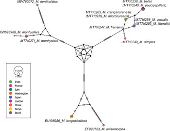
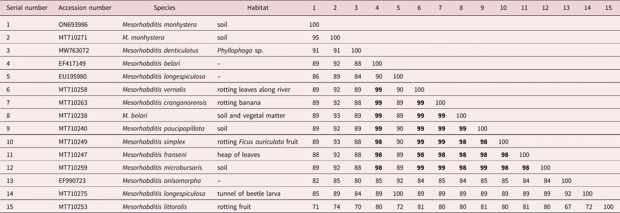
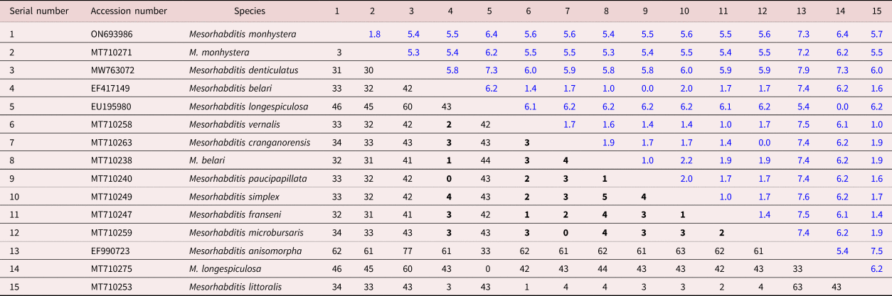
The sequence of the present population of M. monhystera (ON693986) showed 95% similarity and three (3) base divergence with another isolate of M. monhystera (MT710271); however, it showed 91% similarity and thirty-one (31) base divergence with M. denticulatus. In this context, several species of Monhystera-group with sequence deposited in GenBank, were observed to demonstrate high similarity (98–99%) with very little base divergence (0–5 bases) from each other. The sequences of M. belari (EF417149; MT710238) and M. paucipapillata (MT710240) with 99% similarity, 0–1 base divergence along with 0.0–1.0 standard error appeared to be conspecific. Likewise, the sequences of M. cranganorensis (MT710263) and M. microbursaris (MT710259) with 99% similarity and 0 base divergence with 0.0 standard error, and M. simplex (MT710249) and M. franseni (MT710247) with 98% similarity and 1 base divergence with 1.0 standard error also indicated overlap. Although M. vernalis (MT710258) and M. littoralis (MT710253) clustered in one clade with 81% similarity, there was only 1 base difference with standard error (1.0) (tables 2 and 3). The Haplotype network of the isolates of Mesorhabdittis sampled from different geographical locations and inferred using the median-joining network method, revealed three distinct clusters originating from the ancestral stock. Considerable differences based on allele frequency could be noted between the haplotypes of M. denticulatus and M. monhystera and between M. longispiculosa and M. anisomorpha but the cluster representing M. vernalis, M. littoralis, M. belari, M. paucipapillata, M. cranganorensis, M. microbursaris M. simplex and M. franseni showed insignificant genetic deviation and little change in allele frequency (figs 6 and 7). Launay et al. (Reference Launay, Félix, Dieng and Delattre2020), based on cross-breeding experiments, reported the above-mentioned, largely pseudogamous isolates to be true species and emphasized that little genetic variation was sufficient for transition to a new species in the asexual regime, while intraspecific genetic diversity accumulated with sexuality and recombination. However, in view of the bifurcated findings, a revision based on the complete SSU (18S rDNA) and LSU (28S rDNA) is required to resolve the status of the above species in addition to the validation of its identity in the light of original types.
Haplotype network of the isolates of Mesorhabditis inferred using the median-joining network method (Bandelt et al., Reference Bandelt, Forster and Röhl1999) based on the D2/D3 domain of large subunit 28 s rDNA. The network was evaluated using the POPART (Leigh & Bryant, Reference Leigh and Bryant2015). The circle represents the haplotype and its size indicates allelic frequency of the haplotype. Hatch marks between nodes indicate degree of divergence and the colour of the circle shows the geographical location of the isolate of Mesorhabditis. Note: nucleotide diversity (pi) = 0.131852; segregating sites = 89; parsimony-informative sites = 47; Tajima's D statistic (D) = 3.48433; and p (D ≥ 3.48433) = 0.

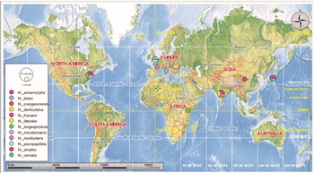
The geographical location of D2/D3 based sequences of the isolates of Mesorhabditis. The sampling location of such isolates was used to place the sequences on the map. The geographical location of the taxa was evaluated using the POPART (Leigh & Bryant, Reference Leigh and Bryant2015). The colour represents different taxa of Mesorhabditis.

Percentage similarity within the species of genus Mesorhabditis based on the D2/D3 domain of the large subunit 28S rDNA sequences along with information on the microhabitat and country-wise location (similarity statistics are as follows: minimum = 67.3; maximum = 100; mean = 90.3; and standard deviation = 7.5).

Bold values indicate close relationship based on high degree of similarity.
Base differences per nucleotide among the sequences of the species of genus Mesorhabditis based on of D2/D3 domain of the large subunit 28S rDNA.

The values below the diagonal indicate the base differences, while those above the diagonal (in blue) indicate standard errors.
Bold values indicate close relationship indicating very less base divergence.
Morphological status of the congeners of Mesorhabditis (Monhystera-group)
The species of Mesorhabditis represent widely distributed, r-selective bacterivores reported from enriched habitats including rotten wood, plant residues and sediment at the shore of freshwater bodies. The salient characters include: rounded, separated lips, each with one thorn-like sensillum; long stoma having glottoid apparatus with two setose denticles at each sector; pharyngeal sleeve absent or very small; zipper-like corpus lumen, swollen metacorpus; mono-prodelphic gonad; female tail mostly elongate conoid, males occasionally rare with well-developed to the reduced bursa, and spicules distally fused and two pairs of precloacal genital sensilla. Owing to the heterogeneity, the members are divided into two species groups viz., Monhystera-groups and Spiculigera-groups (Sudhaus, Reference Sudhaus2011). The differentiation of the groups is mainly based on the reproductive mode and the male features. The Monhystera-group mainly represents small–medium-sized females that are largely hermaphroditic/parthenogenetic or pseudogamous (amictic) with males rare or few while the Spiculigera-group usually contains large-sized, gonochoristic individuals with fair representation of males. The males of the Monhystera-group usually show small spicules with rudimentary bursa not enclosing all genital sensilla which often are inconspicuous or reduced in number. The females usually possess a tail shorter than vulva–anus distance. On the other hand, males of the Spiculigera-group usually possess large-sized spicules and well-developed bursa enclosing 9–10 pairs of genital sensilla including phasmids.
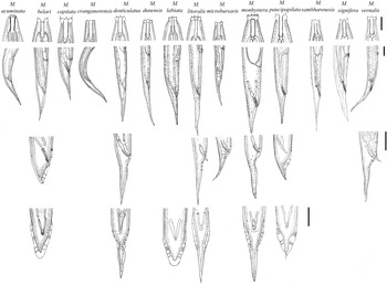
The molecular data of the present population of M. monhystera and the other previously reported isolates (MT710269; ON693986; MT710271) revealed close relationships with M. denticulatus Mahboob and Jahan, Reference Mahboob and Jahan2021; however, the present population of M. monhystera showed closeness to M. denticulatus and M. litoralis Yeates, Reference Yeates1969 in appearance of lip region and labial sensilla, shape and size of the male tail, and similar shape of spicules. However, M. monhystera could be differentiated from the latter two species in having females with conoid (vs. slender) tails and arrangement of genital papillae in five (vs. nine) pairs. Mesorhabditis denticulatus significantly differed from M. litoralis in having relatively smaller females (411–538 μm vs. 600–720 μm); smaller b (3.3–4.5 vs. 5.2–6.0) and V (66–70% vs. 72–82%) values; vulval lips protruded (vs. not protruded); stoma wide (vs. narrow), 4 times (vs. 5–6 times) longer than wide; phasmidial opening posterior (vs. at level of anus); gubernaculum relatively small (vs. large) covering about (50% vs. 70%) of the spicule length; and precloacal genital papillae one pair (vs. two pairs) in M. litoralis apud Yeates (Reference Yeates1969) (fig. 8).
Pictorial key for the comparison of the species of genus Mesorhabditis (Monhystera- group) based on female anterior region (scale bar = 10 μm); female posterior region (lateral view) and male tail region (lateral and ventral view) (scale bar = 20 μm. [The type species except Mesorhabditis monhystera (after Sudhaus & Fitch, Reference Sudhaus and Fitch2001) were redrawn from the original descriptions].

Besides the close morphological relationships of M. littoralis with M. monhystera and M. denticulatus, the molecular analysis showed that M. littoralis positioned with M. vernalis Andrássy, Reference Andrássy1982, but significantly differed from the latter in a combination of characters viz., larger females (600–720 μm vs. 410–560 μm); greater b (5.2–6.0 vs. 3.6–4.4) value; stoma wide (vs. narrow); spicules with indistinguishable (vs. distinguishable) neck; gubernaculum relatively smaller (vs. larger) covering about (50% vs. 70%) of the total spicule length; and genital papillae five (vs. six) pairs with one pair (vs. three pairs) of precloacals in M. vernalis apud Andrássy (Reference Andrássy1982) (fig. 8).
The molecular phylogenetic tree constructed from the existing GenBank sequences for M. belari Nigon, Reference Nigon1949 and M. paucipapillata Paetzold, Reference Paetzold1955 showed close relationships of the two which markedly differed in original description in the type of bursa (peloderan vs. leptoderan), tail shape (with vs. without spike) and the number and arrangement of genital papillae (eight pairs vs seven pairs) with two precloacal pairs (vs. one precloacal pair) in M. paucipapillata apud Paetzold (Reference Paetzold1955) (fig. 8).
Likewise, no congruence could be observed between the molecular data obtained from GenBank for M. cranganorensis Khera, Reference Khera1968 and M. microbursaris (Steiner, Reference Steiner1926) Andrássy, Reference Andrássy1983 and the morphological features of both species in original and subsequent descriptions. Despite being similar on account of body size (405–615 μm) and overlapping morphometric values, M. cranganorensis showed significant differences in having greater a (28–31 vs. 18–24) value; cuticle with fine or smooth (vs. coarse and prominent) annulations; continuous (vs. offset) lip region with reduced (vs. well-developed globular) lips; female tail slender with blunt tip (vs. conoid with fine terminus); and recorded without (vs. with) males in M. microbursaris apud Zeidan & Geraert (Reference Zeidan and Geraert1989) (fig. 8).
Although molecular characterization is regarded as the most reliable tool for identification, morphological characters cannot be undermined. At a time when classical taxonomists are diminishing, the molecular data submitted to GenBank on the name of an old species becomes very critical. It is instead better to give a new name to a molecularly-characterized population if the holotype and paratype of the proposed species could not be compared.
Conflict of interest
None.
Financial support
This work was supported by the Science and Engineering Research Board (SERB), Government of India, New Delhi, India.
Ethical standards
The authors assert that all procedures contributing to this work comply with the ethical standards of the relevant national and institutional guidelines onthe care and use of laboratory animals.