Gestational diabetes mellitus, the most frequently observed metabolic disorder in pregnancy, affects approximately 7% of pregnant women. Reference Setji, Brown and Feinglos1–Reference Schäfer-Graf and Vetter3 Despite significant advancement in perinatal diagnosis, surveillance, and management of complications, gestational diabetes mellitus persists as a prominent contributing factor to fetal teratogenicity. Hypertrophic cardiomyopathy, pericardial effusion, intermittent or persistent bradycardia, and congenital heart anomalies are the most common associated cardiac disorders in fetuses of diabetic mothers. Reference Atiq, Ikram, Hussain and Saleem4 Fetal hyperinsulinemia has been suggested as a cause of fetal myocardial hypertrophy with impaired cardiac function, especially during diastole, as a result of decreased left ventricular distensibility and altered left atrial dynamics secondary to myocardial hypertrophy. Reference Breitweser, Meyer, Sperling, Tsang and Kaplan5–Reference Ullmo, Vial and Di Bernardo7 Myocardial hypertrophy in fetuses of diabetic mothers resolves within the first few months of life after the resolution of fetal hyperinsulinemia. Reference Way, Wolfe, Eshaghpour, Bender, Jaffe and Ruttenberg8 However, neonates may present with cardiomegaly and respiratory distress secondary to poor left ventricular compliance. These findings emphasise the need of adequate prenatal assessment of fetal cardiac function.
Fetal echocardiography has developed over the past 30 years as a crucial non-invasive modality to evaluate fetal cardiac anatomy, function, and non-invasive hemodynamics. However, the assessment of diastolic function is a particular challenge, due to the weak correlation between Doppler echocardiographic parameters and ventricular filling pressures. Reference Nishimura, Appleton, Redfield, Ilstrup, Holmes and Tajik9,Reference Nagueh, Lakkis, Middleton, Spencer, Zoghbi and Quinones10 Previously, isovolumetric relaxation time was found to be a sensitive parameter to detect diastolic dysfunction in poor glycaemic control patients. Reference Atiq, Ikram, Hussain and Saleem4 Several studies have showed that increased left ventricular filling pressure is associated with increased left atrial volumes, with changes in diameter correlating with an increase in diastolic dysfunction. Reference Briguori, Betocchi and Losi11 There are few data reporting the applicability of left atrial shortening fraction in the assessment of fetal diastolic function. Reference Chen, Gui, Ren and Shi12 The purpose of this study was to assess cardiac function (systolic, diastolic, and global myocardial performance) in fetuses of diabetic mothers as compared to the control. We also aimed to assess the difference in left atrial shortening fraction in fetuses of diabetic mothers with or without myocardial hypertrophy, and compared to the control.
Patients and methods
Mothers with a diagnosis of gestational diabetes mellitus referred for fetal cardiac evaluation between 20 and 30 weeks gestation, to the Pediatric Echocardiography Unit at The Aga Khan University Hospital, Karachi, Pakistan from July 2017 to June 2018, were enrolled as case patients in this study. Non-diabetic mothers who had a fetal echocardiogram between 20 and 30 weeks gestation for other indications were enrolled as the control. Exclusion criteria were type 2 diabetes mellitus, inadequate echocardiographic images, fetuses with any additional anatomical abnormality of the heart, arrhythmia or malformation involving other organs, or mothers with systemic pathology other than maternal diabetes, twin or multiple pregnancy, as well as a refusal of the mother to give informed written consent. Ethical approval was obtained from the Ethics Review Committee at The Aga Khan University (ERC No- 4896-Ped-ERC-17).
Sample size was calculated using openEpi version 3 on the two-sided confidence intervals = 95%, power = 80%, mean left atrial shortening fraction of non-exposed group = 0.53+/−0.09, and 0.46+/−0.12 in the exposed group = [15]. Sample size calculated was 37 in each group.
Details of history, maternal and gestational ages, medications, and non-cardiac anomalies detected on routine obstetric ultrasound scans were noted.
Fetal echocardiogram was performed using GE Vivid E9 Ultrasound System (GE Healthcare Life Sciences, Pittsburg, USA) with the curvilinear 4C probe and standard fetal software for analysis. 2D, M-mode, colour flow mapping, pulsed wave Doppler was used to analyse the fetal cardiac structure and function. The study was performed by a single Paediatric Cardiologist by standard method, after obtaining good views and near parallel Doppler alignment with either ventricular wall or interventricular septum, with an incidence angle of less than 25°. No correction for angle was done. Dimensions of cardiac structures, M-mode of tricuspid, and mitral annular plane systolic excursion (in mm) were noted. Tricuspid and mitral inflow velocities were noted as E and A waves depicting passive and active ventricular filling, respectively. Systolic function was calculated as ejection fraction on 2D measurements and when possible with M-mode measurements. Diastolic function was assessed by pulsed Doppler measuring the E/A ratios, isovolumetric relaxation time (defined as the interval of time between aortic valve closure and opening of mitral valve in ms), isovolumetric contraction time (defined as the interval of time between mitral valve closure and opening of aortic valve in ms), ejection time (time interval from opening to closure of aortic valve in ms), and myocardial performance index (Tei index) calculated by the formula: (isovolumetric contraction time + isovolumetric relaxation time)/ejection time. Myocardial hypertrophy was defined as interventricular septum thickness at end-diastole greater than two standard deviations above the norm for gestational age, using previously published nomograms as a reference. Reference Hornberger, Allan and Shasland13 The left atrium was assessed by M-mode sonography, guided by a 2D image, in a section equivalent to the parasternal long axis view, with simultaneous recording of the aortic valve and the left atrium, excluding the left atrial appendage and the confluence of pulmonary veins. The cursor was placed in a perpendicular position in the medial plane of the atrium, determining end-systolic (maximum) and end-diastolic (minimum) diameters. Left atrial shortening fraction was calculated using the formula: (left atrium end-systolic diameter – left atrium end-diastolic diameter)/left atrium end-systolic diameter. These measurements were obtained by standard techniques described earlier. Reference Hernandez-Andrade, Benavides-Serralde, Cruz-Martinez, Welsh and Mancilla-Ramirez14
Statistical analysis
Data were analysed using SPSS version 24 (SPSS Inc., Chicago, IL, USA). Qualitative data were reported as frequency and percentage, while quantitative variables were reported as mean ±standard deviation. Independent t-test was done to assess the differences between the two groups: fetuses of mothers with gestational diabetic mellitus as compared to the control. p < 0.05 was considered to be statistically significant.
Results
A total of 100 mothers were enrolled with 50 each in the gestational diabetes and control groups. In the gestational diabetes group, 18 fetuses had myocardial hypertrophy. The control group was heterogeneous, with indications for fetal echocardiography ranging from previous child with cardiac or non-cardiac malformations, echogenic focus found on routine ultrasound examination, advanced maternal age, previous miscarriages, and increased nuchal translucency.
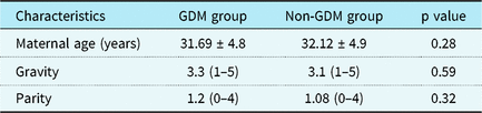
Mean maternal age, number of pregnancies, and parity were similar in both groups (Table 1). The mean (±standard deviation) gestational age at the time of examination in fetuses of gestational diabetic mothers was 23.8 ± 2.4 weeks and in fetuses of the control group was 23.3 ± 2.5 weeks. In the gestational diabetes group, there was no significant difference in drug usage, between those with and those without fetal myocardial hypertrophy.
Demographic characteristics of groups

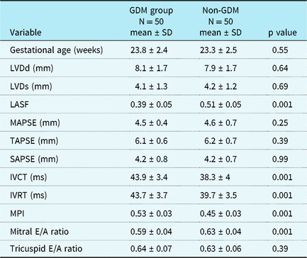
Comparison of variables between fetuses of gestational diabetic and control groups is shown in Table 2. There was no statistical difference in most of the dimensional variables. However, functional variables showed that mitral E/A ratios were significantly lower in the gestational diabetes group as compared to the control (p < 0.001). Isovolumetric relaxation and contraction times were significantly prolonged in fetuses of gestational diabetic mothers (p < 0.001) and myocardial performance index was also significantly abnormal in the gestational diabetic group (p < 0.001) as compared to the control.
Comparison of variables in fetuses of GDM and non-GDM

GDM, gestational diabetic mothers; non-GDM, non-gestational diabetic mothers.
Mean left atrial shortening fraction was statistically significant between fetuses of gestational diabetes mothers (0.39 ± 0.05) and fetuses of the control group (0.51 ± 0.05) (p < 0.001).
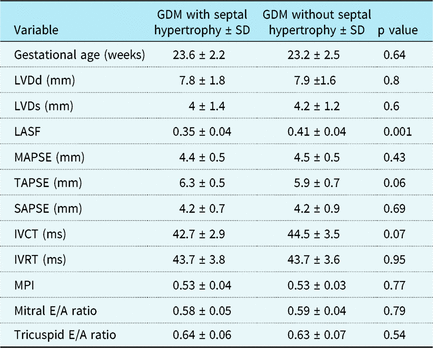
Variables were also compared between fetuses with and without myocardial hypertrophy (Table 3). The only statistically significant difference was in left atrial shortening fraction of fetuses with myocardial hypertrophy compared to those without myocardial hypertrophy (p < 0.001).
Comparison of variables in fetuses of gestational diabetic mothers with and without septal hypertrophy

Discussion
Fetuses of gestational diabetic mothers are at increased risk of structural and functional heart diseases leading to significant perinatal morbidity and mortality. Reference Lisowski, Verheijen and Copel15 Myocardial hypertrophy, as a result of fetal hyperglycemia and hyperinsulinism, is a frequent finding in the fetuses of diabetic mothers, which may lead to increased ventricular stiffness thereby affecting diastolic ventricular filling as well as systolic cardiac function. Reference Palmieri, Simões, Silva, Santos and Ferreira16–Reference Wang, Xu, Fu and Huang18 It is well known that impairment of ventricular function may cause diastolic dysfunction first and subsequently lead to systolic dysfunction in children with heart diseases. However, data in the literature, evaluating specific systolic and diastolic parameters in fetuses, are varied due to variation in methods used by researchers to evaluate heart function. Many authors have reported ventricular diastolic dysfunction in fetuses of diabetic mothers, even in the absence of septal hypertrophy. Reference Balli, Pac, Ece, Oflaz, Kibar and Kandemir19,Reference Hatém, Zielinsky and Hatém20 Therefore, early recognition of subtle changes may help in timely management with improved outcome. The results of this study show that fetuses of gestational diabetic mellitus mothers developed cardiac diastolic dysfunction independent of myocardial hypertrophy.
The myocardial performance index has been reported to be a useful, non-invasive index for global myocardial function and is independent of both heart rate and ventricular geometry. Reference Chuwa and Rodeheffer21,Reference Tei, Dujardin, Hodge, Kyle, Tajik and Seward22 In this study, isovolumetric contraction, isovolumetric relaxation time, and myocardial performance index of the left ventricle were significantly increased in the gestational diabetic group indicating impaired global ventricular function. In our previous study, Reference Atiq, Ikram, Hussain and Saleem4 poor glycaemic control patients had significantly prolonged isovolumetric relaxation time and septal thickness, which might reflect diastolic dysfunction in the poor glycaemic control group. In this study, glycaemic status of the mother was not accounted.
The decreased ventricular diastolic parameters, including mitral and tricuspid inflow patterns, denoting preload characteristics, have been documented in previous studies and are known to change with gestational age. This may be likely due to improvement in ventricular compliance with increasing gestational age. Reference Miyake23,Reference Weiner, Zloczower, Lerner, Zimmer and Itskovitz-Eldor24 In this study, left ventricular diastolic parameters, including mitral E/A ratio, were found to be significantly lower (p < 0.001) compared to the control group, depicting a less compliant left ventricle. However, we did not find statistically significant difference between mitral inflow velocities of fetuses with myocardial hypertrophy compared to those who did not have hypertrophy. The possible explanation of this phenomenon may be the increasing E/A ratio as pregnancy progresses. Reference Kenny, Plappert and Doubilet25 Hence, we can speculate that although the mitral E/A ratio is lower in diabetic fetuses, it did not correlate with the progressive nature of the disease.
Left atrial shortening fraction appears to be dependent on left ventricular preload and to be proportional to ventricular compliance, as suggested by Briguori et al, Reference Briguori, Betocchi and Losi11 and provides a reliable non-invasive assessment of diastolic dysfunction in hypertrophic cardiomyopathy. Therefore, we used left atrial shortening fraction to evaluate fetal diastolic function in addition to Doppler analysis of mitral inflow signals. We found significantly decreased left atrial shortening fraction in fetuses of the gestational diabetes group. This could possibly be due to increased left atrial pressure and decreased left ventricle compliance. Previously, Zielinsky et al Reference Zielinsky, Luchese and Manica26 have shown lower left atrial shortening fraction in pre-gestational diabetic fetuses with or without hypertrophy. However, correlation of conventional mitral E/A ratio with left atrial shortening fraction between the diabetic and control group was not done. We also demonstrated lower left atrial shortening fraction in fetuses with myocardial hypertrophy than in those without myocardial hypertrophy in the gestational diabetes group. Recent studies have shown that good glycaemic control in gestational diabetic mothers may delay the development of hypertrophy in fetuses but does not prevent the degree of hypertrophy. Reference Chen, Gui, Ren and Shi12 In our study, the lower left atrial shortening fraction in diabetic fetuses with septal hypertrophy is possibly due to decreased ventricular compliance and indicates the progressive nature of the disease.
As acknowledged, the limitation of this study is that we do not have complete post-natal follow-up of these fetuses. Moreover, the glycaemic control of the diabetic mothers was not available due to referral from multiple centres across the country. However, the study opens up the potential for further research in detection of parameters predictive of ventricular dysfunction in the infants of diabetic mothers. Furthermore, it would be interesting to see how these parameters work in infants of mothers with poorly controlled diabetes. A further study design, including mothers with tight and poor glycaemic control with post-natal follow-up, would help in advancing our knowledge of their fetal cardiocirculatory status.
Conclusion
The results of this study showed that fetuses of gestational diabetes mothers are at increased risk of cardiac diastolic dysfunction, which can occur even in the absence of septal hypertrophy. We propose that in addition to the routine mitral E/A ratio and global myocardial performance indicators, left atrial shortening fraction be used as a marker of diastolic function in fetuses of diabetic mothers, especially in patients without structural abnormality.
Acknowledgements
The authors acknowledge echocardiographic technical assistance by Ms Hina Ali and Zamzama Rashid.
Financial Support
This research received no specific grant from any funding agency, commercial, or not-for-profit sectors.
Conflicts of Interest
The authors declare no conflict of interest. The corresponding author has done the study with the technical help of echocardiography staff and would shoulder the responsibility of overall integrity of the work. All other authors have collectively contributed to conception, design, collection of data and critical revision.
Ethical Standards
Informed consent was obtained from all individual participants included in the study and study was approved by Ethics review committee Aga Khan University Hospital.