Implications
Fattening lambs consuming high amounts of concentrate are susceptible to sub-clinical acidosis and rumenitis. Naringin is a type of citrus flavonoid with proven antioxidant properties, which might improve animal welfare in lambs with sub-clinical acidosis (e.g. by reducing the discomfort that might be associated with damage to the ruminal wall). Adding naringin to the concentrate at a dose of 0.15% affected ruminal bacteria, although no anti-inflammatory effects were observed in the ruminal wall.
Introduction
Many consumers require high quality (e.g. safe, healthy and nutritious) meat that has been produced from animals reared to high welfare standards. These demands have lead to the search for new substances and additives that could help to reach those objectives within the current production systems. Thus, the number of papers dealing with the beneficial effects of some additives or ingredients supplied at low concentrations in the diet has recently increased. Flavonoids are a type of polyphenols widely concentrated in Citrus fruits, which have received special attention because of their physiological effects in humans, rabbits and rats (Jeon et al., Reference Jeon, Bok, Jang, Lee, Nam, Park, Rhee and Choi2001; Tripoli et al., Reference Tripoli, La Guardia, Giammanco, Di Majo and Giammanco2007; D’Argenio et al., Reference D’Argenio, Mazzone, Tuccillo, Grandone, Gravina, Graziani, Fogliano and Romano2008). In fact, the flavonoids are suggested to be responsible for the prevention of chronic and degenerative diseases in human nutrition (Tripoli et al., Reference Tripoli, La Guardia, Giammanco, Di Majo and Giammanco2007), but ruminant production may also benefit from these compounds. Naringin, one type of grapefruit and citrus flavonoid with proven antioxidant properties (Jeon et al., Reference Jeon, Bok, Jang, Lee, Nam, Park, Rhee and Choi2001) might be a good choice because it has been authorized as a feed additive in ruminant nutrition by the European Commision (Community Register of Feed Additives, 2009).
These compounds have been reported to show beneficial effects on certain inflammatory and immune parameters, particularly under oxidative stress conditions (Hamer, Reference Hamer2007; Tripoli et al., Reference Tripoli, La Guardia, Giammanco, Di Majo and Giammanco2007), so the administration of naringin might be interesting in animals whose immune response might be counteracted as a consequence of the transportation stress. In addition, some of them have antimicrobial activity and other implications for improving gut health (D’Argenio et al., Reference D’Argenio, Mazzone, Tuccillo, Grandone, Gravina, Graziani, Fogliano and Romano2008) that might also result in benefits to animal health and welfare. For example, experimental trials in pigs have demonstrated that administration of polyphenols seems to improve the intestinal microbiota balance, as reflected by a decrease in Clostridium perfringens counts (Hara et al., Reference Hara, Orita and Hatano1995). To consider the suitability of naringin as a supplement for ruminant nutrition, it is important to evaluate the possible effects of naringin on the ruminal microbial community (Tripoli et al., Reference Tripoli, La Guardia, Giammanco, Di Majo and Giammanco2007), since the anti-inflammatory activity of naringin, which could help to mitigate the effects of sub-acute ruminal acidosis (associated with high concentrate diets), might be affected as a consequence of the ruminal bacteria metabolism.
Therefore, the aim of this study was to examine whether naringin had beneficial effects when used as a supplement in a high concentrate diet (80% to 90% concentrate) offered to fattening lambs. For this purpose, the effects of naringin on the oxidative stability, haematological variables and immune parameters of lambs under transportation stress were studied. Finally, animals were slaughtered and ruminal and faeces bacterial communities, together with ruminal wall protection against sub-clinical acidosis, were studied.
Material and methods
Animals and diets
One month before the commencement of the trial, 40 male Assaf lambs were treated with ivermectin (Ivomec, Merial Labs, Barcelona, Spain) and vaccinated against enterotoxaemia (Miloxan, Merial Labs, Barcelona, Spain).
After random stratification on the basis of body weight (average BW, 24.8 ± 1.64 kg), the lambs were allocated to either a control (CONTROL) or a treatment group (NAR). They were then individually penned. All handling practices followed the recommendations of European Council Directive 86/609/EEC for protection of animals used for experimental and other scientific purposes and all the animals were able to see and hear other sheep.
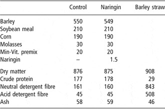
After 7 days of adaptation to the basal diet (barley straw and basal concentrate feed), all the lambs were fed barley straw and the corresponding concentrate feed alone (CONTROL group) or supplemented with 1.5 g/kg of naringin (Sigma-Aldrich, St Louis, MO, USA; NAR group). Ingredients and chemical composition of the concentrates and barley straw are given in Table 1. All the ingredients of each concentrate were mixed and ground in a mill and fed to the animals as dry meal. The straw (200 g/day) and concentrate (30 g/kg BW per day) offered to each lamb were weighed daily. The orts refused (<5% of the total diet) were also weighed daily, and samples were collected for subsequent analyses.
Ingredients (g/kg) and chemical composition (g/kg dry matter) of the experimental concentrates and barley straw

Stress induction and blood sampling
On the last day of the experimental period (day 49, week 7) all the animals were subjected to a 4-h transportation period. Road transportation was used as a quantifiable source of stress (Kent, Reference Kent1997; Kannan et al., Reference Kannan, Terrill, Kouakou, Gazal, Gelaye, Amoah and Samake2000) as well as a means of transporting the lambs to slaughter. The lambs were transported at a space allowance of 0.30 m2 per animal in a truck with a non-slip metal floor that was covered with straw bedding.
Before (0 h) and immediately after a 4-h transportation period (4 h), blood samples were collected by jugular venipuncture into each of four 10 ml vacutainers containing no anticoagulant, sodium heparin, lithium heparin or ethylene diamine tetraacetic acid (EDTA).
Blood samples collected in the sodium heparin tubes were immediately placed in iced water and centrifuged at 1000 × g for 10 min at 4°C. The plasma obtained was stored at −80°C until required for antioxidant analyses. Those samples in tubes with no anticoagulant were allowed to clot for 30 min at room temperature and then centrifuged at 2000 × g for 15 min at 4°C. Thereafter, serum obtained was stored at −20°C until used to measure cortisol and Fe concentrations. Cortisol was measured by a solid-phase, competitive chemiluminescent enzyme immunoassay using a commercial kit (Immulite 1000 Cortisol, Siemens Healthcare, Munich, Germany; inter- and intra-assay coefficient of variation (CV) of 6.7% and 7.3%, respectively). Serum Fe was measured with a commercially available assay kit (Cobas Integra Iron, Roche Diagnostics, Barcelona, Spain; inter- and intra-assay CV of 2.9% and 2.4%, respectively). Vacutainer tubes with EDTA were used for haematological analyses within 1 h from the time of collection. The blood sample collected just before the transportation in a lithium heparin tube was kept at room temperature and processed within 8 h after collection to measure IFNγ production.
Antioxidant parameters in plasma samples
Lipid peroxidation was analysed in the plasma samples using the thiobarbituric acid reactive substances (TBARS) Assay Kit (Cayman Chemical, Ann Arbor, MI, USA). The inter- and intra-assay CV were 5.5% and 5.9%, respectively.
Haematological parameters and IFNγ production
Blood samples for red blood cell (RBC) and white blood cell (WBC) counts (leukocytes, lymphocytes, monocytes and granulocytes), packed cell volume and haemoglobin were assayed in an electronic cell counter (Cellanalyzer CA530, Bromma, Sweden).
The peripheral cellular immune response induced by vaccination against agalactia was assessed by means of an assay for the determination of IFN-γ production. Four 1.4 ml aliquots of each blood sample were dispensed in each of four 1.5 ml Eppendorf safe-lock tubes. Each aliquot of blood was mixed with 100 μl of either sterile phosphate buffered saline solution (PBSS; negative control; PBS, Sigma-Aldrich ) or 100, 250 or 500 μl of a suspension of dead Mycoplasma agalactiae (Laboratorios Ovejero S.A., León, Spain), used for the elaboration of the vaccine inoculated earlier to the animals, at a concentration of 5 × 10⁸ microorganisms/ml. The M. agalactiae preparation was prediluted in PBSS to the working concentration. Blood cultures were incubated for 20 h at 37°C in a humidified atmosphere. After centrifugation of the tubes, plasma was harvested from atop the sedimented RBC and was stored at −20°C. The plasma samples were then assayed in duplicate for IFN-γ using a commercial bovine enzyme immunoassay kit (Bovigam, Prionics AG, Zurich, Switzerland) that reacts with ovine IFN-γ, according to the manufacturer’s instructions. The optical density (OD) results were transformed to an index value by division of mean OD of the plasma from the M. agalactiae stimulated blood by mean OD of the same plasma incubated with PBSS (negative control).
Slaughter procedure
In the morning of day 50, 10 lambs from each group were weighed, stunned, slaughtered by exsanguination from the jugular vein, eviscerated and skinned. Samples of faeces and ruminal content were collected in aseptic flasks and immediately frozen and kept at −80°C until analysis for microbial diversity, whereas two pieces from different locations of ruminal wall (posterior dorsal and anterior ventral areas) were introduced in 10% formalin for further anatomopathological study.
Characterization of microbial diversity in ruminal content and faeces
Terminal restriction fragment length polymorphisms (T-RFLP) is a technique used to rapidly distinguish bacterial communities on the basis of the size of the terminal fragments of 16S rRNA genes following restriction endonuclease digestion. Thus, samples of ruminal content and faeces were freeze-dried, DNA was extracted and purified using the commercial QIAamp® DNA Stool Mini Kit (Qiagen Ltd, Crawley, UK) and then stored at −20°C until analysis for microbial diversity by T-RFLP of genes encoding 16S rRNA (Liu et al., Reference Liu, Marsh, Cheng and Forney1997).
Briefly, a fragment of the 16S rRNA gene was amplified by PCR using bacterial universal primers (Hongoh et al., Reference Hongoh, Yuzawa, Ohkuma and Kudo2003), one of them being a fluorescein-labeled forward primer (FAM-AGAGTTTGATCCTGGCTCAG; CTTGTACACACCGCCCGT). Fluorescent-labeled PCR products were purified on kit GFX PCR DNA and Gel Band Purification (GE Healthcare, Roosendaal, The Netherlands). Thereafter, DNA was quantified and 100 ng of each sample was subjected to a restriction reaction with enzyme HhaI (Takara Bio Inc., Otsu, Shiga, Japan) during 12 h at 37°C. Fluorescent labeled terminal restriction fragments (TRF) were analysed by capillary electrophoresis on an automatic sequence analyzer Megabace 500 (GE Healthcare, Roosendaal, The Netherlands) with internal Et-ROX labeled DNA size standards (Amersham Biosciences, Fairfield, CT, USA). Finally, the matrix generated by the Gene Marker program showing the presence (1) or absence (0) of the peaks (TRF) obtained for each sample was analysed by principal component analysis (PCA).
Anatomopathological characteristics of ruminal wall
Anatomopathological characteristics of ruminal wall were determined as described by Nocek and Kesler (Reference Nocek and Kesler1980). Briefly, the thickness of tunica mucosa and submucosa were measured with the aid of a Wild Heerbrugg dissecting microscope. In addition, triplicate determinations of the number of papillae/cm2 and the average length of six papillae were measured from each piece of ruminal wall.
Statistical analyses
Data of feed intake, IFN-γ and anatomopathological characteristics of ruminal wall samples were subjected to analysis of variance using the GLM procedure of SAS package (SAS, 1999). Data corresponding to cortisol, TBARS and haematological variables were analysed as a repeated measures design using the MIXED procedure of SAS with treatment and day as fixed effects and individual lamb as the experimental unit. Least square means were generated and separated using the PDIFF option of SAS for main or interactive effects, significance being determined at P < 0.05. Finally, as stated beforehand, microbial diversity data obtained for each sample were analysed by PCA.
Results
There was no significant difference between the NAR and the CONTROL groups in the concentrate or barley straw intake (P > 0.05, overall mean values being 723 ± 21.1 and 154 ± 17.8 g/animal per day, respectively). As expected, BW gain was not affected by dietary treatment (173 ± 7.2 g/animal per day).
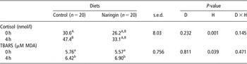
Table 2 summarizes changes in cortisol concentration and lipoperoxidation (TBARS) of plasma samples in response to dietary treatments and transport, both parameters rising after transport (P < 0.05).
Least square means of plasma cortisol and antioxidant parameters thiobarbituric acid reactive substances (TBARS) before the transport period (0 h) and immediately again after a 4-h transportation period (4 h)

P-values for diet (D), hour (H) and their interaction (D × H).
Different superscripts in the same row or column indicate statistical differences (a,bP < 0.05; A,BP < 0.01) among diets or hours.
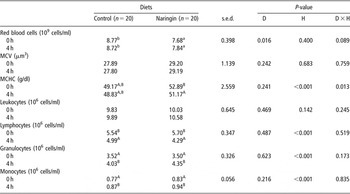
Mean values of haematological variables for CONTROL and NAR groups can be seen in Table 3. The NAR lambs had a lower RBC count than CONTROL lambs before and after transport (P < 0.05) and the NAR lambs had a decreased mean corpuscular haemoglobin concentration after transport (MCHC, P < 0.05). There were no changes in leukocyte counts, but lymphocyte counts decreased and granulocyte and monocyte counts increased after transport in both groups. On the other hand, plasma Fe values were not different between groups, average mean values being 33.5 and 34.2 μmol/l for CONTROL and NAR lambs, respectively (P > 0.05).
Least square means of haematological variables before the transport period (0 h) and immediately again after a 4-h transportation period (4 h)

MCV = mean corpuscular volume; MCHC = mean corpuscular haemoglobin concentration.
P-values for diet (D), hour (H) and their interaction (D × H).
Different superscripts in the same row or column indicate statistical differences (a,bP < 0.05; A,BP < 0.01) among diets or hours.
The cytokine IFN-γ showed no clear behaviour in response to the M. agalactiae antigens and could not be related to the consumption of naringin (data not shown).
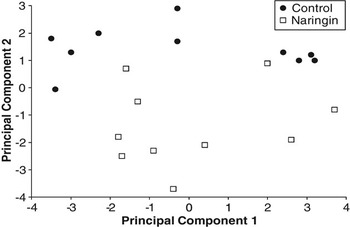
PCA performed on matrix data showing the presence (1) or absence (0) of TRF peaks in ruminal content revealed two clusters corresponding to the lambs having consumed naringin or the control diet (N, NAR group and C, CONTROL group, Figure 1), with 85% samples suitably classified according to PC2. Regarding microbial diversity in faeces, no clusters could be observed according to T-RFLP results (data not shown).
Discriminant analysis on the basis of PCA performed on matrix data showing the presence (1) or absence (0) of peaks detected by T-RFLP of ruminal content for Naringin (□) and Control (●) lambs.

No differences in the thickness of tunica mucosa (mean value of 0.787 mm, P > 0.10) and submucosa (mean value of 0.612 mm, P > 0.10) could be observed under the dissecting microscope. The number of papillae/cm2 and their average length was also similar in both groups (mean values of 44.3 papillae/cm2 and 3.74 mm of papillae length, P > 0.10).
Discussion
Antioxidant parameters
Cortisol concentration is used in the stress assessment of farmed livestock, and regarded as very reliable indicators of acute and long-term stress (Carcangiu et al., Reference Carcangiu, Vacca, Parmeggiani, Mura, Pazzola, Dettori and Bini2008). In this study, we observed a significant rise (42%) in the cortisol concentration after transportation. Nevertheless, Larraín et al. (Reference Larraín, Schaefer, Richards and Reed2008) observed no changes in plasma cortisol concentration of steers consuming a high-tannin shorgum diet. Conversely, flavonoid compounds seem to reduce the stress response in rats because of a reduction of adrenocorticotropic hormone and corticosterone concentration in plasma (Butterweck et al., Reference Butterweck, Hegger and Winterhoff2004). In this study, the lack of statistical difference between treatments could be due, first, to the large individual variability in cortisol concentration between animals, and second, to the low rates of naringin used in this experiment. In fact, Gladine et al. (Reference Gladine, Rock, Morand, Bauchart and Durand2007) observed an increase in plasma antioxidant status when sheep were supplemented with supra-nutritional doses (10%) of naringin.
The stress experienced by the animal when transported to the slaughterhouse exacerbates the lipoperoxidation of polyunsaturated fatty acids (Kannan et al., Reference Kannan, Terrill, Kouakou, Gazal, Gelaye, Amoah and Samake2000). Lipid peroxides can be measured by TBARS procedure because these compounds are unstable and decompose to form reactive carbonyl compounds such as malondialdehyde. Accordingly, all the lambs experienced an increment in plasma TBARS values due to the transportation stress (Table 1, 4 h). Unexpectedly, no effect due to the antioxidant properties of naringin could be observed in TBARS values. However, many authors have described a dose-dependent effect of the flavonoids (Ostrowska et al., Reference Ostrowska, Gabler, Sterling, Tatham, Jones, Eagling, Jois and Dunshea2004; Farinacci et al., Reference Farinacci, Colitti, Sgorlon and Stefanon2008). Therefore, the lack of effect of naringin treatment on TBARS observed in this study does not imply the ineffectiveness of this additive at different rates.
Haematological parameters and IFN-γ production
The lambs in the NAR group had lower RBC counts than those in the CONTROL group. Some authors have suggested a direct effect of naringenin (the aglycone fraction of naringin), which can chelate the ferrous ion needed to form the central core of the haemoglobin, making it unavailable (Gnanamani et al., Reference Gnanamani, Sudha, Deepa, Sudha, Deivanai and Sadulla2008; Renugadevi and Milton Prabu, Reference Renugadevi and Milton Prabu2009). Other authors, with similar results after feeding flavonoids to animals, have suggested a lipoperoxidation of blood cell membranes resulting in haemolysis and removal of damaged erythrocytes by the cells of the reticulo-endothelial system (Munday et al., Reference Munday, Munday and Munday2003; Ostrowska et al., Reference Ostrowska, Gabler, Sterling, Tatham, Jones, Eagling, Jois and Dunshea2004). Nevertheless, in this study, the lack of significant differences in plasma Fe values and MCHC do not support the idea that naringin may cause haemolysis. Moreover, it has been suggested that naringin plays an important role in sparing vitamin E, and thus increasing the plasma antioxidant capacity (Jeon et al., Reference Jeon, Bok, Jang, Lee, Nam, Park, Rhee and Choi2001); however, the TBARS values observed in this experiment do not agree with this theory.
Although no significant differences were observed for leucocyte counts after transportation period (Table 2), the differential leukocyte counts (lymphocytes, granulocytes and monocytes) revealed significant changes after the transport. Thus, all the lambs showed an increase in granulocytes and a decrease in lymphocyte counts after the transport (Table 2). Neutrophilia and lymphopenia are commonly observed in stressed animals and are associated with changes in the WBC trafficking and release from the bone marrow by the elevated concentrations of the glucocorticoids (Kannan et al., Reference Kannan, Terrill, Kouakou, Gazal, Gelaye, Amoah and Samake2000). However, it must be pointed out that these changes in this study seemed to be quite moderate, which would indicate that the transportation may not have been particularly stressful.
This study also suggests that the immune system of the lambs was not affected by the flavonoid (naringin) included in the diet. Thus, no significant modification because of the diet was observed either in the WBC count or in the differential count of WBC. In this respect, contradictory results can be found in the literature describing trials with flavonoids. For example, Marzo et al. (Reference Marzo, Tosar and Santidrian1990), Yen et al. (Reference Yen, Chen and Peng2001) and Gnanamani et al. (Reference Gnanamani, Sudha, Deepa, Sudha, Deivanai and Sadulla2008) showed genotoxic effects (a serious damage to DNA) on WBC when high doses of flavonoids were supplied. On the contrary, Ostrowska et al. (Reference Ostrowska, Gabler, Sterling, Tatham, Jones, Eagling, Jois and Dunshea2004) and Sultan and Hussein (Reference Sultan and Hussein2006) observed an increment of WBC in animals being fed different flavonoids. These apparently divergent results can be explained by a dose-dependent effect of these compounds (Ostrowska et al., Reference Ostrowska, Gabler, Sterling, Tatham, Jones, Eagling, Jois and Dunshea2004; Farinacci et al., Reference Farinacci, Colitti, Sgorlon and Stefanon2008).
The lacking differences in cytokine IFN-γ production are in agreement with those described by Jacob et al. (Reference Jacob, Periago, Böhm and Berrezuelo2008) in humans for a combination of lycopene and vitamin C. On the contrary, quercetin added to cell cultures showed a dose-dependent upregulation of gene expression; thus, increasing IFN-γ production (Nair et al., Reference Nair, Kandaswami, Mahajan, Chadha, Chawda, Nair, Kumar, Nair and Schwartz2002). All these divergent results might be due once again to different doses of flavonoids used in these studies.
Characterization of microbial diversity in ruminal content and faeces
The PCA results highlight variations in the ruminal bacterial community of lambs caused by naringin consumption, and might influence the pattern of ruminal fermentation and, thus, the amount and proportions of volatile fatty acids produced. The antimicrobial properties reported on flavonoids might be partially responsible for this circumstance (Chung et al., Reference Chung, Wei and Johnson1998; Tripoli et al., Reference Tripoli, La Guardia, Giammanco, Di Majo and Giammanco2007). In addition, naringin may be metabolized by ruminal microorganisms, so this fact might also be partially responsible for the variations in bacterial community, enhancing some species, but showing detrimental effects on others. In fact, the glycosidic flavanone naringin must be hydrolyzed by ruminal microorganisms to allow the absorption of the aglycone fraction (naringenin) by the animal (Gladine et al., Reference Gladine, Rock, Morand, Bauchart and Durand2007). After absorption, naringenin is transported to the liver, where it forms glucuronide and/or sulfate or methyl conjugates before being eliminated normally through the urinary and fecal systems (Spencer et al., Reference Spencer, Abd El Mohsen, Minihane and Mathers2008).
The discrimination observed in Figure 1 may have been due to those variables (TRFs) found more eccentric with high loading weights, whereas those with low loading weights could be considered irrelevant. A theoretical assignment of these TRFs to compatible bacterial species could be performed using the tap-tRFLP tool of the Ribosomal Data Project (Castillo et al., Reference Castillo, Martín-Orúe, Nafrarías, Manzanilla and Gasa2007). Thus, it was concluded that most of these TRFs (206, 231, 120, 201, 180 base pairs) were compatible to ruminal clostridia, whose presence might have been modified by the consumption of naringin. However, it was not possible to discriminate between individual genera or species of bacteria associated to these TRFs because of the large individual variability among animals regarding ruminal bacteria. These difficulties can be overcome by in vitro trials, in which the individual variability among animals is canceled in favour of just one ruminal inoculum for all the possible treatments. On the other hand, no clusters could be observed according to T-RFLP results (data not shown) when studying microbial diversity in faeces, which could be because of the metabolism and absorption of naringin in rumen and small intestine (Gladine et al., Reference Gladine, Rock, Morand, Bauchart and Durand2007)
Anatomopathological characteristics of ruminal wall
D’Argenio et al. (Reference D’Argenio, Mazzone, Tuccillo, Grandone, Gravina, Graziani, Fogliano and Romano2008) observed a reduced aspirin-induced injury (less discontinuous surface, dilated glands, haemorrhage or damage to superficial cells) to the rat gastric mucosa in those animals consuming an apple polyphenol extract (APE). These authors showed that this protective effect was independent of inhibition of gastric acid secretion, and seemed at least partially because of the antioxidant activity of APE. However, there is no evidence in the literature regarding the supplementation of the diet with antioxidants or flavonoids that prevent processes related to injury in the rumen mucosa such as rumenitis. Nevertheless, other strategies to prevent rumenitis such as a change in feeding strategy (e.g. providing adequate dietary fibre content and having regular meals, Krause and Oetzel, Reference Krause and Oetzel2006) have been successful.
From the anatomopathological point of view a dark color of the ruminal mucosa, characteristic of parakeratosis, is an unequivocal indicator of acidosis (Nocek and Kesler, Reference Nocek and Kesler1980). In this study, the interior of the rumen wall was grey to dark in all the animals, indicating rumenitis, so the first examination evidenced no protection of naringin against damage caused by subclinical acidosis. This lack of effect of naringin on the inflamation of ruminal ephitelium was corroborated lately by the lack of differences in the thickness of tunica mucosa and submucosa and the anastomosis and number of papillae. The absence of an effect of naringin on signs of ruminal acidosis is consistent with the absence of an effect of naringin on the TBARS values (Table 2).
Conclusions
Supplying naringin at a dose of 0.15% in the concentrate for fattening lambs seems to reduce RBC counts. This dose had clear effects on the ruminal bacterial community, but showed no protection against the ruminal wall damage caused by subclinical acidosis. No effects were observed in immunological parameters. Further experiments, using different doses of naringin might show health benefits of naringin supplementation in lambs, but a clear beneficial effect on health was not readily apparent in this study.
Acknowledgements
Financial support received from ‘Consejería de Educación de la Junta de Castilla y León’ (Project GR158) and CSIC ‘Intramural’ Project 200840I116 is gratefully acknowledged. Raúl Bodas and Nuria Prieto have a JAE-Doc contract from the CSIC under the programme ‘Junta para la Ampliación de Estudios’.