Definition of simulation
Medical simulation may be defined as the creation of an educational environment in which learning occurs through the use of a device, mannequin or team, (Team means, we ask some health care providers to present themselves as patients (simulated patients) with certain symptoms for known disease and the student will take a history and when he goes to examine a certain organ the team patient will tell him about the sign) without the presence of an actual patient.Reference Dawson1 In medical training, simulation offers a safe and realistic environment within which students can repeatedly practise clinical scenarios and team-building skills, without subjecting actual patients to risk. A medical simulator is a physical object that reproduces, to a greater or lesser degree, a real clinical scenario. The simulators available in simulation laboratories may have many life-like attributes that enhance training, such as blinking eyes, exhalation of carbon dioxide, chest movements, crying, coughing and palpable pulses.
History of simulation
Edward Link was the first to use simulation in teaching, with his invention in 1929 of the flight simulator.Reference Dawson1 Today, the aviation industry still leads in the use of simulators, which are utilised to train novice pilots.Reference Rosen2
In recent years, continuous medical education has begun to suffer due to reduced patient contact time. This has been due to shortened hospital stays; moreover, there is now more emphasis on out-patient and minimally invasive therapies. Hospitalised patients are generally more severely ill, and in-patient procedures more complex. For all these reasons, current trainees have little contact with patients, and their procedural experience during residency is reduced.
The use of simulation in medical education started with the teaching of anatomy on physical models, long before the current revolution in computer technology. Demands for increased attention to patient care resulted in clinical education innovations, with a growing interest in the use of simulators for medical training. With the rapid development of computer technology, it is now possible for surgical training to be undertaken using computer screens and haptic (tactile force) devices. Such haptic surgical training provides realistic simulation of clinical situations, created from data obtained from real human bodies, and is superior to traditional methods in many respects, including reduced cost, avoidance of ethical problems, and the facility to repeat training scenarios as required.Reference Zhang, Gu, Li and Fang3 Medical skills simulators can be used not only as training devices but also as assessment tools.Reference Tavakol, Mohagheghi and Dennick4
Types of simulation
The simulation environment may comprise a full patient mannequin, a computer-based procedural simulator, or a hybrid system combining a physical form and a computer interface.Reference Dawson1
Clinical simulators may include the following: part task trainers, computer-based systems, virtual reality and haptic systems, simulated patients, simulated environments, and integrated simulators.Reference Tavakol, Mohagheghi and Dennick4, Reference Maran and Glavin5
Model-based simulators are simple benchtop models, and have many limitations. They only simulate isolated parts of the body, so a complete illusion of reality is impossible. Moreover, they are inanimate models which are unable to give feedback or provide objective assessment of performance. Learning of this kind requires extensive support from expert tutors. However, such models are useful for practising urinary catheterisation, venepuncture, intravenous infusion, wound closure, and relatively simple surgical procedures such as the removal of cysts and lipomas.Reference Kneebone6
Computer-based simulation has emerged as a powerful healthcare training technique. Such simulators utilise a combination of technologies, allowing students to interact efficiently with three-dimensional computerised databases in real time, using their natural senses and skills. Surgeons can now use accurate recreations of individual patients' organs, created from high-resolution scanning data, to practise patient-specific operations within a virtual environment.
Lastly, hybrid simulation combines physical simulation models with computers, often using a realistic interface (such as real diagnostic or surgical instruments) to bridge the gap between the model and the computer.
The overall goal of the collaborative effort of FESS training which includes videotape viewing, cadaver dissection, observation of the operation in the theatre and FESS simulator training is improved patient safety for surgical procedures, using highly sophisticated surgical simulators to improve the screening, education, training, error detection skills, assessment and certification of surgeons. The resulting improvements in technical skills will render operative procedures safer, with fewer medical errors.
Advantages of simulation
With the help of simulators, medical students and junior residents can perform a series of standardised surgical and non-surgical tasks on inanimate models, under the direct observation of an expert tutor.Reference Martin, Regehr, Reznick, MacRae, Murnaghan and Hutchison7 These teaching methods have the following advantages: the risks to both patient and student are avoided, undesired interface such as learning of intravenous injection in children or venous cut down is reduced, tasks can be created as needed, skills can be practised repeatedly, training can be tailored to individuals, retention and accuracy are increased, and transfer of skills from classroom to real life is enhanced.Reference Holzman, Coex and Ewan8
It is anticipated that, by 2010, surgical simulators will have reached a sufficient level of performance to be acceptable as tools for testing and certification.Reference Fried, Sadoughi, Weghorst, Zeltsan, Cuellar and Uribe9 To date, a large variety of haptic surgical trainers have been developed, ranging from a simple simulation of laparoscopic drills, running on a personal computer, to an advanced hepatic surgery simulator, running on a ‘high-end’ graphics workstation. Simulation devices for neurological surgery, ophthalmic surgery, laparoscopic surgery, open abdominal surgery and anastomosis, and knee and shoulder arthroscopy have already been developed.Reference Fried, Sadoughi, Weghorst, Zeltsan, Cuellar and Uribe9
In addition to their application for training and education, surgical simulators can also be used for pre-operative planning. It is anticipated that simulation will be much in demand for surgery involving complex anatomy, such as the craniofacial region.
Applications of ENT simulation
Computer-aided surgical simulation is widely used, and appears to be at least as good as traditional methods used to teach students and residents.Reference San Vicente, Buchart, Borro and Celigueta10, Reference Xia, Philips, Gateno, Teichgraeber, Christensen and Gliddon11
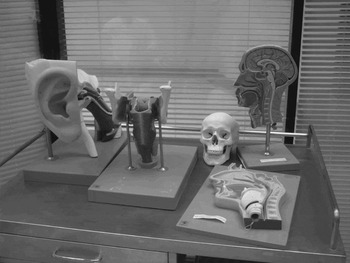
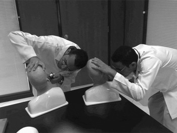
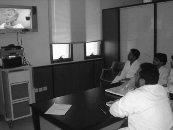
In the ENT field, students, residents and clinicians are currently taught using a variety of methods, including lectures, clinical rounds, physical models (Figures 1 and 2), video tape (Figure 3), cadaver dissections and direct observation of procedures (the first four methods are used at Al-Ahsa College of Medicine). Simulation can be used to develop the surgical (and other) skills of otolaryngology residents prior to application in a clinical environment.Reference Khalid and Murray12 Virtual reality environments, including haptic devices which recreate tactile experiences (used at Al-Ahsa College of Medicine), have made such training possible. Surgical simulation is becoming a powerful tool for teaching, and also for planning surgery and simulating surgical results before performing a procedure.
Physical models used for teaching ENT anatomy in the simulation laboratory.

Students demonstrating the use of physical models for teaching ear pathology. Models incorporate a movable tape of slides showing different middle-ear diseases.

Use of a video recording for teaching ENT examination.

In the field of ENT surgery, there are many surgical procedures which we have begun to teach using simulators. Currently, these comprise bronchoscopy, tracheotomy, functional endoscopic sinus surgery (FESS) and temporal bone dissection.
Bronchoscopy
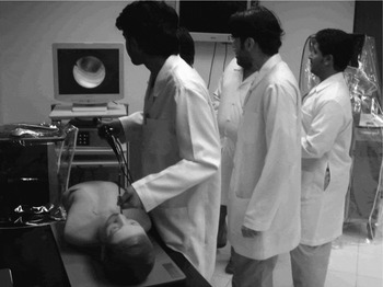
Flexible fibre-optic bronchoscopy is a common procedure performed by anaesthetists, respiratory physicians and otorhinolaryngologists, and the use of simulation has been proven to augment clinical training (Figure 4).Reference Colt, Crawford and Galbraith13 Even a short, focused course of instruction and unsupervised practice using the simulator enables novice trainees to attain a sufficient basic level of manual and technical skill to perform a diagnostic inspection of the tracheobronchial tree. The procedure can be constantly monitored by the trainer, and this allows the trainee to gain quickly the necessary psychomotor skills involved in rigid bronchoscopy.
Students demonstrating the use of a computer-based bronchoscopy teaching system.

During an airway crisis, such as foreign body inhalation with rapidly developing hypoxia and asphyxia, time is of the essence, and interpersonal and communication skills are essential for efficient teamwork. These teamwork skills can be practised in a high fidelity, realistically simulated clinical environment, under the rubric of crisis resource management.Reference Khalid and Murray12
With the availability of virtual reality technology, it has become an ethical imperative to train residents prior to patient contact.
Usage of an animal model has been reported to be beneficial in allowing students to practise inserting a rigid bronchoscope under monitor visualisation, followed by examination of the trachea and direct visualisation of the carina and right and left bronchi.Reference Ram, Oluwole, Blair, Mountain, Dunkley and White14
The flexible fibre-optic bronchoscope simulator, used at Al-Ahsa College of Medicine, provides the student with realistic images as they navigate through the virtual anatomy. Exercises involve an initial introduction to full bronchoscopic examination, with a robotic interface tracking the motions of the flexible bronchoscope and reproducing, via tactile feedback, all the forces felt during an actual bronchoscopic examination.
Tracheotomy
Airway emergencies are challenging, life-threatening crises that can be repeatedly practised in a simulated environment until a specified level of competency has been attained. The acquisition of new skills such as percutaneous tracheotomy (taught at Al-Ahsa College of Medicine using a simulator) requires supervised training and the opportunity for sufficient practice.
Tracheotomy is an airway procedure with little margin for poor surgical skill, and has significant morbidity and mortality in inexperienced hands.Reference Khalid and Murray12 Usage of an animal model has been found to be highly beneficial, as it allows practice inserting the seeker needle, inserting the tracheostomy tube and checking the arrangement endoscopically.Reference Gardiner, White, Carson, Shearer, Frizelle and Dunkley15
Functional endoscopic sinus surgery
Functional endoscopic sinus surgery is one of the most common procedures performed by otolaryngologists, and one that poses significant risk to the surrounding anatomical structures, including the eye and brain.
Otolaryngology residents' training in FESS must include the anatomy and physiology of the sinuses, the relevant disease processes, and the concepts and skills involved in the procedure. Cadaver dissection may be performed, but most training is conducted in the operating theatre under the supervision of an experienced surgeon. As training is dependent upon real patients, it is imperative that the operating theatre environment is kept as risk-free as possible. Even with the use of cadavers for training (which are unrealistic in that they do not bleed), once a single dissection is completed, it cannot be erased for the purpose of repeating the procedure, since the anatomy has been permanently altered.Reference Fried, Satava, Weghorst, Gallagher, Sasaki and Ross16
The FESS training simulator was developed in response to the training needs of novice sinus surgeons. As students advance through the simulator training programme, they are given more complex tasks to perform. The simulator has three levels of difficulty: novice, intermediate and advanced. The novice setting allows basic skills to be developed, whereas the intermediate setting adds paranasal sinus anatomy. The advanced setting also uses virtual anatomy but, unlike the intermediate level, does not label anatomical landmarks.Reference Solyar, Cuellar, Sadoughi, Olson and Fried17
Computer graphics and haptic feedback are used in FESS simulators to create a virtual surgical environment. The FESS simulator is composed of four principal components: a silicon graphics computer, which serves as the simulation host platform; a personal computer based haptic system controller, which provides the requisite high-rate control over a physical instrument handle associated with a set of virtual surgical instruments; a computerised, virtual, voice recognition ‘instructor’ which responds to spoken commands and controls the simulator; and an electromechanical platform, which houses a physical replica of an endoscope, a mechanically linked surgical tool handle, and an external head anatomy mannequin.Reference Fried, Sadoughi, Weghorst, Zeltsan, Cuellar and Uribe9, Reference Fried, Satava, Weghorst, Gallagher, Sasaki and Ross16
The simulator permits the user to perform FESS on a virtual patient, using a wide range of simulated surgical instruments. The surgical trainee explores the virtual anatomy by manipulating the simulated endoscope in the nose of the mannequin, just as they would in a real patient. With the aid of the computerised virtual instructor, the trainee can make audible requests for various virtual instruments, and can see them appear subsequently on the monitor. The surgical field appears as it would in reality, and all instruments, apart from the endoscope, provide haptic feedback through the universal instrument handle. This enables the student to feel the tissue resistance appropriate to the tool (e.g. cutting resistance for scissors, or vibration for a debrider).Reference Edmond, Heskamp, Sluis, Stredney, Sessanna and Wiet18
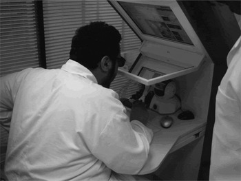
In our institution, we use the Voxel-Man TempoSurg simulator (Spiggle and Theis Medizintechnik, GmbH Diepenbroich IS DE-51491 Overath, Germany); this is mainly designed for temporal bone dissection, but it also has basic software for FESS, which we use to teach medical students.
Temporal bone dissection
Temporal bone dissection is an invaluable exercise for the trainee otologist, as an intimate knowledge of temporal bone anatomy is essential for safe otological surgery. The profusion of temporal bone courses testifies to this principle. It is only by frequent and repeated temporal bone dissection practice that the necessary surgical skills can be acquired, based on a thorough knowledge of temporal bone anatomy and its variants.Reference Golding-Wood19 Nowadays, most units have dissection facilities, and it is almost unthinkable that mastoid surgery should ever be performed without preliminary temporal bone dissection practice.
However, temporal bone dissection has many limitations, including: difficulty in securing specimens; contamination issues; strict disposal protocols; and the possibility of only one dissection (with no facility for correction of errors). For these reasons, simulation has begun to replace dissection courses involving real temporal bones.
Both students and teachers recognise simulation as a powerful teaching tool in temporal bone dissection. Experienced surgeons also use simulators to hone their skills and prepare for delicate procedures. The Voxel-Man TempoSurg simulator is used at Al-Ahsa College of Medicine to teach students the three-dimensional anatomy of the temporal bone, and also to facilitate training in the bone drilling interventions involved in middle-ear surgery. This simulator supplies realistic visual and haptic feedback, and enables the student to perform procedures such as mastoidectomy (Figure 5).Reference Tolsdorff, Petersik, Pflesser, Pommert, Tiede and Leuwer20–Reference Pflesser, Petersik, Tiede, Hohne and Leuwer22 It also enables the student to practise several types of drilling, such as pneumatic mastoid, sclerotic mastoid, cholesteatoma and middle cranial fossa approach.
Temporal bone dissection teaching using a Voxel-Man TempoSurg simulator in the simulation laboratory.

Conclusion
Medical simulation will never replace real life clinical experiences. However, its potential application in the field of medical education is huge, although much development is still required. At Al-Ahsa College of Medicine, we have begun to apply this sophisticated technology for ENT training, and have found it highly beneficial.