Introduction
The nematode genus Strongyloides contains some 50 species of obligate gastrointestinal parasites of vertebrates, of which two species, Strongyloides stercoralis and Strongyloides fuelleborni, infect humans (Grove, 1996). Strongyloidiasis is a soil-transmitted helminthiasis (STH), mainly caused by S. stercoralis, and occurs throughout tropical and warm, temperate regions of the world (Boulware et al., Reference Boulware, Stauffer, Hendel-Paterson, Rocha, Seet, Summer, Nield, Supparatpinyo, Chaiwarith and Walker2007). While infections with S. fuelleborni are known, this species is geographically restricted and zoonotic within non-human primates (Grove, Reference Grove1996; Ashford et al., Reference Ashford, Reid and Wrangham2000).
Diagnosis of strongyloidiasis can, however, be difficult. Parasitological methods are insensitive (Koga et al., Reference Koga, Kasuya and Ohtomo1992; de Kaminsky, Reference de Kaminsky1993; Dreyer et al., Reference Dreyer, Fernandes-Silva, Alves, Rocha, Albuquerque and Addiss1996), serological methods potentially cross-react with other helminth infection(s), especially filarial nematodes (Conway et al., Reference Conway, Atkins, Lillywhite, Bailey, Robinson, Lindo, Bundy and Bianco1993; Grove, Reference Grove1996), or fail in immunocompromised patients as antibody titres are much diminished (Brown et al., Reference Brown, Kizza, Watera, Quigley, Rowland, Hughes, Whitworth and Elliott2004; Sarner et al., Reference Sarner, Fakoya, Tawana, Allen, Copas, Chiodini and Fenton2007). To identify positive cases with confidence, multiple methods of diagnosis may therefore be required. As this can be laborious, time consuming and expensive (Brown et al., Reference Brown, Bukusuba, Hughes, Nakiyingi, Watera, Elliott and Whitworth2003), precise epidemiological information on the occurrence of strongyloidiasis within communities is usually lacking. Globally, however, it is estimated that between 30 million and 100 million people are infected, the majority of whom are asymptomatic (Genta, Reference Genta1989; Grove, Reference Grove1996; Siddiqui & Berk, Reference Siddiqui and Berk2001).
In its chronic, uncomplicated form, strongyloidiasis is primarily a dermatological and gastroenterological disease (Grove, Reference Grove1994). In its severe, complicated form, the disease can progress rapidly, by autoinfection, into a disseminated life-threatening condition. This severe syndrome of hyperinfection is associated with severe immunosuppression, corticosteroid use or sometimes with AIDS (Petithory & Derouin, Reference Petithory and Derouin1987; Cahill & Shevchuk, Reference Cahill and Shevchuk1996; Grove, Reference Grove1996). In sub-Saharan Africa, where the prevalence of HIV is high, chronic strongyloidiasis can be more evident in adults through elevated episodes of clinical diarrhoea (Morales et al., Reference Morales, Atzori, Ludovisi, Rossi, Scaglia and Pozio1995) and is a significant risk factor determining low birthweight of the children of HIV-infected mothers (Dreyfuss et al., Reference Dreyfuss, Msamanga, Spiegelman, Hunter, Urassa, Hertzmark and Fawzi2001). In Uganda, for example, surveys around Entebbe have shown that 12.3% of pregnant women can have parasitologically proven strongyloidiasis (Muhangi et al., Reference Muhangi, Woodburn, Omara, Ornoding, Kizito, Mpairwe, Nabulime, Ameke, Morison and Elliott2007) and in this context of maternal and child health, the general significance of the disease in the younger child is receiving increasing attention (Rice et al., Reference Rice, Skull, Pearce, Mulholland, Davie and Carapetis2003).
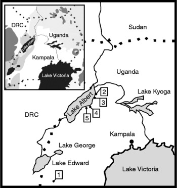
Uganda is one of five African ‘fast-track’ countries to receive support from the US Agency for International Development (USAID) for development of an integrated control programme against neglected tropical diseases (Kolaczinski et al., Reference Kolaczinski, Kabatereine, Onapa, Ndyomugyenyi, Kakembo and Brooker2007). As the broad geographical distribution of the major helminth diseases (e.g. schistosomiasis, onchocerciasis, lymphatic filariasis) is known, integrated preventive chemotherapy control is advancing quickly, with national drug delivery utilizing a variety of distribution channels, e.g. primary schools, mother and child clinics, etc. (Onapa et al., Reference Onapa, Simonsen, Baehr and Pedersen2005a; Kabatereine et al., Reference Kabatereine, Fleming, Nyandindi, Mwanza and Blair2006, Reference Kabatereine, Brooker, Koukounari, Kazibwe, Tukahebwa, Fleming, Zhang, Webster, Stothard and Fenwick2007; Kolaczinski et al., Reference Kolaczinski, Kabatereine, Onapa, Ndyomugyenyi, Kakembo and Brooker2007). Owing to the paucity of relevant epidemiological information, a national policy for control of strongyloidiasis has yet to be formulated as previous field-based surveillance for STH has used Kato–Katz examination (Kabatereine et al., Reference Kabatereine, Brooker, Koukounari, Kazibwe, Tukahebwa, Fleming, Zhang, Webster, Stothard and Fenwick2007) which is unable to detect S. stercoralis larvae (Steinmann et al., Reference Steinmann, Xiao-Nong, Zun-Wei, Jin-Yong, Li-Bo, Xue-Zhong, Lan-Hua, Marti and Utzinger2007). Moreover, previously targeted surveys for Strongyloides have been concentrated on (peri)urban environments (Kabatereine et al., Reference Kabatereine, Kemijumbi, Kazibwe and Onapa1997; Muhangi et al., Reference Muhangi, Woodburn, Omara, Ornoding, Kizito, Mpairwe, Nabulime, Ameke, Morison and Elliott2007), such that the endemicity of strongyloidiasis within the major ecoregions of Uganda (see fig. 1) and associated rural environments is presently unknown.
Sketch map of Uganda with the five sampled villages indicated: 1, Hamukaaka (Kabale district); 2, Bugiogo (Masindi district); 3, Walakuba (Masindi district); 4, Runga (Hoima district); and 5, Tonya (Hoima district). Inset, an ecoregion map of Uganda depicting major ecological zones (taken in modified form from Olson et al., Reference Olson, Dinerstein, Wikramanayake, Burgess, Powell, Underwood, D'Amico, Itoua, Strand, Morrison, Loucks, Allnutt, Ricketts, Kura, Lamoreux, Wettengel, Hedao and Kassem2001).

To shed light on the endemicity of strongyloidiasis in mothers and their infant/preschool children and develop better disease surveillance methods, a field-based epidemiological survey was conducted in western Uganda. A combination of serological and parasitological methods was used: a commercially available enzyme-linked immunosorbent assay (ELISA), Baermann concentration and Koga agar plate. The potential cross-reactivity of the strongyloidid ELISA was first assessed in the laboratory using reference patient sera donated by the Hospital for Tropical Diseases (HTD), UK and, to assess and compare general patterns of STH endemicity and intestinal schistosomiasis with this mother and child group, Kato–Katz faecal smears were also prepared.
Materials and methods
Study area and population
In total, five villages representative of two of five ecoregions within Uganda (Olson et al., Reference Olson, Dinerstein, Wikramanayake, Burgess, Powell, Underwood, D'Amico, Itoua, Strand, Morrison, Loucks, Allnutt, Ricketts, Kura, Lamoreux, Wettengel, Hedao and Kassem2001; Brooker et al., Reference Brooker, Whawell, Kabatereine, Fenwick and Anderson2004) were visited during July 2007 (fig. 1). The villages are located within three Ugandan districts and global positioning system (GPS) coordinates were taken at each village with a hand-held receiver: Hamukaaka, Kabale district (GPS: 01°13.525′S, 029°52.300′E); Bugiogo, Masindi district (01°54.491′N, 031°24.592′E); Walakuba, Masindi district (01°50.533′N, 031°22.964′E); Runga, Hoima district (01°41.102′N, 031°18.514′E); and Tonya, Hoima district (01°35.177′N, 031°05.399′E). All villages were in rural and impoverished environments, there was neither on-site electricity nor functional community-based sanitation facilities.
Prior to the day of survey, mothers and their children were invited to attend a sensitization seminar, where the study objectives were explained and written informed consent was obtained for a target enrolment of 30 mother and child pairs per village. Stool containers were distributed and patients were requested to bring morning stool samples to the field-clinic on the following two consecutive days. Two thick Kato–Katz faecal smears were then made for each specimen (Katz et al., Reference Katz, Chaves and Pellegrino1972). Helminth eggs were counted by microscopy at 100 × magnification and expressed as a tally of eggs per gram (EPG) of stool. This was then classified according to WHO intensity of infection categories: light, medium or heavy (Montresor et al., Reference Montresor, Crompton, Bundy, Hall and Savioli1998). To assess general levels of awareness of strongyloidiasis, as well as ascariasis and schistosomiasis, within the village community, each mother was asked a suite of simple yes/no questions pertaining to the diseases and their reported symptoms.
Field diagnosis of Strongyloides
Three approaches were taken for diagnosis of strongyloidiasis in the field: ELISA (see below), Baermann concentration and Koga agar plate methods. To assess the day-to-day reproducibility of the ELISA, duplicate fingerprick blood was collected and tested on two consecutive days at Hamukaaka and Bugiogo. Owing to logistic and time constraints, duplicate stool samples were examined only in a sub-set of patients at Hamukaaka, Bugiogo and Walakuba villages, with Baermann concentration method and all further stool examinations being examined upon single specimens. Owing to limited resources, the Koga agar plate method was performed only at Hamukaaka, Bugiogo and Walakuba villages.
Strongyloidid ELISA
A microwell ELISA test (IVD Research Inc., Carlsbad, California, USA) for human IgG antibodies against S. stercoralis antigen was used according to the manufacturer's instructions. To test for potential cross-reactivity of the strongyloidid ELISA in the laboratory, an archival panel of 20 anonymous patient sera from the HTD was examined at 1:20 and 1:64 dilutions. The panel contained sera from: 1 patient with parasitologically proven strongyloidiasis, 14 with schistosomiasis (urinary or intestinal), 1 with fascioliasis, 1 with lymphatic filariasis and 3 negative controls.
When in the field, approximately 50 μl of finger-prick blood was collected from each patient using a Gilson pipette, and placed in a 1.5 ml Eppendorf tube. Total blood was clotted then centrifuged for 5 min at 14,000 rpm, after which the supernatant was collected, diluted 1:64 in dilution buffer, and 200 μl applied to an antigen-coated microwell. The sample was incubated for 10 min and then each microwell was rinsed three times with washing buffer. Two drops of peroxidase-conjugated protein A were then added and incubated for 5 min. After three further rinses with washing buffer, 2 drops of chromogen (tetramethylbenzidine) were added and the development reaction was subsequently stopped after 5 min with addition of 2 drops of stop solution (1 m H3PO4). Both positive and negative controls were performed alongside test reactions; when held against a white background, a positive test reaction within the microwell was judged visually as a perceivable yellow colour.
Baermann concentration
The Baermann concentration method was performed on approximately 3 g of stool, according to conditions reported by Dreyer et al. (Reference Dreyer, Fernandes-Silva, Alves, Rocha, Albuquerque and Addiss1996). To facilitate community screening at each village, a wooden box was constructed with an internally fitted 60 W electric light bulb with external plug. The box also had a detachable lid and removable wooden insert into which 15 circular holes were cut. A Baermann glass funnel with silicone tube and Mohr clamp would fit into each hole, enabling 15 stool samples to be processed in parallel. Once assembled the box's lid would be reattached and the electric light switched on, illuminating the Baermann funnels from below. Samples were incubated for 2 h and faecal sediments were harvested by sequential centrifugation in 15 ml test tubes. Pellets were then re-suspended in a total volume of 400 μl of sterile distilled water (SDW), 100 μl of this suspension was placed on to each one of four microscope slides and a double glass cover slip was mounted. The four slides were examined by microscopy at 100 × magnification for strongyloidid larvae and other helminth eggs.
Koga agar plate
On the day of stool collection, Koga agar plates were freshly prepared, following Koga et al. (Reference Koga, Kasuya and Ohtomo1992), in batches of 25 plates, using pre-weighed reagents. Briefly, 700 ml of SDW in a 1-litre Schott bottle was heated to boiling point using a camping gas stove. After cooling to 60°C, reagents were added and the Schott bottle lid closed. The bottle was shaken vigorously and then the solution was again heated to boiling point. The molten mixture was allowed to cool to 45°C, after which the bottle was opened and approximately 25 ml of solution was poured into each plastic Petri dish. Once the agar mixture had solidified, a sample of 3 g of stool was placed in the centre of each plate and the lid was closed. The edge of the Petri dish was then sealed with Parafilm to prevent evaporation and 25 dishes were placed inside a clear, sealable plastic bag containing moistened filter paper to ensure high humidity. Koga plates were incubated at environmental temperature for 48 h before inspection under the dissecting microscope for strongyloidid larvae.
Ethical review and anthelminthic treatments
Ethical permission for the study was granted by the London School of Hygiene and Tropical Medicine (06/45) and the National Council of Science and Technology, Kampala, Uganda. At the end of the study all participants were administered with single tablet of albendazole (400 mg). Those found with egg-patent Schistosoma mansoni infection were offered praziquantel (40 mg kg− 1) and those with S. stercoralis larvae were offered ivermectin (0.2 mg kg− 1).
Results
In the laboratory, the ELISA was shown to have high specificity against the panel of archival patient sera, no cross-reactions were observed at either 1:20 or 1:64 dilutions. The known strongyloidiasis-positive serum was correctly identified, eliciting a strong positive reaction, a golden yellow colour.
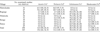
In the field, duplicate Kato–Katz faecal smears were prepared from 158 mothers (mean age 27 years, minimum 17 and maximum 45 years) and 143 children (mean age 3 years, minimum 8 months and maximum 6 years). Mean EPG was calculated across the four faecal smears and this value was graded into light, medium or heavy categories for intestinal schistosomiasis and for each STH. General prevalence and categories of infection intensities for mother and child groups are shown by village in table 1. It is evident that in Hamukaaka village, Ascaris and Trichuris infections were dominant in both mothers and children, intestinal schistosomiasis and hookworm infections were absent. By contrast, intestinal schistosomiasis was dominant in all other villages, while Ascaris, Trichuris and hookworm were at much lower levels throughout.
Prevalence (%) and intensity of soil-transmitted helminthiasis (STH) and intestinal schistosomiasis by village.

n/a, Not applicable.
a Infection intensity categories: light = 1–4999 epg; medium = 5000–49,999 epg; heavy ≥ 50,000 epg.
b Infection intensity categories: light = 1–999 epg; medium = 1000–9999 epg; heavy ≥ 10,000 epg.
c Infection intensity categories: light = 1–99 epg; medium = 100–399 epg; heavy ≥ 400 epg.
d Infection intensity categories: light = 1–1999 epg; medium = 2000–3999 epg; heavy ≥ 4000 epg.
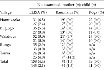
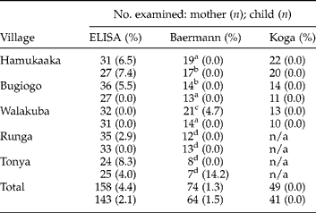
Owing to time and logistical constraints, it was not possible to assemble a complete dataset for each patient for each diagnostic method. The results of ELISA, Baermann and Koga agar plate methods are shown in table 2. In total, seven mothers and four children were found to be sero-positive for Strongyloides. Evidence of infection was found in all villages with the exception of Walakuba village. The putative prevalence of infection estimated by ELISA was slightly higher in mothers than in children, although no sero-positive cases could be corroborated with either Baermann and(or) Koga agar plate methods. Strongyloidid larvae were, however, encountered from two patients using the Baermann method, a mother from Walakuba (single larva) and child from Tonya (three larvae). Both, however, were sero-negative upon ELISA and neither had hookworm. Koga agar plate inspection of the Walakuban mother was negative and, across the survey, no strongyloidid larva was encountered with this method. In addition to retrieval of S. stercoralis, eggs of Ascaris, Trichuris and Schistosoma were often found using the Baermann concentration method.
Prevalence (%) of strongyloidiasis by village for each method of diagnosis.
n/a, Not applicable.
a With 7 duplicate examinations.
b With 9 duplicate examinations.
c With 8 duplicate examinations.
d Duplicate examinations not undertaken.
From questionnaire data, assessments were made of general levels of awareness of schistosomiasis, STH (ascariasis) and strongyloidiasis in mothers. With the exception of Hamukaaka (7%), levels of awareness of schistosomiasis were good: Bugiogo (54%), Walakuba (62%), Runga (45%) and Tonya (82%). Awareness of ascariasis was fair: Hamukaaka (74%), Bugiogo (35%), Walakuba (18%), Runga (33%) and Tonya (19%). Geophagia, or soil eating, by mothers was reported in the following descending order: Hamukaaka (96%), Tonya (93%), Bugiogo (87%), Runga (84%) and Walakuba (68%). General awareness of strongyloidiasis was very poor (5%) with no mother able to describe correctly the signs and symptoms of this infection.
Discussion
Control of neglected tropical diseases in Uganda is progressing quickly as there are national strategies for community control with orally administered anthelminthics (Kabatereine et al., Reference Kabatereine, Fleming, Nyandindi, Mwanza and Blair2006, Reference Kabatereine, Brooker, Koukounari, Kazibwe, Tukahebwa, Fleming, Zhang, Webster, Stothard and Fenwick2007; Kolaczinski et al., Reference Kolaczinski, Kabatereine, Onapa, Ndyomugyenyi, Kakembo and Brooker2007). Moreover Uganda is one of five African countries to receive ‘fast-track’ funding to develop integrated control approaches (Kolaczinski et al., Reference Kolaczinski, Kabatereine, Onapa, Ndyomugyenyi, Kakembo and Brooker2007). For example, Child Health Days (CHDs) operate from static and mobile health units twice a year (in April and October) to accelerate the delivery of maternal and child health community interventions (Kolaczinski et al., Reference Kolaczinski, Kabatereine, Onapa, Ndyomugyenyi, Kakembo and Brooker2007). These include vitamin A supplementation, childhood vaccination, health education and administration of oral benzimidazoles. Across the country the endemic zones of the major helminth diseases are broadly known, including where there is geographic overlap, e.g. schistosomiasis, lymphatic filariasis and onchocerciasis are co-endemic in the West Nile region (Kolaczinski et al., Reference Kolaczinski, Kabatereine, Onapa, Ndyomugyenyi, Kakembo and Brooker2007) and the ongoing mass drug administration initiatives will also have an impact upon other diseases, e.g. strongyloidiasis, especially where ivermectin is administered (Hotez et al., Reference Hotez, Molyneux, Fenwick, Ottesen, Sachs and Sachs2006).
In contrast, the distribution of strongyloidiasis is poorly known and the disease has yet to be fully considered within the national control strategy, as relevant epidemiological information is lacking. From a public health perspective, preferred treatment options would include targeted treatment of high-risk groups with ivermectin, with prior diagnostic screening within the community (Conway et al., Reference Conway, Lindo, Robinson and Bundy1995). Although strongyloidiasis is known to be endemic in the Lake Victoria region – for example, the prevalence of the disease in women of childbearing age is ~12% (Muhangi et al., Reference Muhangi, Woodburn, Omara, Ornoding, Kizito, Mpairwe, Nabulime, Ameke, Morison and Elliott2007) – the distribution of the disease outside this region was far from clear. Other strongyloidids, at least in non-human primates, have been reported from western Uganda (Ashford et al., Reference Ashford, Reid and Wrangham2000) but, like elsewhere in Africa, the ecological range of S. stercoralis will be shaped by environmental determinants (Appleton et al., Reference Appleton, Maurihungirire and Gouws1999) and these will influence the endemicity of helminth diseases (Brooker et al., Reference Brooker, Whawell, Kabatereine, Fenwick and Anderson2004). For example, Kabale is some 1250 m higher than Lake Albert and the climate is generally much cooler, for example, the mean annual maximum and minimum temperatures differ by 6°C and 12°C, respectively. The clear division within the epidemiological landscape is evidenced by the dominance of Ascaris/Trichuris infections in Kabale district and S. mansoni infections in Hoima/Masindi districts.
One immediate hurdle to gathering relevant epidemiological information is the precise disease diagnosis. As excretion of larvae can vary, day-to-day analysis of several stool samples may be needed (de Kaminsky, Reference de Kaminsky1993; Dreyer et al., Reference Dreyer, Fernandes-Silva, Alves, Rocha, Albuquerque and Addiss1996) or, as an alternative approach, multiple methods may be used simultaneously (Siddiqui & Berk, Reference Siddiqui and Berk2001; Brown et al., Reference Brown, Bukusuba, Hughes, Nakiyingi, Watera, Elliott and Whitworth2003). This may not always be practical, given the availability of local resources and infrastructure. Pragmatic, cost-effective disease surveillance methods and protocols are therefore needed not only to monitor the disease within the community but also to better allocate and guide anthelminthic therapies (Marti et al., Reference Marti, Haji, Savioli, Chwaya, Mgeni, Ameir and Hatz1996; Orem et al., Reference Orem, Mayanja, Okongo and Morgan2003).
Choosing the most appropriate method for diagnosis in the field, or in a rural clinic, can be a challenge, for it is an inevitable balance between theoretical and practical issues. A precise interpretation of the present diagnostic results is difficult as, unfortunately, concordant data were lacking. Foremost, although serology can suffer from cross-specific reactions with other helminths, the specificity of the ELISA produced by IVD has been demonstrated to be very good (97%) (van Doorn et al., Reference van Doorn, Koelewijn, Hofwegen, Gilis, Wetsteyn, Wismans, Sarfati, Vervoort and van Gool2007). This was in accordance with our results from the archival patient sera. In the field, both lymphatic filariasis and onchocerciasis are known not to occur within the five surveyed villages, so ELISA positives were unlikely to result from co-infection with these diseases. Similarly, another nematode, Mansonella perstans, occurs throughout Uganda, but it is rarely found in Kabale district (Onapa et al., Reference Onapa, Simonsen, Baehr and Pedersen2005b). It is therefore likely that the ELISA represents true positives. Given the insensitivity of Baermann and Koga methods, it was not surprising that parasitological examinations were unable to confirm the ELISA-positive results (Dreyer et al., Reference Dreyer, Fernandes-Silva, Alves, Rocha, Albuquerque and Addiss1996). In contrast, the ELISA sero-negative individuals who were parasitologically positive present a different scenario. In both cases the numbers of larvae were low, so it may be that this level of infection was insufficient to stimulate a detectable antibody response, or these individuals were unable to mount an immune response because of their immunosuppression (Brown et al., Reference Brown, Bukusuba, Hughes, Nakiyingi, Watera, Elliott and Whitworth2003).
On balance, we have found the performance of the ELISA to be very promising. On the positive side, it was quick ( < 2 h), had the highest screening throughput and was both robust and reliable on a day-to-day basis. On the negative side, it was, however, the most demanding method for necessary resources and equipment. In terms of replenishment costs, the basic price of each ELISA was approximately US$1 and this does not incorporate costs of other necessary items, e.g. lancettes, tubes and tips. Perhaps the greatest obstacle is the need for an electrical microcentrifuge and stable power supply. While it is possible to use the survey vehicle's 12 V DC supply with an electrical adapter, power requirements of the microcentrifuge are over and above this for regular use.
For the parasitological methods, the Baermann and Koga agar plate have several methodological similarities as both require microscopy, and much of the plastic and glassware can be re-used after washing with boiling water alone. Replenishment costs for the Koga agar plate and Baermann have been reported previously, and differ some three- to fourfold in costs due to reagents required within the agar plate (de Kaminsky, Reference de Kaminsky1993). The diagnostic sensitivities of the Baermann and Koga methods have been compared previously against single and multiple stool examinations (Koga et al., Reference Koga, Kasuya and Ohtomo1992; Grove, Reference Grove1996; Steinmann et al., Reference Steinmann, Xiao-Nong, Zun-Wei, Jin-Yong, Li-Bo, Xue-Zhong, Lan-Hua, Marti and Utzinger2007). In our study, the Baermann was slightly more sensitive than the Koga, although only two individuals were encountered and thus a more formal comparison could not be made. For field surveillance, one immediate advantage of the Baermann, or a modification thereof (Orem et al., Reference Orem, Mayanja, Okongo and Morgan2003), is its potential to overlap with much of the necessary equipment for standard Kato–Katz examinations, e.g. compound microscope, glass slides, etc., which are the backbone of schistosomiasis and STH surveillance in Uganda (Kabatereine et al., Reference Kabatereine, Kemijumbi, Kazibwe and Onapa1997, Reference Kabatereine, Fleming, Nyandindi, Mwanza and Blair2006, Reference Kabatereine, Brooker, Koukounari, Kazibwe, Tukahebwa, Fleming, Zhang, Webster, Stothard and Fenwick2007). Indeed, future co-surveillance for strongyloidiasis and these other helminths could present the next logical step in developing a strategic framework for integrated disease management. It should be noted that calculation of optimal patient sample sizes and multiple stool requirements will have to be adjusted accordingly to give a field-team an acceptable workload, given that the daily screening throughput of the Baermann is some threefold lower than that of Kato–Katz.
In conclusion, it appears that strongyloidiasis is endemic in western Uganda but at much lower levels than elsewhere; for example, around Entebbe (Muhangi et al., Reference Muhangi, Woodburn, Omara, Ornoding, Kizito, Mpairwe, Nabulime, Ameke, Morison and Elliott2007). However, better routine epidemiological screening for this disease is needed within the mother and child group, as it can have a negative impact upon neonatal weight (Dreyfuss et al., Reference Dreyfuss, Msamanga, Spiegelman, Hunter, Urassa, Hertzmark and Fawzi2001). It would therefore be advisable to target screening upon women of childbearing age, especially those with HIV, as ivermectin treatment would be beneficial for both mother and baby. In the rural clinic setting, the Baermann concentration method, when used in parallel, appears to be the most immediate and pragmatic option for disease diagnosis.
Acknowledgements
We would like to thank Dr Juerg Utzinger and Hans-Peter Marti of Swiss Tropical Institute, Basel for advice on practical diagnosis of strongyloidiasis. We are grateful to Dr David Lambillotte and Ms Alison Knoth IVD Research Inc., Carlsbad, California, USA for the provision of the ELISA kits free of charge and to Professor Peter Chiodini and HTD for provision of patient sera. For technical assistance in the field we thank Mr Charles Jennison and Ms Nicola Batchelor and are grateful to the Vector Control Division local officers in Kabale, Hoima and Masindi districts. We thank Dr David Rollinson, Ms Claire Standley and referees for improvements made to this manuscript. We extend our sincere thanks to the mothers and children who took part in this survey. The study received umbrella programme support from the European Union FP6 STREP CONTRAST Contract No. 032203.