Implications
Understanding the processes involved in the regulation of skeletal muscle differentiation is necessary to develop strategies for improving the efficiency of meat production. Most information on satellite cells and their regulation during differentiation induction is based on studies performed using rodent animal models. The information available from large farm animals is limited. The present study is the first report on the correlation between skeletal differentiation factors and a number of myomiRNAs during myogenic differentiation in bovine species. The study describes a spatiotemporal pattern of expression for different transcription factors involved in satellite cell development.
Introduction
Satellite cells are adult stem cells that reside between the basal lamina and sarcolemma of the muscle fiber (Mauro, Reference Mauro1961). Under physiological conditions, these cells constitute a population of quiescent mononuclear cells, yet they maintain strong proliferative potential and the capacity to differentiate (Brack and Rando, Reference Brack and Rando2012). In response to environmental factors, satellite cells can restore damaged muscle fibers or even form completely new muscle fibers. Satellite cells are responsible for muscle preservation and growth, and they exhibit limited gene expression and protein synthesis due to low cellular turnover (Brack and Rando, Reference Brack and Rando2012).
MicroRNAs (miRNAs) play essential roles during animal development and in the regulation of stem cell self-renewal and differentiation in various tissues, including skeletal muscle tissue (Crippa et al., Reference Crippa, Cassano and Sampaolesi2012). Many miRNAs are expressed during muscle development. MiRNA-1, miRNA-133 and miRNA-206 are strictly muscle specific and for this reason are also known as myomiRNAs (Boutz et al., Reference Boutz, Chawla, Stoilov and Black2007; McCarthy, Reference McCarthy2008). Both miRNA-1, found in skeletal and cardiac muscle, and miRNA-206, specific to skeletal muscle, have been shown to promote myoblast to myotube differentiation. By contrast, miRNA-133 promotes the proliferation of myoblasts (Chen et al., Reference Chen, Mandel, Thomson, Wu, Callis, Hammond, Conlon and Wang2006; Kim et al., Reference Kim, Lee, Sivaprasad, Malhotra and Dutta2006). Muscle miRNAs are integrated into myogenic regulatory networks and their expression is under the transcriptional and post-transcriptional control of myogenic factors; as a consequence, they exert widespread control of gene expression in muscle tissues (Rao et al., Reference Rao, Kumar, Farkhondeh, Baskerville and Lodish2006).
Most information on the satellite cells and their regulation and behavior during differentiation induction has been gained from studies using rodent animal models, whereas very limited information is available about satellite cells in large farm animals (Dai et al., Reference Dai, Wang, Zhang, Liu, Li, Ding and Guo2016). Understanding the processes involved in the regulation of muscle differentiation is necessary to develop strategies for improving the efficiency of meat production. In the present study, we investigate Holstein bovine satellite cells (BoSCs) during their differentiation and characterize the expression pattern of the main genes and miRNAs involved in this skeletal muscle process.
Material and methods
Holstein bovine satellite cells isolation and culture
Immediately after slaughter, samples of longissimus thoracis muscle weighing ~500 g were collected from six Holstein breed male cattle aged 18 to 23 months. Sample collection was performed with the authorization and under the supervision of representatives of the Veterinary Services of the Italian National Health Service – a section of the Ministry of Health. Samples were immersed in 70% ethanol for 15 min and then placed in sterile phosphate buffer solution with antibiotic/antimyotic solution (Sigma-Aldrich, St. Louis, MO, USA). Muscles were transported to the laboratory within 1 h at room temperature and immediately processed to collect satellite cells in a sterile field under a tissue culture hood. To obtain satellite cells, vessels, fat and connective tissues were removed carefully from the muscle samples. These cleaned muscle tissues were then transferred to a glass Petri dish with Dulbecco’s Modified Eagle’s Medium (DMEM) and cut into small fragments using scissors. After trituration, enzymatic digestion was performed with 1 mg/ml of pronase XIV (Sigma-Aldrich) in DMEM in the presence of antibiotic/antimyotic solution (100 U penicillin/ml, 100 μg streptomycin/ml and 2.5 μg amphotericin/ml) and without serum for 60 min at 37°C with gentle agitation at 15 min intervals. The protease was removed after centrifugation at 400×g for 5 min at room temperature. The pellet was resuspended in DMEM 10% horse serum and the suspension allowed to settle. Without handling the coarse precipitated material, the satellite cell-containing supernatant was retained. This process was repeated twice. The pooled supernatant was filtered using a 40 μm cell strainer (BD Biosciences, San Jose, CA, USA) and then centrifuged at 1000×g for 10 min to recover the cells. Myosatellite cells were then resuspended in growth medium (GM) (DMEM 20% fetal bovine serum (FBS), 10% horse serum, 100 U penicillin/ml, 100 μg streptomycin/ml and 2.5 μg amphotericin/ml) and cultured on plates pre-treated with 2% gelatine (Type A, Sigma-Aldrich). An aliquot of cell sample was suspended in 90% FBS and 10% dimethylsulfoxide and frozen in liquid nitrogen. After 3 days of satellite cell isolation and plating, cells had adhered onto the gelatine-coated dish surface, and on day 6 the culture medium was removed and changed with differentiation medium (DM) (DMEM 2% horse serum, 100 U penicillin/ml, 100 μg streptomycin/ml and 2.5 μg amphotericin/ml) to induce differentiation. At the time of adhesion to the culture plate, analysis of the BoSCs indicated that >95% of the cells were myogenic, as also observed in mouse and human models (Günther et al., Reference Günther, Kim, Kostin, Lepper, Fan and Braun2013), as measured by the immunofluorescent detection of PAX7 (Supplementary Figure S1).
RNA extraction
Total RNA from cultured cells was extracted using 1 ml of TRI Reagent (Sigma-Aldrich) and any residual genomic DNA was removed using a DNAse I Recombinant RNAse free kit (Roche, Mannheim, Germany). The RNA concentration was determined by spectrophotometry (BioPhotometer, Eppendorf, Hamburg, Germany). The ratio of the optical densities measured at 260 and 280 nm was >1.9 for all RNA samples. Complementary DNA (cDNA) was synthesized from 3 μg of total RNA using the RT High-Capacity cDNA Reverse Transcription Kit (Applied Biosystems, Foster City, CA, USA), according to the manufacturer’s protocol. The cDNA was subsequently diluted in nuclease-free water and stored at −20°C.
Quantitative assessment of gene and microRNA expression
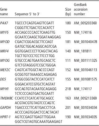
cDNA (3 μg) was prepared in a single run for the real-time quantitative PCR (q-PCR) experiments for all the selected genes. To determine the relative amount of paired box protein pax-7 (PAX7), myogenic factor 5 (MYF5), myogenic differentiation-1 (MYOD), myogenic regulator factor 4 (MRF4), myogenin (MYOG), myocyte enhancer factor 2C (MEF2C), desmin (DES), myosin heavy chain-1 (MYH1) and transmembrane protein 8C (TMEM8C), also called myomaker transcripts, q-PCR was performed using the CFX Connect Real-Time PCR System (Bio-Rad, Hercules, CA, USA). Primers for target and reference genes were designed on Bos taurus GenBank messenger RNA (mRNA) sequences using Primer 3 Software (version 4.0). Oligonucleotides were designed to cross the exon/exon boundaries to minimize the amplification of contaminant genomic DNA and were analyzed using the IDT tool (available at http://www.idtdna.com/scitools/scitools.aspx) for hairpin structure and dimer formation. Primer specificity was verified with BLAST analysis against the genomic NCBI database. Table 1 summarizes the primer information, providing the sequences, gene accession number and amplicon sizes. The dilution method was applied to establish the efficiency of each primer. Primer efficiency was then calculated using CFX Manager (version 3.0; Bio-Rad), employing the linear regression slope of the dilution series. The efficiency of each primer set was found to be ⩽95%. Hypoxanthine phosphoribosyl-transferase 1 (HPRT-1) and glyceraldehyde 3-phosphate dehydrogenase (GAPDH) genes were used as reference genes in order to normalize the transcription data for the target genes.
Genes subjected to quantitative PCR (q-PCR) analysis

PAX7=protein pax-7; MYF5=myogenic factor 5; MYOD=myogenic differentiation-1; MRF4=myogenic regulator factor 4; MYOG=myogenin; MEF2C=myocyte enhancer factor 2C; DES=desmin; MYH1=myosin heavy chain-1; TMEM8C=transmembrane protein 8C; GAPDH=glyceraldehyde 3-phosphate dehydrogenase; HPRT=hypoxanthine phosphoribosyl-transferase 1.
Primer sequences for q-PCR were designed on Bos taurus GenBank messenger RNA sequences. Primer sequences, gene accession number and amplicon sizes are shown.
To quantify the expression of mature miRNA-1, miRNA-133a and miRNA-206, 100 ng of total RNA were reverse transcribed using the TaqMan miRNA Reverse Transcription Kit and subjected to q-PCR using the TaqMan miRNA Assay Kit (Applied Biosystems), according to the manufacturer’s protocol (miR-1 assay ID; miR-133a assay ID 002246; miR-206 assay ID 000510; miR-16 assay ID 000391). MiRNA-16 was used to normalize the results (Miretti et al., Reference Miretti, Martignani, Accornero and Baratta2013). The q-PCR parameters were as follows: cycle 1, 95°C for 3 min; cycle 2, 95°C for 15 s, 60°C for 30 s for 44 cycles. Each reaction was run in triplicate and a no template control was included using water instead of cDNA.
Data analysis
To evaluate mRNA and miRNA expression, data were obtained as quantification cycle values (C
q
). C
q
values of technical replicates were averaged then used to determine ∆C
q
values (∆C
q
=C
q
of the target gene/miRNA−C
q
of the reference gene/miRNA) (Theil et al., Reference Theil, Sørensen, Nissen and Oksbjerg2006a). The
 $$2^{{{\minus}\Delta \Delta C_{q} }} $$
method was used to analyze the data, and results were expressed as fold changes compared with control samples (Pabinger et al., Reference Pabinger, Thallinger, Snajder, Eichhorn, Rader and Trajanoski2009). To exclude potential bias generated through use of the
$$2^{{{\minus}\Delta \Delta C_{q} }} $$
method, statistic elaboration was performed at the ∆C
q
stage (Theil et al., Reference Theil, Sørensen, Therkildsen and Oksbjerg2006b).
Immunocytochemistry
Myoblasts or myotubes were fixed with a suitable volume of acetone/methanol solution (1 : 1 proportion) at room temperature for 30 s, followed by washing twice with Tris buffer saline at room temperature for 5 min. Non-specific antigen sites were blocked with 10% goat serum at room temperature for 30 min. Subsequently, fixed cells were incubated at 4°C overnight with the following antibodies: 1 : 100 diluted anti-PAX7 (supernatant), 1 : 10 diluted anti-myosin heavy chain antibody (MF20 supernatant; Developmental Studies Hybridoma Bank, University of Iowa, Iowa City, IA, USA), 1 : 500 diluted anti-myogenin (F5D ascites; Developmental Studies Hybridoma Bank) and 1 : 500 diluted anti-Ki67 (DakoCytomation, Glostrup, Denmark). After extensive washing with Tris buffer saline, the fixed cells were incubated with secondary antibodies: goat anti-mouse AlexaFluor®488 (1 : 150) (Invitrogen, Eugene, OR, USA). Negatively stained controls were obtained for each antigen by replacing the primary antibody with a suitable isotype (normal mouse IgG from Santa Cruz Biotechnology Inc., Santa Cruz, CA, USA) at the same concentration. After washing twice with Tris Buffer saline at room temperature for 5 min, the nuclei of the cells were stained with 4′,6-diamidino-2-phenylindole (DAPI; Thermo Scientific, Rockford, IL, USA). The number of positive cells for each antibody was scored in 10 randomly chosen fields. The percentage of both positive nuclei and cells was calculated relative to the total number of nuclei.
Statistical analysis
Statistical significance between ∆C
q
values of genes expression of culture in GM v. cultures in DM (days 7 to 10, DM) was assessed using the Kruskal–Wallis non-parametric test as the data were not normally distributed, assessed using the Kolmogorov–Smirnov test. Analogous comparison was done for miRNAs. To exclude potential bias by use of
$$2^{{{\minus}\Delta \Delta C_{q} }} $$
method, statistic elaboration was performed on ∆Cq values. The Spearman correlation non-parametric test was applied to correlate each miRNA expression and all gene mRNA expression data. Data were expressed as means±SD. Differences were considered significant if P⩽0.05. All statistical analyses were performed using GraphPad InStat software (version 3.10).
Results
We examined BoSC preparations from different Holstein male animals and analyzed their capacity to differentiate reproducibly into multinucleated myotubes in cell culture. The growth and the differentiation of the cells were followed by time-lapse microscopy (images acquired over 7 days; 10× magnification) using a computer controlled Leica AF6000LX (Leica Microsystem, Wetlar, Germany) inverted microscope equipped with a motorized stage controlled by LAS AF software (Supplementary Material S1). All satellite cell preparations consistently demonstrated myotube differentiation.
Messenger RNA expression of skeletal muscle differentiation markers in bovine satellite cells
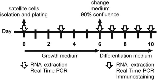
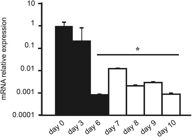
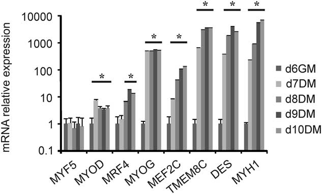
After having evaluated the reproducibility of cell differentiation into myotubes in vitro, BoSCs were further analyzed for the expression of several mRNA markers of skeletal muscle cells: PAX7, MYF5, MYOD, MRF4, MYOG, MEF2C, TMEM8C, DES and MYH1. BoSCs can begin to differentiate when cells are confluent, even in the presence of GM. However, no myotube formation was observed using phase-contrast microscopy during the days in which cells were cultured under proliferation conditions (days 0 to 6), or on day 7 following their transfer to DM (data not shown). In the GM condition (days 0 to 6), BoSCs expressed the satellite cell lineage marker, PAX7, as well as transcripts for myogenic regulatory factors (MYF5, MYOD, MRF4 and MYOG) and other genes associated with skeletal muscle tissue (MEF2C, DES, MYH1, TMEM8C). The mRNA levels of most of the genes analyzed were similar throughout the last 3 days of culturing under proliferative conditions (days 4 to 6; data not shown), suggesting that BoSCs had not yet started to differentiate despite reaching 90% confluence (Figure 1). PAX7 is recognized as a lineage marker required for embryonic and fetal maintenance of the vast majority of satellite cells (Lepper et al., Reference Lepper, Conway and Fan2009). Under the growth promoting conditions of the present study, the PAX7 mRNA levels in BoSCs dramatically decreased between 3 and 6 days post isolation (P<0.01) even though differentiation had not been induced and other genes, such as MYOD, were not yet up-regulated (Figures 2 and 3). MYF5, the earliest marker of myogenic commitment and a member of the muscle-specific determination genes, plays an important role in skeletal muscle development (Hopwood et al., Reference Hopwood, Pluck and Gurdon1991). In this bovine model, we did not detect any significant difference in MYF5 gene expression during satellite cell activation and differentiation. MYOD mRNA levels increased on day 7, once cells had been transferred to the DM condition (P<0.02), indicative of the gradual activation of the myogenic gene program; they then decreased, starting on day 8. Over the course of 4 days culturing in the low serum condition (DM; days 7 to 10), the mRNA levels of several analyzed genes increased (MRF4, MYOG, MEF2C, DES and MYH1), starting on day 7 (P<0.05), and these levels remained constant until the end of the experimental model (Figure 3). The novel muscle-specific gene TMEM8C, also named myomaker, was recently described in mouse and zebrafish models and demonstrated to promote myoblast fusion (Millay et al., Reference Millay, O’Rourke, Sutherland, Bezprozvannaya, Shelton, Bassel-Duby and Olson2013; Landemaine et al., Reference Landemaine, Rescan and Gabillard2014). In BoSCs, TMEM8C mRNA expression was significantly up-regulated (P<0.05), starting on the 1st day of cultivation under differentiating conditions (day 7) and until day 10, by which time the myoblasts had fused (Figure 3).
Experimental timeline for bovine satellite cell cultures. Cells were cultured from days 0 to 6 in growth medium such that 90% confluence was obtained. Cells were then transferred to a differentiation medium for 4 days (days 7 to 10).

PAX7 messenger RNA (mRNA) expression during bovine skeletal muscle cell differentiation. Skeletal muscle cells cultured in vitro as myoblasts and induced to differentiate were harvested at various time points for the analysis of gene expression. Black bars: under proliferative conditions (days 0, 3 and 6 (growth medium)); white bars: following differentiation induction (days 7 to 10 differentiation medium). Relative mRNA levels are shown on a log-10 scale. Differences in gene expression are shown as fold changes (mean±SD). *P<0.05 compared with expression values as assessed on day 0.

Muscle-specific gene expression levels in cultured bovine satellite cells during the differentiation program. Gene expression in bovine satellite cells was determined at various time points during proliferation (growth medium (GM)) and differentiation (differentiation medium (DM)). Relative messenger RNA (mRNA) expression levels are shown on a log-10 scale. Differences in gene expression are shown as fold changes (mean±SD). *P<0.05 compared with expression values assessed on day 6 (GM). MYF5=myogenic factor 5; MYOD=myogenic differentiation-1; MRF4=myogenic regulator factor 4; MYOG=myogenin; MEF2C=myocyte enhancer factor 2C; TMEM8C=transmembrane protein 8C; DES=desmin; MYH1=myosin heavy chain-1.

MyomiRNAs are up-regulated in bovine satellite cells during differentiation
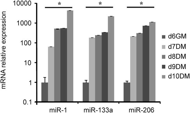
We have already reported that miRNA-27b is involved in the post-transcriptional regulation of gene expression during the skeletal muscle differentiation process (Miretti et al., Reference Miretti, Martignani, Accornero and Baratta2013). Therefore, we examined the expression of miRNA-1, miRNA-133a and miRNA-206, collectively known as myomiRNAs; miRNA-16, a ubiquitously expressed miRNA, was also assessed as an internal control. We profiled miRNA expression during the course of BoSC proliferation and differentiation. These experiments were performed on low-passage primary cultures using our defined models of BoSC proliferation and differentiation (Figure 1). Although the expression of the main genes involved in the differentiation process increased, on the 1st day of culture under differentiation conditions (day 7, DM) miRNA-1, miRNA-133a and miRNA-206 were also strongly up-regulated (P<0.001) (Figure 4). Spearman’s correlations indicated a highly significant positive correlation between the expression of the three myomiRNA’s and MEF2C and MYH1 gene expression (P<0.001); a highly significant positive correlation between miRNA-1, miRNA-133a and MRF4, TMEM8C and DES gene expression (P<0.001); and a negative correlation between these same miRNAs and MYOD (P<0.05).
MyomiRNA expression levels in cultured bovine satellite cells during the differentiation program. MiRNA-1, miRNA-133a and miRNA-206 were screened in quantitative PCR and expression was normalized to miRNA-16, used as an endogenous control. Differences in gene expression are shown as fold changes (mean±SD). *P<0.05 compared with expression values on day 6 (growth medium (GM)). DM=differentiation medium.

Skeletal muscle protein expression analysis
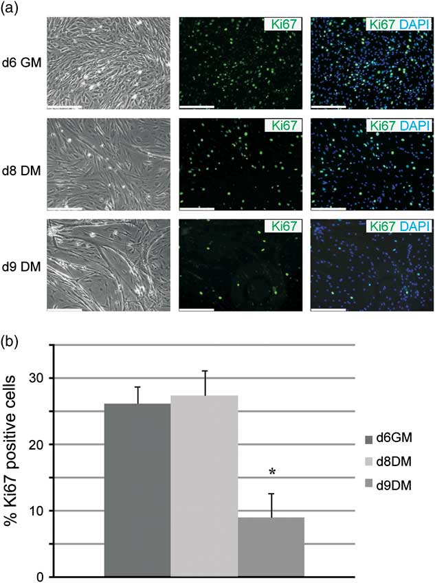
The isolation process triggers the activation of satellite cells, which are normally quiescent. This activation process involves cell proliferation and results in a temporary expansion of the satellite cell pool followed by their differentiation into myoblasts (Collins et al., Reference Collins, Olsen, Zammit, Heslop, Petrie, Partridge and Morgan2005). Skeletal muscle terminal differentiation is a two-step process, starting with irreversible cell cycle withdrawal followed by the activation of the muscle genetic program, in turn, resulting in cell fusion and the formation of multinucleated myotubes (Braun and Gautel, Reference Braun and Gautel2011). We assessed the proportion of active satellite cells in our model by means of Ki67 staining – a marker of cell proliferation that is expressed during all phases of the active cell cycle under both proliferating and differentiation conditions (Figure 5a). Figure 5 shows that the percentage of active satellite cells observed during the culturing of BoSCs in GM was about 26% and this value remained stable at day 8 (DM), with a mean value of 27%. On day 9 (DM), the percentage of Ki67 positivity dramatically decreased to 8.9% (P<0.05); as reported in other satellite cell models (Günther et al., Reference Günther, Kim, Kostin, Lepper, Fan and Braun2013). Immunocytochemical detection of Ki67 demonstrated that the number of proliferating cells only decreased after 3 days of culture in DM (P<0.05) (Figure 5b).
Immunostaining analysis to determine the portion of active satellite cells during differentiation. (a) Representative 10× phase-contrast and dark-field images of Ki67 immunostained cells (green) after 6 days in growth medium (GM) (phase-contrast image) or 3 days in differentiation medium (DM) (dark-field images; days 8 to 9 DM). Cell nuclei are stained blue by 4′,6-diamidino-2-phenylindole (DAPI). (b) Percentage of Ki67 positive cells in bovine satellite cell cultures following 6 days of proliferation (day 6 GM), and 2 and 3 days of differentiation (days 8 and 9 DM, respectively). Results are means±SD. *P<0.05 compared with expression values at day 6 (GM). Bars=250 μm.

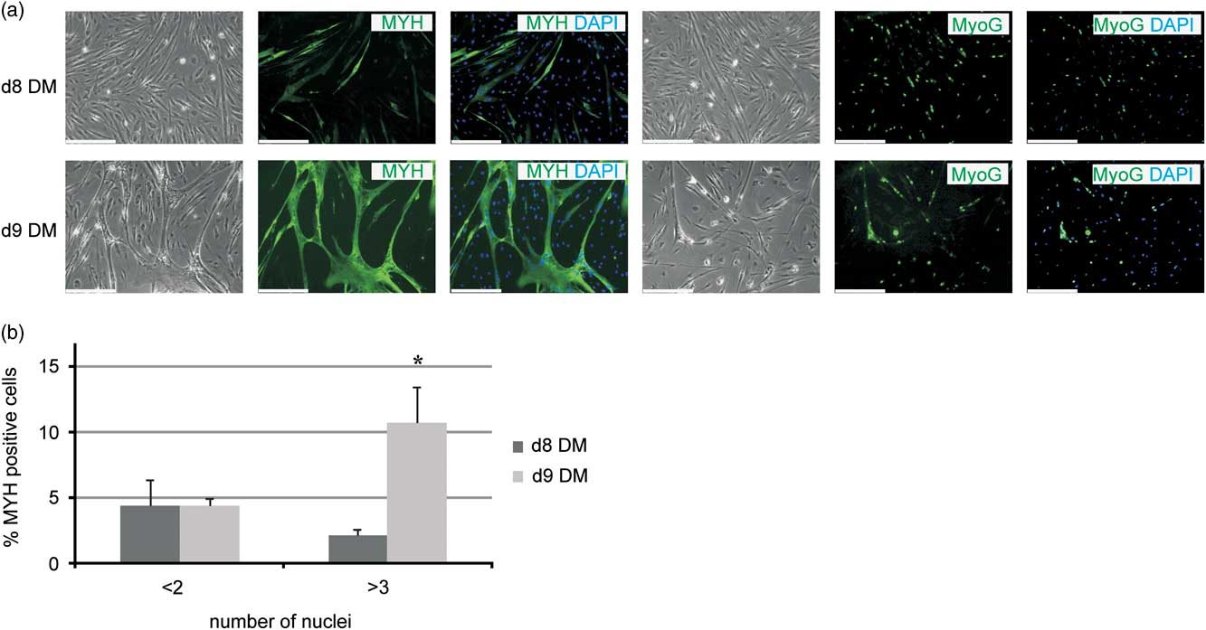
In the present work, MYH and MYOG were used as indicators of late muscle differentiation. Undifferentiated cells cultured in GM did not express MYH, although occasional MYH-positive cells were seen in growing cultures, possibly resulting from cell confluence associated with contact-dependent cell differentiation. To minimize the occurrence of MYH expression, cells were carefully cultured such that confluence was not obtained and cell differentiation not induced. All skeletal muscle samples differentiated into MHY-positive multinucleated myotubes within 2 days after their transfer to DM (Figure 6a). The presence of MYH in cell cultures was assessed by immunohistochemistry during proliferation and differentiation using anti-MYH (clone MF20), a monoclonal antibody that recognizes all skeletal myosin heavy chain isoforms (Figure 6a). No MYH expression was observed in cells cultured in GM (days 0 to 6) or on day 7 (1st day in DM) (data not shown). On day 8 (DM), about 5% of mononucleated cells showed a strong reaction with anti-MYH. On day 9 (DM), the number of mononucleated cells showing positive staining for MYH decreased (2.1%) (P<0.05), whereas the number and size of myotubes increased (Figure 6b). The percentage of cells positive for MYH remained stable during day 10 (DM) (data not shown). The combined staining of nuclei with DAPI and anti-MYH was used to measure the fusion index (Figure 6b). This index reached a mean of 11% after 3 days of differentiation.
Immunostaining analysis of myosin heavy chain (MYH) and myogenin (MYOG) expression in differentiating bovine satellite cells (BoSCs). (a) Satellite cells after 2 and 3 days of differentiation (days 8 and 9, respectively). Fixed cells were immunostained using antibodies against MYH and MYOG. Cell nuclei are stained in blue by 4′,6-diamidino-2-phenylindole (DAPI). (b) Percentage of MYH-positive cells in BoSC cultures following 2 and 3 days of differentiation (days 8 and 9, respectively). Changes in the fusion index during satellite cell differentiation at days 8 and 9 (differentiation medium (DM)) were expressed as the number of total nuclei located in myotubes (at least three nuclei) stained in green. Results are means±SD. *P<0.05 compared with expression values at day 8 (DM). Bars=250 μm.

MYOG is a protein necessary for the formation of new myofibrils. Here, MYOG expression was detected by immunofluorescence technique in cells following 2 days of cultivation in DM and was thereafter found to increase linearly until the end of the experiment. It was noted that the organization of nuclei within the myotubes tended to be arranged as single or small groups in linear arrays. Representative immunocytochemical images of myoblasts and myotubes are shown in Figure 6.
Discussion
The objective of this study was to describe and define the temporal changes in the expression of mRNAs and miRNAs closely associated with bovine skeletal muscle tissue during the proliferation and differentiation of myogenic progenitor cells in vitro. The majority of satellite cells, defined as myogenic stem cells, express PAX7, whereas the progenitor subpopulation expresses both PAX7 and myogenic regulatory factors, such as MYOD or MYF5 (Kuang et al., Reference Kuang, Charge, Seale, Huh and Rudnicki2006). In our study, satellite cells are shown to express PAX7 mRNA and protein on the day of isolation. After 3 days cultivation in the GM condition, when cell adhesion is complete, PAX7 mRNA levels start to decrease, reaching levels 10-fold lower at 6 day post isolation compared with day 0. Curiously, during the 1st day of differentiation, myoblasts showed an opposite trend in PAX7 mRNA expression: levels increased, although they did not return to those detected on the isolation day. Although, most BoSCs isolated from male Holstein adult animals are quiescent, satellite cell progeny proliferate extensively. The number of cells obtained from adult muscle samples is relatively low compared with satellite cells achievable from neonatal animals and the isolation process itself is known to trigger activation (Collins et al., Reference Collins, Olsen, Zammit, Heslop, Petrie, Partridge and Morgan2005). Therefore, we can hypothesize that immediately after cell isolation, myogenic stem cells change their status to myoblasts expressing PAX7 and MYOD, although a fraction of activated satellite cells do not undergo myogenic differentiation and self-renew in order to restore the pool of quiescent satellite cells. In addition, Ki67 staining was able to show the percentage of activated myoblast cells when cultured under proliferative conditions (26%). This percentage of activation seems to persist during the 1st day in DM until day 8. At this time point, we observed a down-regulation of PAX7 and MYOD mRNA, but Ki67 positivity (27%) remained unchanged compared with BoSCs at day 6 (GM; 26%).
The transcript for the endogenous MYF5 gene was present, but levels were not found to change during satellite cell activation and differentiation. This result is probably due to the low transcriptional activity in quiescent satellite cells, making a quantitative comparison complicated, but it is consistent with the results of other studies performed on mouse adult satellite cells (Beauchamp et al., Reference Beauchamp, Heslop, Yu, Tajbakhsh, Kelly, Wernig, Buckingham, Partridge and Zammit2000; Tajbakhsh and Buckingham, Reference Tajbakhsh and Buckingham2000; Crist et al., Reference Crist, Montarras and Buckingham2012). The proliferative ability and temporal expression of mRNA by satellite cells seems to be dependent on the host and the muscle from which they are collected (Powell et al., Reference Powell, McFarland, Cowieson, Muir and Velleman2014). Additional levels of heterogeneity are known to exist between satellite cells populations, as well as different extracellular matrix protein compositions, growth kinetics, myoblast fusion capacities, transcription factor profiles, motility kinetics and migratory properties (Biressi and Rando, Reference Biressi and Rando2010; Boldrin et al., Reference Boldrin, Muntoni and Morgan2010; Li et al., Reference Li, Gonzalez, Walker, Hersom, Ealy and Johnson2011; Li and Johnson, Reference Li and Johnson2013). There are increasing evidence through gene expression profiling and cell-surface marker analysis that satellite cells are a heterogeneous population (Motohashi and Asakura, Reference Motohashi and Asakura2014). It remains possible that satellite cells themselves undergo self-renewal or a minor population myogenic precursor cells de-differentiate and returns to quiescent satellite. In our work, we investigated gene expression related to a bulk population of satellite cells obtained from adult animals (18 to 23 months). Future study will be performed to explore about presence of different satellite cell populations in this bovine model.
Myomaker is a muscle-specific transmembrane protein shown to exhibit important roles in promoting myoblast fusion in mice, zebrafish and chicken (Millay et al., Reference Millay, O’Rourke, Sutherland, Bezprozvannaya, Shelton, Bassel-Duby and Olson2013; Landemaine et al., Reference Landemaine, Rescan and Gabillard2014; Luo et al., Reference Luo, Li, Nie and Zhang2015). However, its expression pattern and roles in bovine species have not been investigated. In this study, we reveal, for the first time, the gene expression pattern of myomaker during the process of myoblast differentiation in adult BoSCs. It is widely recognized that the mRNA expression pattern of a gene may correlate with gene function. Therefore, myomaker may also be fundamental during myotube formation in cattle, but its regulatory mechanisms remain to be determined.
Through the use of miRNAs microarrays, thousands of miRNAs have been identified in cattle and other farm animals. A large number of miRNAs are expressed in a spatiotemporal-specific manner, indicating that miRNAs play critical roles in specific tissue types or at specific developmental stages, as well as in regulation of muscle cell proliferation and differentiation (Muroya et al., Reference Muroya, Taniguchi, Shibata, Oe, Ojima, Nakajima and Chikuni2013; Wang et al., Reference Wang, Gu and Jiang2013; Wang et al., Reference Wang, Ding, Dai, Liu, Guo and Zhang2015). Interestingly, here we identify a correlation between the expression of differentiation factors, such as MEF2C, MYH1, MYOD, MRF4, TMEM8C and DES, and myomiRNAs during the cell differentiation progression. In previous studies, we have already focused on the expression of miRNAs in skeletal muscle samples of Piedmontese and Holstein cattle in order to outline a more precise genetic signature able to elucidate differences in muscle conformation (Miretti et al., Reference Miretti, Martignani, Taulli, Bersani, Accornero and Baratta2011). Our studies have revealed specific expression patterns of myomiRNAs in Piedmontese and Holstein cattle according to muscle phenotype and sex; furthermore, we have demonstrated that MSTN is a specific target of miRNA-27b in bovine species. These results reveal that miRNAs help explain additive phenotypic hypertrophy in Piedmontese cattle selected for the MSTN gene mutation (Miretti et al., Reference Miretti, Martignani, Accornero and Baratta2013). MyomiRNAs may also play important roles in bovine myogenic differentiation processes by regulating related target genes and signaling pathways. To understand the functions of miRNAs, it is necessary to identify their mRNA targets. Previous studies, on the C2C12 murine myogenic cell line, proved that MEF2 exerts its control over the programs for muscle development through direct and indirect mechanisms by coordinating the regulation of mRNAs and miRNAs (Braun and Gautel, Reference Braun and Gautel2011). Our results about MEF2C and myomiRNAs positive correlation seems to suggest that also in bovine specie the activity of MRFs and MEF2C can be regulated via miRNAs. Gagan et al. demonstrated that NOTCH3 normally inhibits differentiation through inhibition of MEF2C. NOTCH3 is targeted by miRNA-1 and miRNA-206. The subsequent inhibition of NOTCH3 by miR-1 and miR-206 is therefore critical for differentiation. Interestingly, NOTCH3 and MEF2C set up a mutually antagonistic network that is dependent on the actions of the miRNAs (Gagan et al., Reference Gagan, Dey, Layer, Yan and Dutta2012). Furthermore, Chen et al. (Reference Chen, Mandel, Thomson, Wu, Callis, Hammond, Conlon and Wang2006) reported that histone deacetylase 4 (HDAC4), a negative regulator of differentiation and a repressor of the MEF2 transcription factor, as the first described miRNA-targeted genes in the context of muscle development is repressed by miRNA-1, which in turn promotes myogenic differentiation and decreases cell proliferation. Recently, Dai et al. (Reference Dai, Wang, Zhang, Liu, Li, Ding and Guo2016) demonstrated that miRNA-1 and miRNA-206 promote bovine skeletal muscle satellite cell myogenic differentiation and limit their proliferation by down-regulating PAX7 and HDAC4 expression. Through the use of deep sequencing technology, it has also been found that miRNA-2400, a bovine-specific miRNA, is involved in the differentiation of satellite cells and directly targets the 3′ untranslated regions of MYOG mRNA (Zhang et al., Reference Zhang, Tong, Sun, Hu, Yang, Li, Yan and Li2015). At present, no other evidence exists in the literature on the regulatory relationships between bovine myomiRNAs and the other genes tested in our study. The present study is focused on providing a detailed description of a BoSC model by simultaneously analyzing myomiRNAs and skeletal muscle transcription factors during myogenic differentiation in vitro. We also identified a correlation between myomiRNAs and the expression of genes involved in the differentiation progression. This work will be expanded in the near future by elucidating exactly which miRNAs regulate the bovine myogenic differentiation process. These studies also hold the potential to provide knowledge about new mechanisms underlying muscle hypertrophy. Knowledge about the post-transcriptional changes correlating with the growth and differentiation of skeletal muscle fibers is of high importance for the improvement of meat production in livestock.
Acknowledgments
This work was financially supported by PRIN2010-2011 and University of Torino grant 2013. The authors thank Dr Daniele Nucera for helpful discussions regarding data analysis and Cristina Cecere for her technical assistance.
Conflicts of Interest
The authors declare there to be competing interests.
Supplementary Material
To view supplementary material for this article, please visit http://dx.doi.org/10.1017/S1751731116001488