INTRODUCTION
Morphological variation among individuals of the same species is the product of interactions between genetic differences and environmental influences. In gastropods, variation in shell morphology caused by extrinsic factors has been particularly well-studied (Vermeij, Reference Vermeij1987, Reference Vermeij1993). Phenotypic variation in snail shells results from the action of several biotic and abiotic factors. For instance, exposure to heavy surf, predation pressure from shell-crushing crabs, interspecific competition, and food abundance can all affect the morphometry and ornamentation of snail shells (e.g. Fenchel, Reference Fenchel1975; Reimchen, Reference Reimchen1982; Currey & Hughes, Reference Currey and Hughes1982; Trussell, Reference Trussell1996; Boulding et al., Reference Boulding, Holst and Pilon1999; Irie, Reference Irie2006; Preston & Roberts, Reference Preston and Roberts2007). The vast majority of studies to date have compared different populations of the same snail species, or samples from the same population but from different and spatially distinct habitats. Variation in shell morphology also occurs among conspecific individuals living side by side, without necessarily resulting from genetic differences. Recently, parasitism has been shown to alter shell growth (see below), and thus shell morphology, in gastropods; it may thus be an important source of phenotypic variation among snails on small spatial scales.
Trematode parasites use gastropods as their first intermediate hosts, in which they multiply asexually to produce large quantities of infective stages that leave the snail to penetrate the next host in the life cycle (Kearn, Reference Kearn1998). In the process, trematodes typically castrate the snail host by either destroying its gonads or preventing their development. An associated phenotypic effect of infection by trematodes is gigantism, i.e. infected snails often attain larger sizes than uninfected conspecifics (Gorbushin & Levakin, Reference Gorbushin and Levakin1999; Sorensen & Minchella, Reference Sorensen and Minchella2001; Poulin, Reference Poulin2007). This is often interpreted as a side-effect of castration, with the energy normally allocated to reproduction being channelled instead into growth, although an alternative interpretation is that gigantism is a host adaptation allowing the snail to outlive the infection with the possibility of achieving reproductive status thereafter (see Sousa, Reference Sousa1983; Mouritsen & Jensen, Reference Mouritsen and Jensen1994; Gorbushin & Levakin, Reference Gorbushin and Levakin1999; Sorensen & Minchella, Reference Sorensen and Minchella2001). Interestingly, it is not only the actual size of snails that is affected by trematode infection, but also the morphology of their shell. Recent studies have shown that trematode parasitism changes the morphometrics of the shell, i.e. the ratio of length to maximum width, as well as the degree of its ornamentation, such as the length of spines (McCarthy et al., Reference McCarthy, Fitzpatrick and Irwin2004; Levri et al., Reference Levri, Dillard and Martin2005; Hay et al., Reference Hay, Fredensborg and Poulin2005; Lagrue et al., Reference Lagrue, McEwan, Poulin and Keeney2007). Since the snail host is castrated, often permanently, it is difficult to reconcile these changes with potential fitness benefits for the host; instead, they may represent phenotypic manipulation by the parasites to increase the shell volume available for their own development (McCarthy et al., Reference McCarthy, Fitzpatrick and Irwin2004; Levri et al., Reference Levri, Dillard and Martin2005). The fact that different trematode species induce different morphological changes in the same snail species also suggest that these are parasite adaptations (Hay et al., Reference Hay, Fredensborg and Poulin2005; Levri et al., Reference Levri, Dillard and Martin2005); if altered morphology was a mere host response to castration, then we would expect this response to be the same for any castrating parasite species.
Trematode-induced changes in morphology are potentially very important, especially in snail populations where prevalence of infection is high. They can lead to an overestimation of the intrinsic variability within a population, and cause confusion in snail taxonomy. More importantly, snails with phenotypes altered by trematodes can play ecological roles that differ from those filled by uninfected snails in the same population. For instance, Miura et al. (Reference Miura, Kuris, Torchin, Hechinger and Chiba2006) found that a large proportion of individuals in the intertidal snail species Batillaria cumingi are infected by trematodes, and that infected snails not only display a different shell morphology and microhabitat preference, but also consume different resources compared to uninfected conspecifics.
Here, we investigate the effect of trematode parasitism on the morphology of the whelk Cominella glandiformis (Gastropoda: Buccinidae), a common predator and scavenger on New Zealand intertidal mudflats (Morton & Miller, Reference Morton and Miller1968). The only described trematode species parasitic in C. glandiformis is the echinostomatid Curtuteria australis (Allison, Reference Allison1979). However, recent surveys have revealed other trematode species. We compare two populations, one from a highly-sheltered inlet and the other from a less sheltered bay, to see how trematode infections and other environmental variables combine to affect shell shape. We also determine whether different trematode species induce different morphological changes in whelks, together acting to generate substantial variability in shell shapes within whelk populations.
MATERIALS AND METHODS
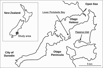
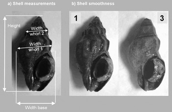
Approximately 1300 individuals of Cominella glandiformis were sampled at two localities on the Otago Peninusula, New Zealand: (1) Papanui inlet (highly sheltered); and (2) Lower Portobello Bay (less sheltered) (Figure 1). At both sites we randomly collected over 600 snails >13 mm from the sediment surface in the mid-intertidal. In the laboratory, we measured shell height, shell width at the base and shell width at whorl 1 and whorl 2 with callipers to the nearest 1/10 mm (Figure 2). In addition, we determined the smoothness of the shell on a scale from 1 (ridges prominent) to 3 (smooth shell without any prominent features) (Figure 2). After measurements were taken, the tissue of each snail was removed from the shell and dissected under a dissection microscope and the infection status recorded.
Sampling localities on the Otago Peninsula, Dunedin, New Zealand.

(A) Shell morphological measurements taken from each snail, and (B) examples for shells with a shell smoothness level of 1 (left) and 3 (right).

To estimate the contribution of parasites to the variation in shell morphology between and within the sites we used two-factorial ANOVA designs with site and infection status as fixed factors and shell height and the other three shell morphology metrics as dependent variables. Site was considered to be a fixed factor in all statistical designs as it was not randomly chosen. Instead we chose the two sites to be representative for the local snail distribution and parasite infection levels. To account for the height-dependency of the three shell width metrics we calculated residuals from linear regression analyses of the three metrics against shell height. Regressions over the total sample size were used for the two-factorial analyses (site versus infection status) while for comparisons of infected and uninfected snails within sites site-specific regressions were used. The resulting residuals were used as dependent variables in the ANOVA analyses. To account for the unbalanced design due to the different numbers of replicates among the groups we used un-weighted type III SS in all analyses (Shaw & Mitchell-Olds, Reference Shaw and Mitchell-Olds1993). Visual inspections of residuals plotted against predicted values were used to verify acceptable normal distributions and homogeneity of variances. To calculate the percentage of variance explained by the main factors in the different models we used SS of the factors only (SSTotal without residuals−SSfactor*100) as the large number of replicates naturally resulted in extremely high SS of the residual terms in the models. As we were only interested in the magnitude of the contribution of the main factors we concentrated on these only. Differences in the smoothness of the shells between sites and between infected and uninfected individuals within a site were tested with G-tests.
RESULTS
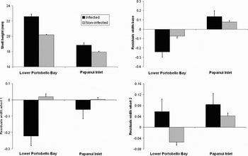
From the 631 snails dissected from Lower Portobello, 58 were infected with parasites (9.1%). The majority of snails was infected with Curtuteria australis (52), followed by undescribed strigid (5) and opecoelid (1) species. At Papanui Inlet, 47 snails out of the 639 snails dissected were infected (7.4%). Again, the majority of snails was infected with Curtuteria australis (33), followed by undescribed microphallid (10), opecoelid (3) and strigid (1) species. Snails from the two different sites differed in most shell morphological metrics. In general, snails from Lower Portobello Bay were longer and thinner (Table 1; Figure 3) and their shells were smoother than the ones from Papanui Inlet (G-test: G = 145.9, P < 0.001).
Effect of site (Lower Portobello Bay and Papanui Inlet) and infection status (Curtuteria australis infected and non-infected) on shell height and shell width at base, whorl 1 and whorl 2. For the latter three measures we used size-corrected data obtained from regressions of raw values against shell height. N = 52 infected and 579 non-infected snails at Lower Portobello Bay, N = 33 infected and 592 non-infected snails at Papanui Inlet.

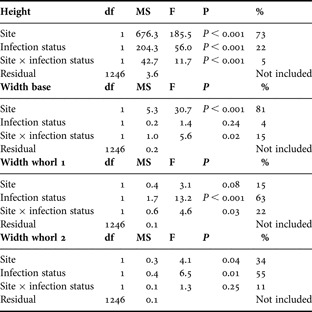
Results of two-factorial ANOVA analyses with site (Lower Portobello Bay and Papanui Inlet) and infection status (infected with Curtuteria australis and non-infected) as fixed factors and shell height or residuals from regressions of shell width at base, whorl 1 and whorl 2 versus shell height as dependent variables. %Variance explained was calculated as SSTotal without residuals−SSfactor*100. For N see text.

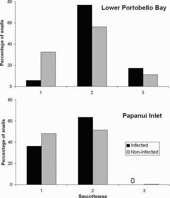
Parasites contributed strongly to differences in all four shell morphological metrics both between and within the two sites (Table 1; Figure 3). In general, snails infected with Curtuteria australis were narrower at their base and whorl 1 but wider at whorl 2 compared to non-infected snails. Due to a significant anti-directional interaction of the two factors, there was no significant effect of infection status on shell width at the base (Table 1; Figure 3). However, when analysed separately within each site infected snails were significantly narrower than non-infected snails at Lower Portobello Bay (F1,623 = 5.9, P = 0.01) while there was no difference in shell width at the base between infected and non-infected snails from Papanui Inlet (F1,623 = 0.9, P = 0.34). Infection status of the snails explained between 20 and 60% of the variance of the main factors in the ANOVA models (Table 1). In general, the magnitude of variation caused by parasites was often larger than the one associated with the site of origin of the snails (Figure 3), also indicated by the larger variance explained in some of the models (Table 1). Parasitism was also related to the smoothness of the snail shells but with different strength at the two sites (Figure 4). While there was no detectable effect of infection status on shell smoothness at Papanui Inlet (G = 2.0, P = 0.36), shell smoothness of infected and non-infected snails was significantly different at Lower Portobello Bay with infected snails tending to be smoother than uninfected snails (G = 20.9, P < 0.001).
Percentage of snails with different smoothness levels of the shell (1–3) depending on infection status (Curtuteria australis infected and non-infected) at the two different sites, Lower Portobello Bay and Papanui Inlet. For N see Figure 3.

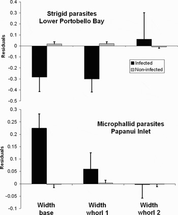
Only two other parasite species occurred in sufficient numbers for further analysis. Infections with the strigid species at Lower Portobello Bay showed a similar pattern with the shell being generally higher (20.4±0.5 mm in infected and 20.2±0.1 mm in uninfected snails), slimmer at the base and wider at the top when infected (Figure 5). However, these differences were not significant (height: F = 0.07, P = 0.79; residuals width base: F = 1.0, P = 0.17; residuals width whorl 1: F = 2.8, P = 0.06; residuals width whorl 2: F = 0.33, P = 0.57). Snails infected with a microphallid species at Papanui Inlet had a higher shell (18.8±0.5 mm in infected and 17.9±0.1 mm in uninfected snails) but were generally wider at the base and whorl 1 (Figure 5). However, only the difference in the width at the base was significant (height: F = 1.9, P = 0.16; residuals width base: F = 5.0, P = 0.03; residuals width whorl 1: F = 0.39, P = 0.5; residuals width whorl 2: F = 0.005, P = 0.98). Shell smoothness did not differ between infected and uninfected snails in strigid infections at Lower Portobello Bay (G-test; G = 3.9, P = 0.14) and microphallid infections at Papanui Inlet (G-test; G = 3.47, P = 0.18).
Effect of different parasite species at Lower Portobello Bay and Papanui Inlet on shell width at base, whorl 1 and whorl 2. Data are size-corrected residuals obtained from regressions of raw values against shell height at the different localities. N = 5 for strigid infections at Lower Portobello Bay and N = 10 for microphallid infections at Papanui Inlet.

DISCUSSION
Our results show that parasitism can be involved in intra- as well as inter-site variation of gastropod shell morphology in terms of size, shape and ornamentation. Snails infected with Curtuteria australis generally had a higher shell and were wider at whorl 2 but narrower at the base and whorl 1 compared to their non-infected conspecifics. In addition, infected snails were also smoother than their non-infected conspecifics. That infected snails attain larger sizes than uninfected conspecifics is a well known phenomenon (gigantism: see Sousa, Reference Sousa1983; Mouritsen & Jensen, Reference Mouritsen and Jensen1994; Gorbushin & Levakin, Reference Gorbushin and Levakin1999; Sorensen & Minchella, Reference Sorensen and Minchella2001). Alterations of shell shape induced by parasites have also been observed in other gastropods, mostly with infected snails having an elongated and narrower shell compared to non-infected conspecifics (McCarthy et al., Reference McCarthy, Fitzpatrick and Irwin2004; Lagrue et al., Reference Lagrue, McEwan, Poulin and Keeney2007). This shell shape alteration results in a larger space available for the trematodes that utilize the snails' gonads which are located in the upper part of the shells (McCarthy et al., Reference McCarthy, Fitzpatrick and Irwin2004; Lagrue et al., Reference Lagrue, McEwan, Poulin and Keeney2007). This could be a fortuitous side-effect of infection but it may also be an adaptation by the parasites to increase the volume of available shell space. Similarly, the observed tendency for shells of infected snails to be smoother than those of their non-infected conspecifics might be a side-effect of infection, also observed in another snail species (Levri et al., Reference Levri, Dillard and Martin2005; Lagrue et al., Reference Lagrue, McEwan, Poulin and Keeney2007), or it might be the result of an adaptive strategy of the parasites if a smoother shell increases their transmission by enhanced predation on the hosts. However, in the case of Curtuteria australis this is unlikely as the down-stream hosts are bivalves and transmission occurs via free-living cercarial stages and not via predation on, for example, encysted metacercariae. Finally, smooth shells of infected snails may not have anything to do with parasites at all as they may result from the fact that older snails are often more likely to be infected and also have the smoothest shells due to wear and tear. As age determination is impossible in our snails, snail age may be a confounding factor regarding shell smoothness. In contrast, the observed differences in shell shape should be independent of age, especially as we corrected for size (correlated with age) in the analyses. However, it may be that narrower and smoother snails are more likely to become infected with C. australis in the first place, or that wider snails with prominent ridges are more resistant to parasite infections. These two alternatives cannot be ruled out in our investigation due to the purely correlative approach. However, other studies have found evidence for the actual involvement of parasites in shell variation (Krist, Reference Krist2000; McCarthy et al., Reference McCarthy, Fitzpatrick and Irwin2004) and there is no reason why this mechanism should be different in our study system.
Parasites were not only correlated with intra-site variation in the shell morphology of the snails but they also explained a large share of the inter-site variation in shell morphology between the two sites, with infection status accounting for 20 to over 60% of the variance of the main factors in the ANOVA models. The magnitude of the effect associated with parasites was generally more pronounced than those caused by site alone (Figure 3), suggesting that parasites are dominant drivers of inter-site variation of shell morphologies, at least in this study. However, the various interaction terms in the model indicate that the effect of parasites was not independent of site with generally less pronounced effects of infection status within Papanui Inlet (Figure 3). Hence, the factor site also contributed to the observed inter-site variation in shell morphology and several environmental variables may differ between the sites, accounting for the observed differences. Both sites differ in their exposure and as gastropod shell size and shape is known to vary with exposure (Trussell et al., Reference Trussell, Johnson, Rudolph and Gilfillan1993; Boulding et al., Reference Boulding, Holst and Pilon1999), this alone may account for the differences in shell morphology observed between the two sites. However, other environmental factors are also known to affect gastropod shell morphology and may vary, along with exposure, between the two sites: presence of predators (Appleton & Palmer, Reference Appleton and Palmer1988; Trussell & Smith, Reference Trussell and Smith2000), substrate types and diet (Lindberg & Pearse, Reference Lindberg and Pearse1990) and food availability (Kemp & Bertness, Reference Kemp and Bertness1984; Martin-Mora et al., Reference Martin-Mora, James and Stoner1995). In addition to environmental factors, inter-site variations in shell morphologies may also have a genetic component (Palmer, Reference Palmer1985; Kyle & Boulding, Reference Kyle and Boulding1998). Hence, the factor site possibly integrates a complex interplay of various environmental and genetic factors. However, our data indicate that parasites can be one of the main drivers in this complex interplay.
Although the effects on shell morphology observed in snails infected with C. australis were noticeable and in line with previous findings, not all parasites infecting whelks may have the same effect. Infections with a strigid species at Lower Portobello Bay generally showed a similar trend, with shells of infected snails being higher and narrower at the bottom while wider at the top. However, these differences were not statistically different, probably due to the low sample size of just five infected snails. The shell of snails from Papanui Inlet infected with a microphallid species were again higher than the ones from non-infected conspecifics but, contrary to our findings above, the shells of infected snails were wider at the base and at whorl 1, although only differences in width at the base were significant. Differential effects of parasites on shell morphologies of their gastropod hosts have been observed in other parasite–host systems (Hay et al., Reference Hay, Fredensborg and Poulin2005) and host snails have been observed to become wider at the shell base in a few cases (Levri et al., Reference Levri, Dillard and Martin2005). Such differences among parasite species may result from differential side-effects or adaptation mechanisms related to the life histories of the parasites. However, whatever the actual direction of the effect, parasites are responsible for intra-site variation in shell morphologies in all these cases even if the direction of the alterations in shell morphology differs among parasite species.
In conclusion, parasites may be an important factor in explaining intra- and inter-site variation in shell morphologies of gastropods. The effects we observed in our study were even detectable at low infection levels (<10% of the local populations being infected). It is easy to imagine that in highly infected snail populations parasites may account for most of the observed variation in shell morphology both within and among sites. Studies concerned with intra- and inter-site variation of gastropod shell morphology should take this into account.
ACKNOWLEDGEMENT
D.W.T. acknowledges support by a fellowship from the German Research Foundation (DFG) (Th 1361/1-1).