Introduction
Despite rapid technological advancements in cancer diagnosis and therapeutics, cancer still remains as a major global health threat (Ref. Reference Dagenais1), with approximately 19.3 million new cancer cases and almost 10.0 million cancer-related death events reported worldwide in 2020 (Ref. Reference Sung2). Transforming growth factor-β (TGF-β) plays important and differential roles in cancer initiation and development (Ref. Reference Derynck, Turley and Akhurst3) besides its known regulatory functionalities in cell growth, differentiation, death and immune response, among others, under physiological conditions (Refs Reference McPherron, Lawler and Lee4, Reference Viel5). For example, TGF-β suppresses the growth of normal and premalignant epithelial cells in the early stages of cancer development (Ref. Reference Gal6). On the contrary, the TGF-β/mothers against decapentaplegic (SMAD) pathway induces epithelial to mesenchymal transition (EMT) to promote tumour progression in the late stage of cancers (Ref. Reference Moon7). Therapeutics involving TGF-β, such as TGF-β monoclonal antibodies (mAb), antisense oligonucleotides (ASOs), ligand traps and small-molecule inhibitors of TβR kinase, have shown great promise in preclinical and clinical studies (Ref. Reference Teicher8). For example, combining galunisertib (LY21557299, Eli Lilly and Company), a TGF-βRI kinase inhibitor, with gemcitabine (a chemotherapy) improves the overall survival of stages II–IV unresectable pancreatic cancer patients from 7.2 to 10.9 months as compared with those treated with gemcitabine and placebo in a clinical trial (NCT01373164, phase II, completed, Eli Lilly and Company) (Ref. Reference Melisi9). Despite successful stories relying on TGF-β for onco-therapeutics, failed cases have been consecutively reported. For example, the maximum dose tolerance of LY3022859, an anti-TGF-βRII mAb, was not determined in a trial composed of 14 advanced solid tumour patients (NCT01646203, phase I, completed, Eli Lilly and Company) because of negative symptoms observed such as uncontrolled cytokine release (Ref. Reference Tolcher10). This failure is likely related to a possible off-target effect of LY3022859, as its target TGF-βRII is ubiquitously expressed including the immune cells (Ref. Reference Tolcher10). Therefore, delineating the relationship between TGF-β and cancer is of critical importance to our understandings on the functionalities of TGF-β under both physiological and pathological conditions for improved onco-therapeutic design.
After briefly introducing some fundamentals of TGF-β including its structure and signalling, this paper focuses on the relationship of TGF-β with cancer hallmarks, as well as canonical and emerging therapeutic strategies utilising TGF-β. We conclude with our insights on how we portrait the roles of TGF-β in cancer and future research directions taking advantages of TGF-β for cancer management.
Fundamentals of TGF-β
Growth factors are major growth-stimulatory molecules, the loss or decreased requirement of which is a common feature of transformed cells and may lead to a growth advantage. Cytokines are a subtype of growth factors produced by haematopoietic and immune cells. They are major regulators of innate and adaptive immunity that enable communications among immune cells over short distances. Cytokine has been used to activate the immune system of cancer patients as an important onco-therapeutic modality such as the approval on the use of interferon alpha (IFN-α) in treating completely resected high-risk melanoma and several refractory malignancies (Ref. Reference Tarhini11). However, cytokines are often associated with severe toxicities capable of, among others, triggering the secretion of immune inhibitory factors such as TGF-β (Ref. Reference Sckisel12). TGF-β belongs to a superfamily of cytokines (namely the TGF-β superfamily) including activins, inhibins, bone morphogenetic proteins, growth differentiation factors and glial cell line neurotrophic factors. These molecules play deterministic roles in the landscape of the organisational development structure of metazoan including, among others, embryonic patterning, cell fate determination and dynamic movements (Refs Reference Lee, Choi and Joo13, Reference Sorre14).
TGF-β structure, biosynthesis and activation
TGF-β is a 25-kD dimeric protein with three isoforms (TGF-β1, TGF-β2 and TGF-β3) in humans (Ref. Reference Lyon, Rushton and Gallagher15). TGF-βs are synthesised as precursor molecules containing large amino-terminal prodomains that are necessary for proper folding (Ref. Reference Shi16). The TGF-β1 homodimer interacts with a latency-associated peptide, forming an inactive form that is secreted into the extracellular matrix (ECM) (Ref. Reference Hyytiainen, Penttinen and Keski-Oja17). Then the complex binds to the latent TGF-β-binding protein localised within ECM, or leucine-rich repeat proteins (or related molecules) localised on cell surface (Ref. Reference Derynck, Turley and Akhurst3). Latent TGF-β1 is cleaved into the N-terminal latency-associated peptides and C-terminal mature peptides by certain proteases to generate active TGF-β (Ref. Reference Khalil18). Although the biosynthesis of TGF-β1 homodimers has been actively investigated, relatively little is reported on the activation of TGF-β2/β3 homodimers or TGF-β heterodimers (Ref. Reference Robertson and Rifkin19).
The receptor of TGF-β has three forms, that is, TGF-βRI, TGF-βRII and TGF-βRIII. TGF-β binds to a hetero-tetramer of TGF-βRI and TGF-βRII that contains serine/threonine protein kinases in the cytoplasmic domain (Ref. Reference Wrana20). TGF-βRIII has no kinase activity but functions as a co-receptor to increase the binding affinity of TGF-β to TGF-βRII (Ref. Reference Rotzer21).
TGF-β signalling
Canonical TGF-β/SMAD signalling
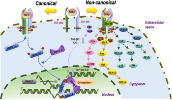
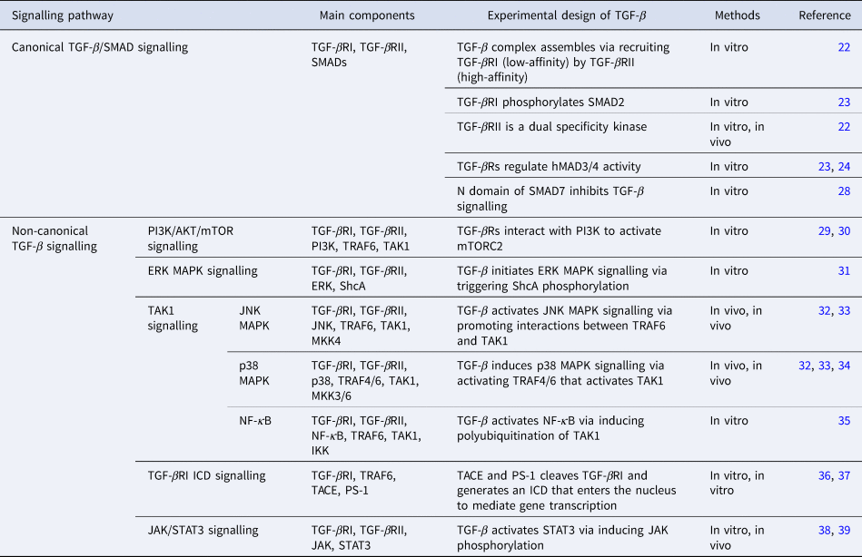
Active TGF-β binds to TGF-βRI and TGF-βRII complex followed by TGF-βRI phosphorylation on specific serine and threonine residues (Ref. Reference Groppe22). Then, the receptor complex activates receptor regulated-SMADs (R-SMADs) through C-terminal serine residue phosphorylation (Refs Reference Zhang23, Reference Piek24). Phosphorylated R-SMADs (SMAD2, SMAD3) recruit SMAD4 that is translocated into cell nucleus and binds transcriptional complexes to regulate the transcription of target genes (Ref. Reference Matsuzaki25). After that, the SMAD2/3/4 complex is moved out of the nucleus by activated TGF-β receptors for cyclical use or degraded through ubiquitination (Refs Reference Xiao26, Reference Wang27). Alternatively, the activity of TGF-β signalling can be tempered by the inhibitory activity of SMAD7. Specifically, SMAD7 can block TGF-β-mediated transcriptional events via binding to DNA in the cell nucleus, form a complex with E3- ubiquitin ligases and translocate from cell nucleus to the plasma membrane for TGF-β receptor ubiquitination, or inhibit TGF-β signalling via blocking interactions between R-SMADs and the TGF-β receptor complex (Ref. Reference Hanyu28) (Fig. 1, Table 1).
Canonical and non-canonical TGF-β signalling pathways. TGF-β signalling is composed of both canonical and non-canonical pathways. The canonical pathway is SMAD-dependent, where active TGF-β binds heterotetrameric complexes of TGF-β type II (TGF-βRII) and type I (TGF-βRI) receptors on cell surface (with TGF-βRIII being the co-regulatory molecule) followed by activation of Smad2/3 and recruiting of Smad4 for cell nucleus translocation and target gene regulation. The non-canonical pathway is SMAD-independent, where TGF-β activates MAPKs, NF-κB, Ras, TRAF6, TAK1-p38/JNK and PI3K kinases, leading to various biological effects. Although TGF-β-induced PI3K/AKT/mTOR and MAPK-ERK signals are initiated from TGF-β receptor homodimers, TGF-β-induced MAPK-JNK and MAPK-p38 signallings are initiated from TGF-β receptor heterodimers. AKT, protein kinase; ERK, extracellular signal-regulated kinase; IKK-2, IκB kinase 2; ICD, intracellular domain; JAK, Janus kinase; JNK, c-JUN N-terminal kinase; MAPK, mitogen-activated protein kinase; MKK, MAP kinase kinases; NF-κB, nuclear factor kappa-light-chain-enhancer of activated B cells; PI3K, phosphatidylinositol 3-kinase; PS-1, membrane-embedded protease presenilin-1; STAT, signal transducers and activators of transcription; TAK1, TGF-β-activated kinase 1; TRAF6, tumour necrosis factor receptor-associated factor 6; TACE, TNF-α converting enzyme; TGF-β, transforming growth factor-β.

Canonical and non-canonical TGF-β signallings

Non-canonical TGF-β signalling
Although SMADs play a vital role in TGF-β signalling, TGF-β also transduces signals independent of SMADs that involves factors such as phosphoinositide 3-kinase/protein kinase (PI3K)/AKT/mTOR, extracellular signal-regulated kinase (ERK), mitogen-activated protein kinase (MAPK), TGF-β-activated kinase 1 (TAK1) that is an upstream factor of p38 MAPK, c-JUN N-terminal kinase (JNK) MAPK and nuclear factor kappa-light-chain-enhancer of activated B cells (NF-κB) signallings, Janus kinase (JAK)/signal transducers and activators of transcription 3 (STAT3) signalling, TGF-βRI intracellular domain signalling. TGF-βRs can trigger PI3K/AKT signalling via directly interacting with PI3K (Ref. Reference Yi, Shin and Arteaga29) that activates mammalian target of rapamycin complex 2 (mTORC2) (Ref. Reference Lamouille30). TGF-βRI can activate the Ras GTPase by phosphorylating the adaptor protein ShcA that leads to the sequential activation of c-Raf, MAP kinase/ERK kinase (MEK) and ERK1/2 kinases (Ref. Reference Lee31). Upon ligand binding, TGF-βRs activate TAK1 via interacting with the tumour necrosis factor receptor-associated factor 6 (TRAF6), an E3 ubiquitin ligase of TAK1 (Ref. Reference Yamashita32); activated TAK1 can initiate p38 MAPK and JNK MAPK signallings via activating MAP kinase kinases (MKKs), where MKK3 and MKK6 are upstream regulators of p38 and MKK4 activates JNK (Ref. Reference Lin33). P38 signalling can also be triggered by TRAF4-mediated TAK1 polyubiquitylation (Ref. Reference Zhang34). Besides, TRAF6-mediated TAK1 polyubiquitylation can also activate NF-κB signalling via activating IκB kinase 2 (IKK-2) (Ref. Reference Hamidi35). Moreover, TGF-β also promotes JAK1/2 phosphorylation that activates STAT3 signalling (Refs Reference Dees38, Reference Tang39). Interestingly, TNF-α converting enzyme (TACE) and membrane-embedded protease presenilin-1 (PS-1) can cleave TGF-βRI and generate an intracellular domain (ICD) that enters the nucleus to mediate gene transcription (Refs Reference Liu36, Reference Gudey37) (Fig. 1, Table 1).
TGF-β and cancer hallmarks
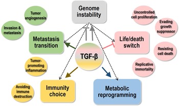
The hallmarks of cancers, firstly proposed in 2000 (Ref. Reference Hanahan and Weinberg40), improved in 2011 (Ref. Reference Hanahan and Weinberg41) and updated in 2022 (Ref. Reference Hanahan42) characterise cancers into 10 properties. Among these hallmarks, ‘uncontrolled cell proliferation’, ‘evading growth suppressor’, ‘resisting cell death’, ‘replicative immortality’ represent the ‘life/death switch’ of cancer cells, ‘inducing tumour angiogenesis’ and ‘activating invasion & metastasis’ reflect the decision of cancer cells on ‘metastasis transition’, ‘tumour-promoting inflammation’ and ‘avoiding immune destruction’ are associated with the ‘immunity choice’ of cancer cells, ‘deregulated cellular energetics’ determines cancer cell ‘metabolic reprogramming’, and ‘genome instability & mutation’ enables the other hallmarks of cancers (Fig. 2).
Involvement of TGF-β signalling in the manifestation of cancer hallmarks. The 10 cancer hallmarks could be categorised into five critical transition events, that is, life/death switch, metastasis transition, metabolic reprogramming, immunity choice and genome instability, where genome instability is the enabling hallmark of the other four traits. In general, TGF-β suppresses tumour proliferation, promotes metastasis, aids in metabolic reprogramming, facilitates cancer immunity evasion and maintains genome integrity under the normal conditions but loses this tumour-suppressive function after the initiation of carcinogenesis.

TGF-β and cancer cell life/death switch
TGF-β in cell proliferation
TGF-β suppresses the growth of normal and premalignant epithelial cells (Ref. Reference Nagaraj and Datta43) (Fig. 2). TGF-β inhibits epithelial cell proliferation by causing cell cycle arrest at the G1 phase that involves three transcriptional events, that is, inducing cyclin-dependent kinase (CDK) inhibitors (Ref. Reference Zhang, Alexander and Wang44), suppressing c-Myc expression (Refs Reference Chen, Kang and Massague45, Reference Seoane46, Reference Seoane, Le and Massague47) and attenuating inhibitor of DNA-binding 1 (ID1), ID2 and ID3 (Refs Reference Je48, Reference Selesniemi, Albers and Brown49). In the cell cycle, the G1/S transition requires the activation of CDKs including CDK2, CDK4 and CDK6, where CDK2 binds cyclin E and CDK4/6 pairs with cyclin D to phosphorylate their substrates (Ref. Reference Zhang, Alexander and Wang44). There are several CDK inhibitors such as P16INK4A, P15INK4B, P21CIP1 and P27KIP1, which control cell cycle progression by pairing with CDK/cyclin complexes (Refs Reference Karimian, Ahmadi and Yousefi50, Reference Abbastabar51, Reference Zhang52). TGF-β downregulates the expression of c-Myc, a known oncogene that enables uncontrolled cell proliferation (Ref. Reference Chen, Kang and Massague45), with the aid of transcription factors P107, E2F4/E2F5 and C/EBP (Refs Reference Chen53, Reference Frederick54). Interestingly, c-Myc inhibition not only directly affects cell cycle progression, but also promotes the expression of several important CDK inhibitors in response to the same TGF-β signal (Refs Reference Seoane46, Reference Seoane, Le and Massague47).
The functionalities of TGF-β on cancer cells change as tumour microenvironment (TME) undergoes drastic changes during cancer progression (Ref. Reference Nagaraj and Datta43). Most cancer cells become insensitive to TGF-β-mediated growth inhibition, which attributes to the loss of function of TGF-βRI/II or SMAD2/4. For example, loss of 18q21 chromosome that harbours SMAD4 was reported in pancreatic and colorectal cancers (Ref. Reference Liu55). Genetic alterations were not observed in some cancers, implicative of other mechanisms for acquiring resistance to TGF-β-mediated growth inhibition (Ref. Reference Papageorgis56). Some tumour cells evade growth restriction by activating Myc/SMAD3 and PI3K/AKT pathways or suppressing p15/p21/WAF1/CIP1 signalling (Ref. Reference Zhou57). The P38/MAPK pathway also participates in the process of circumventing TGF-β-induced growth restrictions, with the specific molecular mechanism remaining to be explored (Ref. Reference Bakkebo58).
TGF-β in programmed cell death
TGF-β and apoptosis. TGF-β has been reported as an apoptosis inducer in most studies (Ref. Reference Zhang, Alexander and Wang44) (Fig. 2). In SMAD-dependent signallings, several apoptotic target genes are regulated by SMAD transcriptional complexes such as death-related protein kinases (DAPK), Src homology inositol phosphatase (SHIP) and TGF-β-induced early gene 1 (TIEG1) (Ref. Reference Yang59). For example, TGF-β upregulates the expression of DAPK towards elevated levels of caspase activity and apoptosis in hepatocytes (Ref. Reference Jang60), enhances SHIP transcription to trigger apoptosis through decreasing Akt phosphorylation in haematopoietic cells (Ref. Reference Valderrama-Carvajal61) and induces TIEG1 expression to promote apoptosis via suppressing Bcl2 in epithelial Mv1Lu cells (Ref. Reference Chalaux62) or inducing oxidative stress in an in vivo mouse model (Ref. Reference Kammoun63). In SMAD-independent pathways, TGF-β-mediated apoptosis involves many other genes, such as Bcl-2 family members, BCL-XL and X-linked inhibitor of apoptosis that are anti-apoptotic, and caspase 3/8 and Bcl-2-interacting killer that are pro-apoptotic (Refs Reference Francis64, Reference Huang65). TGF-β-induced reactive oxygen species (ROS) promote apoptosis via regulating various Bcl-2 family members such as Bcl-2 modifying factor and Bcl-2 interacting mediator in a series of mouse cancer cell lines including AML12 (liver cancer), NMuMG (breast cancer), CMT170 (pulmonary system adenocarcinoma) and A20 (lymphoma) (Ref. Reference Ramjaun66). TGF-β promotes apoptosis via inhibiting the expression of Krüppel-like factor 5 that cooperates with SOX4 during oncogenesis in pancreatic ducal adenocarcinoma cells (Ref. Reference David67).
The effect of TGF-β on tumour growth is double-edged, as it prevents apoptosis under certain conditions despite its functionality as an apoptosis inducer, with the specific mechanism remaining to be investigated (Refs Reference Ehata68, Reference Schlapbach69). In particular, TGF-β induces the expression of Dec1, an apoptosis inhibitor, in mouse mammary carcinoma cells (Ref. Reference Ehata68), and elevates c-FLIP level that prevents Fas-triggered activation of caspase-8 and caspase-3 for cell apoptosis evasion in microglia (Ref. Reference Schlapbach69).
TGF-β and other programmed cell death. Autophagy is an evolutionarily conserved pathway through which cytoplasmic substrates, such as damaged organelles and cytoplasmic proteins, are delivered to lysosomes for degradation (Ref. Reference Wu, Won and Rubinsztein70). It has been reported that TGF-β can activate autophagy in hepatocellular carcinoma and mammary carcinoma cells (Ref. Reference Kiyono71), increase the expression of autophagy-related genes such as Beclin-1, Atg5, Atg7 and DAPK in hepatoma cells (Ref. Reference Suzuki, Kiyono and Miyazono72), trigger autophagosome accumulation and convert microtubule-associated protein 1 light chain 3 (LC3) to its lipidated form, LC3-II (participating in autophagosome formation), in HuH7 human hepatocellular carcinoma cells (Ref. Reference Suzuki, Kiyono and Miyazono72). In addition, silencing SMAD2/3/4 or DAPK, or inhibiting JNK all suppress TGF-β-induced autophagy (Ref. Reference Suzuki, Kiyono and Miyazono72), suggestive of the involvement of both SMAD-dependent and SMAD-independent TGF-β pathways.
Ferroptosis is an iron-dependent form of regulated cell death driven by excessive lipid peroxidation (Ref. Reference Chen73), and TGF-β1 represses SLC7A11 (a marker inhibiting ferroptosis) expression via activating SMAD3 and enhancing lipid peroxidation in hepatocellular carcinoma cells (Ref. Reference Kim74).
Pyroptosis is an inflammatory form of programmed cell death that is critical for amplifying protective immunity during infection (Ref. Reference Jorgensen and Miao75), the impact of which on tumours varies with the type and stage of transformed cells (Ref. Reference Xia76). The roles of TGF-β1 in pyroptosis vary with the cancer type and developmental stage. For instance, TGF-β1 induces pyroptosis in colorectal cancers (Ref. Reference Jiang77) but inhibits pyroptosis in triple negative breast tumours (Ref. Reference Tamura78).
Studies on TGF-β and non-apoptotic cell death are still in the nascent stage. With our incremental knowledge on the roles of TGF-β in controlling the life/death switch of cancer cells, and growing number of novel forms of programmed cell death events being discovered, we foresee increased attention in this field towards improved harness over TGF-β signalling and cancer control.
TGF-β and genome instability
Enhanced genome instability has been recognised as an enabling hallmark of cancer (Ref. Reference Hanahan and Weinberg41). TGF-β is a tumour suppressor that contributes to the maintenance of genome stability under normal conditions because of its regulatory functionalities on cell cycle (Fig. 2). Yet, its role in maintaining the genome integrity gets lost once carcinogenesis initiates (Ref. Reference Elston and Inman79) (Fig. 2).
Renowned as the guardian of the genome, the central role of p53 in suppressing tumour initiation and development is well known. It develops crosstalk with TGF-β by sharing the use of SMADs for signalling, with SMAD2/3 being the bridge that allows for the synergistic activation of TGF-β and p53 (Refs Reference Cordenonsi80, Reference Dupont81, Reference Cordenonsi82). For example, TGF-β target genes such as p21, p15, PAI-1, matrix metalloproteinase 2 (MMP2) require p53 for activation (Refs Reference Cordenonsi80, Reference Cordenonsi82). On the other hand, p53 inactivation contributes to the loss of the tumour suppressive role of TGF-β (Ref. Reference Atfi and Baron83) and gain-of-function for promoted carcinogenesis such as the trigger of TGF-β-induced metastasis by mutant p53 (Ref. Reference Adorno84). Mutations in p53 commonly occur in tumours. Specifically, p53 mutants affect numerous stages of the TGF-β pathway via, for example, repressing the gene that encodes TGF-βRII, delaying or reducing SMAD2 phosphorylation by TGF-βRI, and interfering with the interactions between SMAD2/3 and SMAD4 towards suppressed nucleus translocation (Ref. Reference Kalo85).
TGF-β and cancer metastasis transition
TGF-β in tumour angiogenesis
Tumour growth and metastasis require a rich blood supply to provide nutrients and oxygen, leading to angiogenesis once the tumour size reaches more than 1–2 mm in diameter (Ref. Reference Carmeliet and Jain86). Tumour cells secrete growth factors such as TGF-β to induce tumour angiogenesis towards accelerated cancer development (Ref. Reference Roberts87) (Fig. 2). Endothelial cells with higher cell proliferation, progression and migration rate play a vital role in angiogenesis (Ref. Reference Tian and Schiemann88). TGF-β induces pro-angiogenic growth factors such as vascular endothelial growth factor (VEGF) and connective tissue growth factor to directly stimulate EC towards tumour angiogenesis and accelerated cancer progression (Refs Reference Shimo89, Reference Fang90). TGF-β can significantly increase the expression of VEGF and promote tumour angiogenesis in RAS-transformed epithelial tumours (Ref. Reference Rak91). TGF-β and VEGF form a feedback loop through the semaphorin 3A/Neuropilin-1-expressing monocytes (NEM) axis to avoid over-activated TGF-β signalling (Ref. Reference Groppa92). Moreover, TGF-β signalling can increase the expression of miR29a and angiogenic regulatory enzymes such as MMPs (MMP-2, MMP-9, MMP-10, etc.) that contribute to neovascularisation, or inhibit tissue inhibitors of metalloproteinases (Refs Reference Wang93, Reference Miyazono, Ehata and Koinuma94). Besides, TGF-β promotes tumour vasculature by elevating the pericyte–endothelium association, where tumour-associated fibroblasts enhance tumour blood capillaries through TGF-β-dependent signalling (Ref. Reference Zonneville95).
TGF-β in cancer invasion and metastasis
Most cancers can escape from their primary sites, evade into the bloodstream or lymphatic system and metastasise to the lymph nodes and distant organs such as lung, bones, brain and liver (Ref. Reference Hoshino96). Tumour metastasis, a progressive, multifactorial and multistep dynamic process, is the primary cause of cancer lethality (Ref. Reference Lambert, Pattabiraman and Weinberg97). EMT is the crux of early metastasis that is a multi-step process characterised by altered levels of three prominent biomarkers (E-cadherin, vimentin and N-cadherin), decreased cell adhesion, loss of polarity and tight junctions, as well as gained spindle-shaped and fibroblast-like morphology (Refs Reference Chiang and Massague98, Reference Tsai and Yang99).
The TGF-β/SMAD pathway induces EMT via regulating the expression of E-cadherin, N-cadherin, Snail and vimentin (Ref. Reference Lamouille, Xu and Derynck100) (Fig. 2). Knocking down SMAD4 significantly inhibits bone metastasis in a nude mice model of human breast cancers (Ref. Reference Deckers101). Concomitantly silencing SMAD2 and SMAD3 in mouse hepatocytes suggests the essential role of SMAD3 in EMT (Ref. Reference Ju102). Several molecular axes have been proposed to support the promotive role of TGF-β/SMAD signalling in cancer EMT and metastasis including, for example, the LEF/PDGF axis in colorectal cancers (Refs Reference Neuzillet103, Reference Principe104), the circ-OXCT1/miR-136/SMAD4 axis in gastric cancers, the DCST1-AS2/ANXA1 axis in triple negative breast cancer cells and HCK signalling in glioblastoma cells (Refs Reference Liu105, Reference Tang106, Reference Wang107).
In addition to SMAD signalling, TGF-β can induce EMT through the non-SMAD pathway. Early studies demonstrate that TRAF6/TAK1/JNK, TRAF6/TAK1/p38 and PI3K/AKT-mTOR signallings are required in non-SMAD TGF-β-mediated EMT (Refs Reference Landstrom108, Reference Song and Landstrom109). Rho-like GTPases including RhoA, RAC and CDC42 also regulate TGF-β-induced EMT (Ref. Reference Lin110). Integrin αVβ6 mediates an autocrine TGF-β-induced EMT via associations with p38 MAPK and RhoA signallings (Ref. Reference Lindsey and Langhans111). Other factors such as Fibulin-5, glutaredoxin 1 and MEK/MAPK mediate EMT in a non-SMAD dependent manner (Refs Reference Lindsey and Langhans111, Reference Lee112).
Lastly, it is worth to mention the critical roles of TGF-β signalling in fostering cancer stem cells that drive cancer invasion and metastasis. For instance, TGF-β signalling maintains cancer stemness via elevating the expression of SOX4 in gastric cancers (Ref. Reference Peng113) and that of FOXP2 in prostate cancers (Ref. Reference Nong114); plays a CSC-promotive role in pancreatic cancers through macrophage-expressed CD51 (Ref. Reference Zhang115), in glioma via KDM2A-mediated signalling (Ref. Reference Wang116) and in liver cancers through the PJA1 molecular axis (Ref. Reference Chen and Gingold117).
TGF-β and cancer immunity choice
TGF-β in tumour-associated inflammation
Tumour-associated inflammation is cancer-promotive (Ref. Reference Candido and Hagemann118) by secreting numerous cytokines including TGF-β (Ref. Reference Hanahan and Weinberg41) (Fig. 2). TGF-β1 inhibits the expansion of CD8+ cytotoxic T cells and promotes the development of both pro-inflammatory CD4+ T helper (Th) 17 cells and regulatory T cells (Tregs) (Ref. Reference Landskron119).
TGF-β in immune evasion
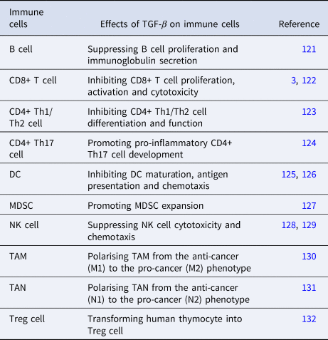
The immune system is the main defense system that recognises and clears foreign antigens including tumour cells (Ref. Reference Chen and Ten Dijke120). However, tumour cells develop varied strategies to evade the immune surveillance including TGF-β-induced immune suppression (Fig. 2). TGF-β may inhibit the proliferation and functionality of various innate and adaptive immune cells such as T cells, B cells, dendritic cells (DCs), natural killer (NK) cells, macrophages, neutrophils, monocytes and granulocytes (Ref. Reference Sanjabi, Oh and Li121) (Table 2).
Effects of TGF-β on immune cells

DC, dendritic cell; NK, natural killer; TAM, tumour-associated macrophage; TAN, tumour-associated neutrophil; MDSC, myeloid-derived suppressor cell; Th, helper T cell; Treg, regulatory T cell.
In innate immunity, DCs are antigen-presenting cells capable of inducing adaptive T cell response and sensitising the anti-tumour immunity (Ref. Reference Diamond133), where TGF-β leads to systemic immunosuppression by downregulating key factors in DC maturation or activation (Ref. Reference Papaspyridonos125). NK cells suppress tumour progression during the anti-cancer immunity by directly recognising tumour cells and initiating cytotoxic reactions (Ref. Reference Caligiuri134). TGF-β1 suppresses NK cells by reducing the production of interleukin-15 (IL-15) and repressing the expression of natural killer group 2 member D (NKG2D), a major NK cell-activated receptor (Refs Reference Friese128, Reference Lazarova and Steinle129). TGF-β can convert tumour-associated macrophages (TAMs) from the tumour-suppressive (M1) to the cancer-promotive (M2) phenotype (Ref. Reference Gong130) according to markers expressed on cells. Similarly, TGF-β polarises tumour-associated neutrophils (TANs) from a cancer-suppressive N1 to a tumour-promotive N2 phenotype, contributing to tumour growth via immunosuppression (Ref. Reference Fridlender131). TGF-β can lower the production of ROS and nitric oxide (NO) that are essential for the cytotoxicity of macrophages against cancer cells under certain conditions (Ref. Reference Siegert135). In addition to affecting TAM/TAN polarisation, TGF-β regulates immune cell functionalities by, for example, increasing PD-L1 in DCs towards T cell anergy against hepatocellular carcinoma cells (Ref. Reference Song126), and driving myeloid suppressor cell differentiation towards immunosuppression if coupled with IL-6 (Ref. Reference Eriksson127). Besides, TGF-β regulates the generation of natural and induced Tregs by, for example, transforming human thymocytes into natural Treg cells ex vivo in combination with elevated IL-2 secretion and ectopic FOXP3 expression (Ref. Reference Gallego-Valle132).
In adaptive immunity, immune-mediated tumour elimination requires cytotoxic CD8+ T cells that kill malignant cells by producing perforin, granzyme A, granzyme B, IFN-c and Fas ligand (FASL) (Ref. Reference Wu and Lanier136), and provides long-term immunity against key tumour epitopes (Ref. Reference Grosser137). TGF-β represses CD8+ T cells through both direct inhibition on CD8+ T cells and indirect effects on accessory cells such as Tregs that regulate CD8+ T cell functions (Ref. Reference Derynck, Turley and Akhurst3). For example, CD4+ and CD8+ T lymphocytes are more effective in clearing thymoma and melanoma cells in TGF-βRII knockout mice than the wild-type mice (Ref. Reference Gorelik and Flavell138). In addition, TGF-β inhibits the proliferation and activation of CD8+ cytotoxic T-cells by regulating the generation of Tregs (Ref. Reference Fu122), suppresses the proliferation of CD4+ Th1/Th2 cells through reducing the expression of their master transcription factors (Ref. Reference Ludviksson123) and induces Th17 lineage development via modulating an IL-12 cytokine family member IL-23 (Ref. Reference Mangan124).
TGF-β and cancer metabolic reprogramming
Extensive metabolic changes have been observed during cancer progression such as high aerobic glycolysis (Ref. Reference Gatenby and Gillies139), elevated tricarboxylic acid (TCA) cycle during EMT (Ref. Reference Zadran140) and abnormal lipid metabolism (Refs Reference Santos and Schulze141, Reference Luo142).
Increased expression or activity of key glycolytic enzymes is one representative alteration of elevated glycolysis. TGF-β increases the activity of multiple glycolytic enzymes such as glucose transporter type 1 (GLUT1), hexokinase 2 (HK2), 6-phosphofructo-2-kinase/fructose-2,6-biphosphatase 3, pyruvate kinase M2, lactate dehydrogenase type A, leading to elevated level of glycolysis in cancer cells (Refs Reference Altenberg and Greulich143, Reference Li144, Reference De Bock145, Reference Rodriguez-Garcia146) (Fig. 2).
The TCA cycle plays a vital role in energy production to sustain cell proliferation and provide important intermediates for amino acid, nucleic acid and lipid biosynthesis (Ref. Reference Guerra147). TGF-β-induced EMT is accompanied by activated TCA cycle for sufficient adenosine triphosphate (ATP) to become motile (Ref. Reference Zadran140) (Fig. 2). Yet, the role of TGF-β in regulating TCA intermediates remains to be fully explored.
Abnormal lipid metabolism induces cancer invasion and metastasis (Refs Reference Santos and Schulze141, Reference Luo142). Altered lipid catabolic metabolism drives tumour metastasis by producing tumourigenic metabolites and molecules that are integrated into signal transduction networks between cancer cells and supporting host cells in the TME. Although cancer cells develop mitochondrial biogenesis, supporting cells fuel cancer cell mitochondria and create an environment favouring metastasis (Ref. Reference Martinez-Outschoorn, Sotgia and Lisanti148). TGF-β is capable of decreasing fatty acid synthase and acetyl-CoA carboxylase, the main enzymes involved in de novo fatty acid synthesis, towards promoted cancer metastasis (Refs Reference Beloribi-Djefaflia, Vasseur and Guillaumond149, Reference Menendez and Lupu150).
Besides rewiring the metabolism of tumour cells, TGF-β also promotes carcinogenesis by regulating the metabolism of cells residing in the TME such as tumour-infiltrating immune cells and cancer-associated fibroblasts (CAFs) (Ref. Reference Angioni151). Specifically, TGF-β inhibits the basal and ATP-coupled oxygen consumption rates, and the functions of complex V and ATP synthase that lead to impaired IFN-γ production by CD4+ cells (Ref. Reference Dimeloe152). Also, TGF-β enables a high oxidative metabolism of Treg cells via reducing the expression of glycolytic genes such as GLUT1 and HK2, and provides intermediates for the TCA cycle through enhancing fatty acid oxidation (Ref. Reference Priyadharshini153). TGF-β-mediated CAF metabolic reprogramming largely relies on an unbalanced oxidative framework because of its regulatory roles on genes responsible for redox homoeostasis maintenance; and this enables the manifestation of a senescence-associated secretory phenotype for CAFs that is featured by a strong release of IL-6/-1/-8, GROα/β (Ref. Reference Coppe154).
Current onco-therapeutics employing TGF-β
Canonical anti-cancer strategies employing TGF-β
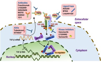
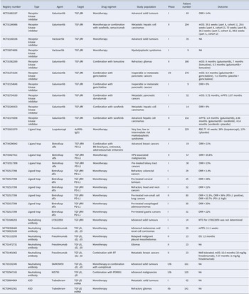
Patients with high TGF-β1 expression have been positively associated with increased tumour progression, lymph infiltration and low disease-free survival, rendering the TGF-β pathway a clinically relevant onco-therapeutic target (Refs Reference Ding155, Reference Roy, Poirier and Fortin156). Receptor kinase inhibitors, ligand traps, neutralising antibodies and ASOs have been dominating this field (Refs Reference Akhurst157, Reference Kim158) (Fig. 3, Table 3).
Roles of TGF-β in onco-therapeutic design. Current onco-therapeutic strategies employing TGF-β mainly consider it as an onco-target. Four types of therapeutics have been developed accordingly, that is, antibodies, ligand traps, ASOs and kinase inhibitors. Representative antagonists been or being evaluated in clinical trials from each canonical therapeutic category are listed. ASOs, antisense oligonucleotides.

Completed clinical trials targeting TGF-β signalling as onco-therapeutics

ORR, overall response rate; mOS, median overall survival; mPFS, median progression-free survival; RBC-TI, red blood cell transfusion independence; MTD, maximum tolerated dose.
Receptor kinase inhibitors
Several TGF-β receptor kinase inhibitors have been designed to interact with the ATP binding site of the catalytic domain and inhibit the ATP-dependent kinase activity of TGF-β receptor towards the blockage of the downstream signalling cascade (Ref. Reference Huynh, Hipolito and Ten Dijke159). Inhibitors including galunisertib (LY2157299) for prostate cancer treatment (NCT02452008, phase II, recruiting, Eli Lilly and Company), LY3200882 for treating solid tumours (NCT02937272, phase I, active, Eli Lilly and Company) and vactosertib (TEW-7197) for curing myeloproliferative neoplasm (NCT04103645, phase II, recruiting, MedPacto) have reached the stage of clinical trials (Refs Reference Jin160, Reference Fujiwara161, Reference Yap162). However, these inhibitors have poor pharmacokinetic and pharmacodynamic properties that do not generate desirable therapeutic effects, as well as poor target (TGF-β receptors) specificity that also inhibit several other TGF-β-related proteins or kinases such as activin, nodal, myostatin and MAPK-p38 (Refs Reference Inman163, Reference Yingling164).
Ligand traps
Ligand traps are soluble and have a high-affinity to TGF-β receptors and, thereby, can block the binding of TGF-β to its receptors (Refs Reference Yang165, Reference Hinck and O'Connor-McCourt166). Ligand traps comprise Fc-stabilised dimers of TGF-ΒRII extracellular domains by design for sequestering TGF-β1 and TGF-β3, which prevent them from binding to TGF-βRII (Ref. Reference Lan167). These agents were firstly used to inhibit tumour dissemination in mice carrying a genetically engineered TGF-β ligand trap (Ref. Reference Yang165). Distinct TGF-β traps have been developed that differ in their ligand binding specificities (Refs Reference Zwaagstra168, Reference Qin169). For example, AVID200 (Formation Biologics), an engineered TGF-β ligand trap that is 1000 times more selective in neutralising TGF-β1 and TGF-β3 than TGF-β2 (Ref. Reference Kim158), has been predominantly applied in haematopoiesis, bone marrow fibrosis and myelofibrosis (Ref. Reference Tremblay and Mascarenhas170). The efficacy of AVID200 is being evaluated in patients carrying malignant solid tumours (NCT03834662, phase I, active but not recruiting, Formation Biologics). M7824 (MSB0011359C, EMD Serono/GlaxoSmithKline/Merck KGaA) is a bifunctional fusion protein targeting PD-L1 and TGF-β that shows encouraging efficacy and manageable tolerability in platinum-treated non-small cell lung cancer (NSCLC) patients in a clinical trial (NCT02517398, phase I, completed, EMD Serono Research & Development Institute, Inc.) (Ref. Reference Strauss171). Luspatercept (Luspatercept-aamt, Acceleron), a recombinant fusion protein that binds TGF-β ligands towards reduced SMAD2/3 signalling, decreases the severity of anaemia in patients with lower-risk myelodysplastic syndromes (Ref. Reference Fenaux172). However, because of the pleiotropic activities of TGF-β and lack of appropriate biomarkers, patient selection criteria and optimal dosing regimens of these ligand traps are yet to be determined (Ref. Reference Kim158).
Neutralising antibodies
Antibodies are used to antagonise TGF-β activity and block the binding of TGF-β ligand to its cognate receptor (Ref. Reference Kim158). Fresolimumab (GC1008, Genzyme) is a human anti-TGFβ IgG4k mAb that shows promising therapeutic efficacy against malignant melanoma and renal cell carcinoma in a clinical trial (NCT00356460, phase I, completed, Sanofi) (Ref. Reference Morris173). LY3022859 is an anti-TGF-βRII mAb that fails in a trial composed of 14 advanced cancer patients (NCT01646203, phase I, completed, Eli Lilly and Company) because of the onset of uncontrolled cytokine-release syndrome at the higher doses examined prior to the observation of any therapeutic benefits (Ref. Reference Tolcher10). There also exist several other antibodies being examined in pre-clinical or clinical trials. For instance, 264RAD (AstraZeneca, pre-clinical) reduces pharyngeal tumour cell growth and metastasis in vivo (Ref. Reference Eberlein174), 1D11 (Genzyme Corp., Sanofi, pre-clinical) suppresses breast cancer metastasis in vivo (Ref. Reference Nam175), the clinical efficacy of SAR439459 is currently being evaluated in malignant solid neoplasm (NCT04729725, phase I, active, Sanofi) (Ref. Reference Bedinger176) and NIS793 is being examined in metastatic pancreatic ductal adenocarcinoma (NCT04390763, phase II, recruiting, Novartis Pharmaceuticals) (Ref. Reference Greco177). Unfortunately, some antibodies show serious side effects such as uncontrolled cytokine-release syndrome at a high dose (Ref. Reference Derynck, Turley and Akhurst3).
Antisense oligonucleotides
ASOs can be used to halt cancer progression by specifically targeting genes that are difficult to inhibit by small molecules or mAbs (Ref. Reference Gleave and Monia178). However, ASO-based therapeutics are normally instable, where nanoparticles and chemical modifications have been developed to improve their delivery and stability (Refs Reference Dias and Stein179, Reference Tibbitt, Dahlman and Langer180). Trabedersen (AP12009, Antisense Pharma GmbH/Isarna) is a TGF-β2-specific phosphorothioate ASO that has been under clinical development for treating patients carrying refractory high-grade glioma, with the median survival after recurrence exceeding that after receiving chemotherapy without observing life-threatening toxicities (Ref. Reference Hau181). A next-generation TGF-β2-selective ASO, ISTH0047 (Isarna Therapeutics GmbH), has been reported to inhibit lung metastasis in syngeneic orthotopic renal cell carcinoma and breast cancer (Ref. Reference Huber-Ruano182).
Combinatorial anti-cancer strategies employing TGF-β
Although targeting TGF-β has demonstrated remarkable potential for cancer treatment, the functional complexity and pleiotropic effects of TGF-β impose a great clinical challenge to therapies targeting TGF-β (Ref. Reference Kim158). Specifically, TGF-β is secreted by nearly every type of cells that regulates numerous critical physiologic processes in healthy cells (Ref. Reference Massague183). Thus, therapies targeting TGF-β may develop both on- and off-target toxicities (Ref. Reference Colak and Ten Dijke184). Therefore, efforts have been devoted to combine TGF-β-targeted therapies with other anticancer strategies such as immunotherapies and chemo-/radiotherapies.
Combinatorial strategies involving TGF-β and immunotherapy
The suppressive role of TGF-β in human innate and adaptive immune systems has led to the feasibility of combining therapeutics against TGF-β with anticancer immunotherapies (Ref. Reference Arteaga185). For instance, TGF-β attenuates tumour response to PD-L1 blockade by contributing to T cell removal, and drive immune evasion in genetically reconstituted colon cancer metastasis (Refs Reference Mariathasan186, Reference Tauriello187), suggestive of the potential synergy between TGF-β inhibition and immunotherapy. As an investigational product candidate, SRK181-mIgG1 (Scholar Rock), a potent and highly selective inhibitor of the TGF-β1 isoform, is developed to overcome drug resistance to checkpoint inhibitors such as anti-PD-(L)1 mAbs for treating bladder cancers (Ref. Reference Martin188). Another example is the anti-TGFβ1/2 antibody NIS793 that is being tested in a dose-escalation trial together with the anti-PD-1 antibody spartalizumab in patients carrying advanced-stage cancers (NCT02947165, phase I/Ib, completed, Novartis Pharmaceuticals). The safety and efficacy of vactosertib (TEW-7197), an inhibitor of TGF-β/SMAD signalling, is evaluated in combination with durvalumab (Imfinzi, Medimmune, AstraZeneca), a mAbs blocking PD-L1, in a phase Ib/IIa clinical trial comprised of 60 metastatic NSCLC and urothelial carcinoma patients (NCT03732274, phase Ib/IIa, active but not recruiting, MedPacto).
Combinatorial strategies involving TGF-β and oncolytic viruses
By altering the phenotype of immune-excluded or immune-desert tumours towards an immune-infiltrated phenotype, perturbating TGF-β signalling (largely through inhibition) is expected to enhance the therapeutic efficacy of oncolytic viruses in treating solid tumours (Ref. Reference Groeneveldt189).
Herpes simplex virus (HSV) is commonly used in oncolytic virotherapy and has been combined with approaches interrogating TGF-β signalling for cancer treatment. For instance, HSV1716 is combined with TGF-β receptor 1 (TGF-βR1) inhibitor A8301 to treat syngeneic models of murine rhabdomyosarcoma, where TGF-β inhibition augments the immunotherapeutic efficacy of HSV1716 (Ref. Reference Hutzen190). HSV1, combined with TGF-βR1 inhibitors SB431542, LY2109761, LY2157299, shows remarkable efficacy in arresting recurrent glioblastoma via an inhibitor-triggered MAPK/JNK blockage (Ref. Reference Esaki191). On the other hand, HSV synergises with TGF-β in glioblastoma treatment, where TGF-β pretreatment sensitises cancer cells to the oncolytic efficacy of HSV by attenuating the innate immune responses mediated by NK cells and macrophages (Ref. Reference Han192).
Another frequently reported oncolytic virus used to create synergies with modalities modulating TGF-β signalling is adenovirus. A selective expression of a soluble form of TGF-β receptor II fused with Fc (sTGFβRIIFc) is combined with an adenoviral vector (namely Ad.sTbetaRFc) to treat cancers that results in over 85% reduced breast cancer progression in vivo (Ref. Reference Seth193), and significantly suppresses prostate tumour burden and bone metastasis (Ref. Reference Hu194). Another combinatorial strategy that relies on adenovirus-based oncolytic virus expressing sTGFβRIIFc, called TAd.sTβRFc, shows comparable efficacy in inhibiting prostate cancer metastasis with Ad.sTbetaRFc (Ref. Reference Hu194). A clinical trial featuring AdAPT-001 (NCT04673942, phase I, recruiting, EpicentRx), a type 5 adenovirus that carries a TGF-β trap transgene, is ongoing, where AdAPT-001 was shown safe and well-tolerated at clinically relevant doses in tumour-bearing mice (Ref. Reference Larson195).
Combinatorial strategies involving TGF-β and chemo-/radiotherapy
TGF-β signalling promotes acquired chemotherapy resistance as a result of induced EMT in cancer cells (Ref. Reference Zhang196), and plays key roles in p53/ATM-mediated DNA mismatch repair as a protective mechanism against radiation-induced damage (Ref. Reference Ewan197). Thus, TGF-β inhibition has been coupled with chemo-/radiotherapies towards enhanced therapeutic responses and activated TME (Refs Reference Zhang196, Reference Wang198). The TGF-β/SMAD inhibitor LY3200882 has been shown as a safe monotherapy and combinatorial therapy once coupled with gemcitabine and nab-paclitaxel in pancreatic cancer patients (NCT02937272, phase I, active but not recruiting, Eli Lilly and Company) (Ref. Reference Yap162). However, coupling TGF-β inhibitors with radiotherapy does not achieve desirable therapeutic outcome. For example, a clinical trial fails in combining focal radiotherapy and TGF-β blockage via fresolimumab (GC1008, Genzyme) in the treatment of patients carrying metastatic breast cancers despite the observed overall survival extension among patients receiving a high dose fresolimumab (NCT01401062, phase II, completed, Weill Medical College of Cornell University) (Ref. Reference Formenti199).
Cold atmospheric plasma: an emerging anti-cancer strategy employing TGF-β
The existing anti-cancer strategies all rely on inhibiting TGF-β signalling given the promotive role of TGF-β on cancer angiogenesis and metastasis. However, its roles in suppressing tumour growth and triggering cancer cell death have not been fully utilised regarding onco-therapeutic design.
Cold atmospheric plasma (CAP), partially ionised gas created at the room temperature and atmospheric pressure, has showcased its anti-cancer efficacy since 2007 and been proposed as a promising therapeutic choice for cancer treatment (Ref. Reference Dai200). CAP is produced and confined within a gap separating the electrodes (or the electrode and the surface of the tissue) using a processing gas that can be air, argon, helium or a mixture of argon/helium with a certain percentage of oxygen. Typical configurations generating CAP include plasma jet, plasma torch and dielectric barrier discharge (DBD) (Ref. Reference Dai200). Although CAP is generated between electrodes and a controlled flow of processing gas carries CAP into the ambient in the form of a plume that propagates following the flow direction in plasma jet or torch, CAP is generated by applying high sinusoidal voltages in the kilohertz frequency range to plate electrodes covered by insulators such as quartz and glass in a DBD configuration. Several CAP sources have been certified for medical use given their clinically verified safety and efficacy including, for example, the plasma jet kINPen MED (neoplas GmbH, Greifswald, Germany) (Ref. Reference Breathnach201), the plasma torch SteriPlas (ADTEC, Hunslow, UK) (Ref. Reference Maisch202), the DBD plasma generator PlasmaDerm VU-2010 device (CINOGY GmbH Duderstadt, Germany) (Ref. Reference Brehmer203) and the Canady Helios Cold Plasma™ Scalpel (US Medical Innovations, LLC) (Ref. Reference Gjika204).
CAP has demonstrated its selectivity against many types of transformed cells such as triple negative breast cancers (Refs Reference Xiang205, Reference Zhou206), prostate cancers (Ref. Reference Hua207), bladder cancers (Ref. Reference Wang208), with varied molecular mechanisms being reported. It belongs to the fourth state of matter following solid, liquid and gas, and is composed of a cocktail of multiple reactive oxygen and nitrogen species such as singlet oxygen (O), hydroxyl radical (OH&), superoxide (O2−), nitric oxide (NO&), hydrogen peroxide (H2O2), ozone (O3), anionic (OONO‒) and protonated forms of peroxynitrite (ONOOH). These species, together with those generated through collision, interact with cancer cell surface to produce a series of signallings that selectively arrest cancer cell growth via inducing, for example, non-immunogenic cell death programmes such as cell cycle arrest (Ref. Reference Hua207), senescence (Ref. Reference Schneider209), apoptosis (Ref. Reference Xiang205) and immunogenic cell death events (Ref. Reference Lin210) such as autophagic cell death (Ref. Reference Miao211), necrosis (Ref. Reference Virard212), ferroptosis (Refs Reference Furuta, Shi and Toyokuni213, Reference Bekeschus214) and immunogenic apoptosis (Ref. Reference Xiang205) in a dose-dependent manner (Ref. Reference Dai215). Such a selective toxicity of CAP against cancer cells is not only caused by the high basal redox level of malignant cells, but also determined by the membrane characteristics of cancer cells such as high aquaporin expression that fosters enhanced H2O2 uptake (Ref. Reference Yan216) and high local catalysis that enables secondary O generation for avalanched redox signalling (Ref. Reference Bauer217). The selectivity of CAP against cancer cells implicates its fundamental roles in controlling the life/death switch of cancer cells, and thus offers therapeutic opportunities if coupled with TGF-β inducers towards synergistic benefits.
On the other hand, CAP has been proposed capable of inducing cancer stemness (Ref. Reference Dai218) and activating the TME (Ref. Reference Dai219), rendering CAP capable of selectively halting cancer cell migration especially the EMT process (Ref. Reference Wang208) and rewiring drug resistance (Ref. Reference Park220). Thus, it is possible to combine CAP with TGF-β inhibitors towards arrested cancer metastasis and improved therapeutic sensitivity.
In addition, the safety of CAP for medical use has been witnessed for decades as evidenced from its wide applications in, for example, ulcer treatment, wound healing and blood coagulation (Refs Reference Miyamoto221, Reference Stratmann222). Our previous pre-clinical efforts have demonstrated its safety as a promising anti-cancer modality (Ref. Reference Zhou206). The first clinical trial examining the efficacy and safety of CAP as an onco-therapy in treating a panel of stage IV malignant solid tumours has been successfully completed by April 2021 (NCT04267575, clinical phase is not applicable, active but not recruiting, Jerome Canady) (Ref. Reference Mark223). Thus, it is promising to combine CAP with existing combinatorial strategies involving TGF-β inhibitors towards reduced drug dose, enhanced therapeutic outcome and improved safety.
Although promising, the road towards the clinical translation of CAP into a first line or adjuvant onco-therapy is impeded by several obstacles. These, among others, include how to identify appropriate CAP doses for treating cancers of different types and at different developmental stages, how to effectively deliver CAP to the tumour loci towards optimal therapeutic outcome and, in particular, when combined with TGF-β therapeutics.
Conclusion
TGF-β has been canonically used as the drug target in onco-therapeutic design despite its double-edged roles in cancer initiation and development. By characterising the functionalities of TGF-β in cancer according to cancer hallmarks, we portrait TGF-β as tumour-suppressive in triggering programmed cell death and tumour-promotive in driving the other cancer hallmarks. Importantly, TGF-β suppresses uncontrolled cell growth that characterises the initial stage of carcinogenesis, but promotes cancer metastasis, tumour-associated inflammation, immune evasion, metabolic reprogramming and genome instability that are featured cancer traits developed at later carcinogenetic stages. However, current onco-therapeutic strategies unanimously rely on its oncogenic traits that sometimes does not reach desirable therapeutic outcome. Thus, how to optimally utilise the roles of TGF-β played during tumourigenesis, for example, being a cell death inducer, in the onco-therapeutic design warrants intensive investigations. We propose, among other therapeutic options, the possibility of combining TGF-β modulators with CAP, an emerging onco-therapeutic capable of selectively inducing cancer cell death, halting cancer metastasis and rewiring drug resistance, for cancer management. That is, we foresee the benefits of combining CAP with TGF-β inducers for treating cancers at the early stage, and coupling CAP with TGF-β inhibitors for managing metastatic malignancies or tumours having already developed drug resistance.
Author contributions
XD conceived the idea, supervised and financed this study. XD and XXL drafted the manuscript, prepared the figures and conducted literature searching. DH co-supervised this study. All authors have read and approved the content of the manuscript.
Financial support
This work was supported by the National Natural Science Foundation of China (Grant No. 81972789), Fundamental Research Funds for the Central Universities (Grant No. JUSRP22011) and Technology Development Funding of Wuxi (Grant No. WX18IVJN017). The funding bodies played no role in the design of the study and collection, analysis and interpretation of data and in writing the manuscript.
Conflict of interest
The authors declare no conflict of interest.