Childhood maltreatment significantly increases risk for behavior problems (Carr, Martins, Stingel, Lemgruber, & Juruena, Reference Carr, Martins, Stingel, Lemgruber and Juruena2013; Hickman et al., Reference Hickman, Jaycox, Setodji, Kofner, Schultz and Barnes-Proby2013; Kim-Spoon, Cicchetti, & Rogosch, Reference Kim-Spoon, Cicchetti and Rogosch2013; Mills et al., Reference Mills, Scott, Alati, O'Callaghan, Najman and Strathearn2013; Norman et al., Reference Norman, Byambaa, De, Butchart, Scott and Vos2012), which can be identified as early as 12–48 months of age and are strongly correlated with the development of significant psychopathology (Briggs-Gowan, Carter, Bosson-Heenan, Guyer, & Horwitz, Reference Briggs-Gowan, Carter, Bosson-Heenan, Guyer and Horwitz2006; Mesman & Koot, Reference Mesman and Koot2001; Roza, Hofstra, van der Ende, & Verhulst, Reference Roza, Hofstra, van der Ende and Verhulst2003). Childhood maltreatment is also significantly associated with adult psychopathology (Brown & Anderson, Reference Brown and Anderson1991; Bryer, Nelson, Miller, & Krol, Reference Bryer, Nelson, Miller and Krol1987; Kendler, Kessler, Neale, Heath, & Eaves, Reference Kendler, Kessler, Neale, Heath and Eaves1993; Lewinsohn, Hoberman, & Rosenbaum, Reference Lewinsohn, Hoberman and Rosenbaum1988); and among those with disorders, a history of maltreatment predicts greater severity, worse prognosis, and multiple comorbidities (Nanni, Uher, & Danese, Reference Nanni, Uher and Danese2012; Widom, DuMont, & Czaja, Reference Widom, DuMont and Czaja2007). A number of biological mechanisms have been proposed to mediate risk for psychopathology following exposure to maltreatment. Alterations of neural structure and function and other biological changes have been observed in children and adults with a history of maltreatment (Cicchetti, Reference Cicchetti2015; Cicchetti, Handley, & Rogosch, Reference Cicchetti, Handley and Rogosch2015; Hart & Rubia, Reference Hart and Rubia2012; Philip et al., Reference Philip, Sweet, Tyrka, Carpenter, Albright and Price2015; Ridout et al., Reference Ridout, Parade, Seifer, Price, Gelernter and Feliz2014, Reference Ridout, Ridout, Kao, Carpenter, Philip and Tyrka2015; Toth, Gravener-Davis, Guild, & Cicchetti, Reference Toth, Gravener-Davis, Guild and Cicchetti2013; Tyrka, Burgers, Philip, Price, & Carpenter, Reference Tyrka, Burgers, Philip, Price and Carpenter2013; Tyrka, Parade, Price, et al., Reference Tyrka, Parade, Price, Kao, Porton and Philip2015). Identifying these mediating factors is critical for understanding how early childhood maltreatment contributes to risk of psychopathology in order to prevent long-term sequelae.
Epigenetic modifications to the genome are of major interest in this effort because they can stably alter gene expression in response to environmental exposures. There is a growing body of literature to support the importance of epigenetic changes in mediating the effects of childhood maltreatment on psychopathology and on biological systems involved in the development of psychopathology (Guintivano & Kaminsky, Reference Guintivano and Kaminsky2014; Januar, Saffery, & Ryan, Reference Januar, Saffery and Ryan2015; Lutz, Almeida, Fiori, & Turecki, Reference Lutz, Almeida, Fiori and Turecki2015). Epigenetic modifications alter gene expression but do not change the DNA sequence and thus allow for elaboration of the genome beyond what is determined by the DNA (Szyf, Reference Szyf2015). Accumulating evidence indicates that childhood maltreatment may exert long-lasting effects through epigenetic changes (Lutz & Turecki, Reference Lutz and Turecki2014; Szyf, Reference Szyf2011; Tyrka, Parade, Eslinger, et al., Reference Tyrka, Parade, Eslinger, Marsit, Lesseur and Armstrong2015). DNA methylation, involving the addition of a methyl group at sites where a cytosine nucleotide is joined to a guanine nucleotide by a phosphodiester bond (CpG dinucleotides), is thought to be the most stable form of epigenetic alteration (Reik, Reference Reik2007). Methylation can block the binding of transcription factors or interfere with gene expression through other mechanisms (Klengel, Pape, Binder, & Mehta, Reference Klengel, Pape, Binder and Mehta2014; Lee et al., Reference Lee, Tamashiro, Yang, Purcell, Huo and Rongione2011; Yang et al., Reference Yang, Han, De Carvalho, Lay, Jones and Liang2014). Consistent with this, genes that are highly expressed typically have low levels of promoter methylation (Sasaki, de Vega, & McGowan, Reference Sasaki, de Vega and McGowan2013; Wagner et al., Reference Wagner, Busche, Ge, Kwan, Pastinen and Blanchette2014; Zhang, Labonte, Wen, Turecki, & Meaney, Reference Zhang, Labonte, Wen, Turecki and Meaney2013).
Maltreatment and the Development of the Physiologic Stress Response
Research examining the biologic underpinnings of the associations between childhood maltreatment and psychopathology highlights the importance of the physiologic stress response system, and in particular, the hypothalamus–pituitary–adrenal (HPA) axis. In response to stressful stimuli, glucocorticoids are released and exert cellular responses by binding at the intracellular glucocorticoid receptor (GR; Tyrka et al., Reference Tyrka, Burgers, Philip, Price and Carpenter2013). GRs are distributed throughout the body and brain where they regulate basal physiologic function and effect changes in various organ systems and tissues that promote adaptive responding to acute stressors (de Kloet, Joels, & Holsboer, Reference de Kloet, Joels and Holsboer2005; Kadmiel & Cidlowski, Reference Kadmiel and Cidlowski2013). Activation of the GR through cortisol binding also involves a negative feedback mechanism that inhibits release of cortisol and prevents damaging effects of chronic activation (Herman, McKlveen, Solomon, Carvalho-Netto, & Myers, Reference Herman, McKlveen, Solomon, Carvalho-Netto and Myers2012; Laryea, Muglia, Arnett, & Muglia, Reference Laryea, Muglia, Arnett and Muglia2015). Thus, changes in GR number or function can influence the activity of the stress response system and, consequently, the biological adaptation to stressful or traumatic experiences.
Studies examining the development of the stress response system reveal that early environment is linked to abnormalities in HPA axis function, with exaggerated or attenuated basal cortisol and stress-induced cortisol concentrations (Carpenter et al., Reference Carpenter, Carvalho, Tyrka, Wier, Mello and Mello2007, Reference Carpenter, Tyrka, Ross, Khoury, Anderson and Price2009; Dozier et al., Reference Dozier, Manni, Gordon, Peloso, Gunnar and Stovall-McClough2006; Elzinga et al., Reference Elzinga, Roelofs, Tollenaar, Bakvis, van Pelt and Spinhoven2008; Gonzalez, Reference Gonzalez2013; Gunnar & Vazquez, Reference Gunnar and Vazquez2001; Heim, Newport, Bonsall, Miller, & Nemeroff, Reference Heim, Newport, Bonsall, Miller and Nemeroff2001; Heim, Newport, et al., Reference Heim, Newport, Heit, Graham, Wilcox and Bonsall2000; Klaassens et al., Reference Klaassens, van Noorden, Giltay, van Pelt, van Veen and Zitman2009; McCrory, De Brito, & Viding, Reference McCrory, De Brito and Viding2010; Tyrka et al., Reference Tyrka, Price, Gelernter, Schepker, Anderson and Carpenter2009, Reference Tyrka, Price, Kao, Porton, Marsella and Carpenter2010). Excessive activation of the HPA axis over time or in response to extreme stress may result in downregulation of this system (Fries, Hesse, Hellhammer, & Hellhammer, Reference Fries, Hesse, Hellhammer and Hellhammer2005; Heim, Ehlert, & Hellhammer, Reference Heim, Ehlert and Hellhammer2000; McEwen, Reference McEwen2007; Pryce et al., Reference Pryce, Ruedi-Bettschen, Dettling, Weston, Russig and Ferger2005; Tyrka et al., Reference Tyrka, Wier, Price, Ross, Anderson and Wilkinson2008). For example, in comparison with children who were not maltreated, maltreated children have greater variance in cortisol levels at baseline, and those with high baseline cortisol levels are more likely than nonmaltreated children to have blunted cortisol levels over time (Doom, Cicchetti, & Rogosch, Reference Doom, Cicchetti and Rogosch2014). Several studies of adults with a history of early life stress show attenuated cortisol response to stress (Carpenter et al., Reference Carpenter, Carvalho, Tyrka, Wier, Mello and Mello2007, Reference Carpenter, Tyrka, Ross, Khoury, Anderson and Price2009; Carpenter, Shattuck, Tyrka, Geracioti, & Price, Reference Carpenter, Shattuck, Tyrka, Geracioti and Price2011; Elzinga et al., Reference Elzinga, Roelofs, Tollenaar, Bakvis, van Pelt and Spinhoven2008; Klaassens et al., Reference Klaassens, van Noorden, Giltay, van Pelt, van Veen and Zitman2009), suggesting that the physiologic effects of maltreatment are long lasting.
The HPA Axis and Risk for Psychopathology and Other Medical Conditions
Converging lines of evidence indicate that excessive exposure to glucocorticoid activity may be involved in the pathogenesis of depressive and anxiety disorders. Stimulation of the HPA system through chronic glucocorticoid treatment produces anxiety- and depressive-like behaviors and causes dendritic atrophy, pyramidal cell loss, and suppression of hippocampal neurogenesis (Cameron & Gould, Reference Cameron and Gould1994; Dachir, Kadar, Robinzon, & Levy, Reference Dachir, Kadar, Robinzon and Levy1993; Gould, Woolley, & McEwen, Reference Gould, Woolley and McEwen1991; Magarinos, Orchinik, & McEwen, Reference Magarinos, Orchinik and McEwen1998; Watanabe, Gould, & McEwen, Reference Watanabe, Gould and McEwen1992; Woolley, Gould, & McEwen, Reference Woolley, Gould and McEwen1990). Altered cortisol responses are associated with internalizing behaviors (Beauchaine, Crowell, & Hsiao, Reference Beauchaine, Crowell and Hsiao2015; Hartman, Hermanns, de Jong, & Ormel, Reference Hartman, Hermanns, de Jong and Ormel2013), externalizing behaviors (Hartman et al., Reference Hartman, Hermanns, de Jong and Ormel2013; Marsman et al., Reference Marsman, Swinkels, Rosmalen, Oldehinkel, Ormel and Buitelaar2008), suicidal ideation (Beauchaine et al., Reference Beauchaine, Crowell and Hsiao2015), and posttraumatic stress disorder (PTSD; de Kloet et al., Reference de Kloet, Vermetten, Geuze, Kavelaars, Heijnen and Westenberg2006, Reference de Kloet, Vermetten, Lentjes, Geuze, van Pelt and Manuel2008; Golier, Caramanica, Makotkine, Sher, & Yehuda, Reference Golier, Caramanica, Makotkine, Sher and Yehuda2014), and depression (Ciufolini, Dazzan, Kempton, Pariante, & Mondelli, Reference Ciufolini, Dazzan, Kempton, Pariante and Mondelli2014; Colich, Kircanski, Foland-Ross, & Gotlib, Reference Colich, Kircanski, Foland-Ross and Gotlib2015; de Kloet et al., Reference de Kloet, Vermetten, Lentjes, Geuze, van Pelt and Manuel2008; Suzuki, Belden, Spitznagel, Dietrich, & Luby, Reference Suzuki, Belden, Spitznagel, Dietrich and Luby2013). The HPA axis directs the secretion of glucocorticoids, which potentiate systemic inflammation and oxidative stress, and advanced cellular aging. These factors may underlie the association of stress, psychopathology, and somatic conditions (Bauer, Jeckel, & Luz, Reference Bauer, Jeckel and Luz2009; Wolkowitz, Epel, Reus, & Mellon, Reference Wolkowitz, Epel, Reus and Mellon2010). Evidence suggests that children with certain genetic alleles that alter the function of the HPA axis may be at a higher risk of developing psychopathology after experiencing maltreatment and that childhood maltreatment exerts its effects on HPA axis function through epigenetic modifications of key regulatory genes (Klengel & Binder, Reference Klengel and Binder2015a; Nugent, Tyrka, Carpenter, & Price, Reference Nugent, Tyrka, Carpenter and Price2011; Turecki & Meaney, Reference Turecki and Meaney2014; Tyrka, Price, Marsit, Walters, & Carpenter, Reference Tyrka, Price, Marsit, Walters and Carpenter2012).
Genetic and Epigenetic Factors Influencing the HPA Axis
There are multiple levels of regulation and control that can help determine the function of the HPA axis in response to stress (Booij, Wang, Levesque Tremblay, & Szyf, Reference Booij, Wang, Levesque, Tremblay and Szyf2013; Stankiewicz, Swiergiel, & Lisowski, Reference Stankiewicz, Swiergiel and Lisowski2013). These include genetic variants such as single nucleotide polymorphisms (SNPs) that influence levels of gene expression as well as epigenetic regulation of HPA axis-involved genes (Booij et al., Reference Booij, Wang, Levesque, Tremblay and Szyf2013; Gillespie, Phifer, Bradley, & Ressler, Reference Gillespie, Phifer, Bradley and Ressler2009; Lutz & Turecki, Reference Lutz and Turecki2014; Stankiewicz et al., Reference Stankiewicz, Swiergiel and Lisowski2013). For example, the GR is regulated by a number of mechanisms allowing for tissue-specific control of its expression and adaptation to stress exposure (Binder, Reference Binder2009; Lutz et al., Reference Lutz, Almeida, Fiori and Turecki2015; Spijker & van Rossum, Reference Spijker and van Rossum2012; Vandevyver, Dejager, & Libert, Reference Vandevyver, Dejager and Libert2014), altering the feedback on and function of the HPA axis. Early environmental experiences are associated with epigenetic changes to the promoter region of the GR gene (nuclear receptor subfamily 3, group C, member 1 [NR3C1]) that influence GR expression and potentially alter HPA-axis set points and responses to stress (Turecki & Meaney, Reference Turecki and Meaney2014; van der Knaap, Oldehinkel, et al., Reference van der Knaap, Oldehinkel, Verhulst, van Oort and Riese2015). NR3C1 methylation is associated with a greater risk of internalizing symptoms and disorders (Radtke et al., Reference Radtke, Schauer, Gunter, Ruf-Leuschner, Sill and Meyer2015; van der Knaap, van Oort, et al., Reference van der Knaap, van Oort, Verhulst, Oldehinkel and Riese2015). Our group recently reported work with the present sample showing that higher NR3C1 methylation is associated with childhood maltreatment and other adversities in preschool-aged children (Tyrka, Parade, Eslinger, et al., Reference Tyrka, Parade, Eslinger, Marsit, Lesseur and Armstrong2015).
The Role of FKBP5 in GR Regulation and the HPA Axis
FKBP5 protein is an important regulator of the GR. In response to stressors, cortisol is released, travels systemically through the blood, and moves into the cytoplasm of cells (Kadmiel & Cidlowski, Reference Kadmiel and Cidlowski2013). Cortisol binding to the GR in the cytosol induces translocation of the GR into the nucleus, where it binds to specific regions of the genome called glucocorticoid response elements. The bound GR serves as a transcription factor at cortisol-responsive genes, and thereby activates systems necessary to cope with stressors (Magee, Chang, Stormo, & Milbrandt, Reference Magee, Chang, Stormo and Milbrandt2006). The GR also provides negative feedback to the brain to shut down the HPA stress response. FKBP5 mediates an additional negative feedback loop on glucocorticoids. GR activation results in rapid induction of FKBP5 that binds to the GR and decreases its ability to bind cortisol and to translocate to the nucleus. Thus, FKBP5 decreases systemic sensitivity to cortisol and reduces GR-mediated negative feedback modulation of the HPA axis (Binder, Reference Binder2009; Cioffi, Hubler, & Scammell, Reference Cioffi, Hubler and Scammell2011; Schmidt et al., Reference Schmidt, Buell, Ionescu, Gassen, Holsboer and Cox2015; Tatro, Ererall, Kaul, & Achim, Reference Tatro, Everall, Kaul and Achim2009).
Genetic variation in the FKBP5 gene confers altered GR function and a poorly regulated neuroendocrine response to stress (Zannas & Binder, Reference Zannas and Binder2014). An SNP in FKBP5 (C to T SNP in intron 2, rs1360780) increases the ability of the GR to bind to the glucocorticoid response elements and induce FKBP5 expression (Zannas & Binder, Reference Zannas and Binder2014). This “risk” T allele is associated with GR resistance (Hohne et al., Reference Hohne, Poidinger, Merz, Pfister, Bruckl and Zimmermann2015; Ising et al., Reference Ising, Depping, Siebertz, Lucae, Unschuld and Kloiber2008; Menke et al., Reference Menke, Klengel, Rubel, Bruckl, Pfister and Lucae2013) and has been linked with PTSD, depressive and anxiety symptoms and disorders, and suicide (Leszczynska-Rodziewicz et al., Reference Leszczynska-Rodziewicz, Szczepankiewicz, Narozna, Skibinska, Pawlak and Dmitrzak-Weglarz2014; Suzuki et al., Reference Suzuki, Matsumoto, Sadahiro, Enokido, Goto and Otani2014; Szczepankiewicz et al., Reference Szczepankiewicz, Leszczynska-Rodziewicz, Pawlak, Narozna, Rajewska-Rager and Wilkosc2014; Van Zomeren-Dohm, Pitula, Koss, Thomas, & Gunnar, Reference Van Zomeren-Dohm, Pitula, Koss, Thomas and Gunnar2015; Zannas & Binder, Reference Zannas and Binder2014).
Recent groundbreaking work by Klengel et al. (Reference Klengel, Mehta, Anacker, Rex-Haffner, Pruessner and Pariante2013) examined the rs1360780 SNP and methylation of functional glucocorticoid response elements in FKBP5 in blood in relation to childhood maltreatment. In comparison with controls, adults with a history of childhood trauma had lower levels of methylation in regulatory regions of intron 7 of FKBP5 in those with the rs1360780 risk allele, and this was associated with decreased GR sensitivity. Moreover, glucocorticoid treatment of human hippocampal progenitor cells also induced long-lasting demethylation of this regulatory region (Klengel et al., Reference Klengel, Mehta, Anacker, Rex-Haffner, Pruessner and Pariante2013). One other study assessed FKBP5 methylation and found effects of maltreatment in saliva DNA from children ages 5–14 (Weder et al., Reference Weder, Zhang, Jensen, Yang, Simen and Jackowski2014), but this involved a 450K methylation array that did include the region studied by Klengel et al. No prior work has examined early childhood, and this may be a sensitive period for the development of these epigenetic effects (Klengel et al., Reference Klengel, Mehta, Anacker, Rex-Haffner, Pruessner and Pariante2013). The aim of the present study was to examine FKBP5 methylation, and the role of the FKBP5 genetic variant, in relation to childhood maltreatment in a sample of impoverished preschool-aged children.
Methods
One hundred seventy-four children were included in this study, including 69 with child welfare documentation of moderate to severe maltreatment in the past 6 months. Children ranged in age from 3 to 5 years (M = 49.87 months, SD = 8.4 months), were racially and ethnically diverse (38 White non-Hispanic, 81 Hispanic, 29 Black, and 26 other races), and 84 were male. Most caregivers (n = 161) were biological mothers. Thirty-one caregivers had less than a high school degree, 65 completed high school, 61 had some postsecondary education, 16 had a bachelor's degree, and 1 did not provide education information. Ninety-eight caregivers were unemployed and 156 of the families qualified for public assistance. No more than 1 child per family was included.
Procedure
Families with a maltreated child were identified from the local child welfare agency and an emergency maltreatment assessment service. Families of children with no indicated case of maltreatment within the past 6 months were recruited at a pediatric medical clinic during a well-child visit and at childcare centers. Based on review of available medical records and parent report, children with a chronic medical or neurologic condition, medication use, obesity, and failure to thrive were excluded. Those with acute illness or medication use were included no less than 2 weeks following resolution of illness and medication use.
Families completed two home visits and questionnaires between the visits. The first home visit, during which caregivers completed interviews on child stress exposure and a saliva sample for DNA isolation was collected from the children, is the focus of the current report.
Measures
Socioeconomic adversity
Indicators of socioeconomic adversity (parental education ≤ high school degree, parental unemployment, and single parenthood) were summed.
Child maltreatment status
All families consented to examination of child welfare records to determine maltreatment status. Trained research staff coded the records using the System for Coding Subtype and Severity of Maltreatment in Child Protective Records (Barnett, Manly, & Cicchetti, Reference Barnett, Manly, Cicchetti, Cicchetti and Toth1993). Five maltreatment subtypes and severity scores ranging from 1 (least severe) to 5 (most severe) were derived. Children with a case of moderate to severe levels of maltreatment (score of 3–5) within the last 6 months were considered as part of the maltreated group (n = 69). Eight children had substantiated cases of physical abuse, 13 sexual abuse, 8 physical neglect/failure to provide, 21 physical neglect/lack of supervision, and 47 emotional maltreatment. Three of the maltreated children were removed from the home and were in the care of their maternal grandmother. The comparison group included children who had never had a substantiated case of maltreatment. In addition 5 children had an episode of moderate maltreatment that occurred at least 18 months prior to participation. Results were consistent whether these children were in the maltreatment or comparison group. Because n = 5 is insufficient for a separate analysis, these children were included in the comparison group.
Contextual stress interview
Caregivers completed a semistructured interview developed in our laboratory to assess the child's experience of contextual stressors in the past month and in the child's lifetime. Categories included death of a caregiver, separation from a caregiver, frequent change of residence or homelessness, inadequate food or clothing, and other events including witnessing neighborhood violence or parental arrest. Each domain was scored positive if at least one episode occurred, and domains were summed for past month and lifetime. Possible scores ranged from 0 (no stressors) to 5 (stressors in all five domains) for each summary scale. Past-month contextual stressors ranged from 0 to 3, with mean of 0.64 and SD of 0.84, and lifetime contextual stressors ranged from 0 to 4 with mean of 1.39 and SD of 1.24.
Traumatic life events
The Diagnostic Infant and Preschool Assessment (Scheeringa & Haslett, Reference Scheeringa and Haslett2010) interview was conducted with caregivers to assess child experiences of traumatic life events. Interviews were conducted by trained clinical social workers and PhD-level psychologists, reviewed in a group supervision format, and scored based upon group consensus. The number of types of traumas experienced in the child's lifetime was summed. Categories included experiencing an accident, animal attack, man-made disaster, or natural disaster; witnessing violence; accidental burning; medical emergency/hospitalization/invasive medical procedure; kidnapping; and other events such as a near drowning. Physical and sexual abuse were not included in this variable because they were assessed as maltreatment (above). Possible scores ranged from 0 to 9, and in the present sample ranged from 0 to 4 with mean of 0.93 and SD of 1.03.
Adversity composite
The number of types of maltreatment experienced, the number of lifetime contextual stressors, and the number of other traumatic life events were summed to create an adversity composite. Possible scores ranged from 0 to 18, and in the sample ranged from 0 to 9 with a mean of 2.93 and a SD of 2.47.
Genotyping
Saliva samples were obtained using the Oragene DISCOVER kits (OGR-575) for Assisted Collections (DNA Genotek, Kanata, Ontario, Canada), and DNA was isolated following the manufacturer's instructions. Samples were genotyped for the SNP rs1360780 through an allelic discrimination assay using predesigned Taqman primers (part #C_8852038_10, Life Technologies) and Taqman universal master mix (Life Technologies) via established protocols as directed by the manufacturer on a Bio-Rad CFX connect. Genotype data were available for 156 children for this report.
FKBP5 and NR3C1 methylation
Sodium bisulfite modification was performed with 500 ng of DNA using the EZ DNA methylation Kit (Zymo Research, Irvine, CA, USA). PyroMark Assay Design software version 2.0.1.15 (Qiagen) was used to design the pyrosequencing assays. The PyroMark (Qiagen) polymerase chain reaction (PCR) kit and forward and reverse primers were used to amplify FKBP5 intron 7 and regions of the NR3C1 promoter, and PCR products were sequenced using a PyroMark MD system (Qiagen) as previously described (Paquette et al., Reference Paquette, Lester, Koestler, Lesseur, Armstrong and Marsit2014; Tyrka, Parade, Eslinger, et al., Reference Tyrka, Parade, Eslinger, Marsit, Lesseur and Armstrong2015). The percentage of DNA methylation at each CpG locus was quantified with PyroMark CpG software, version 1.0.11 (Qiagen). All procedures were performed following manufacturer's protocols. Each sequencing run contained no-template and genomic DNA negative controls, as well as methylated and unmethylated controls (Epitect). A bisulfite conversion control within the assay sequence was used to assess conversion efficiency. PCR, sequencing primers (Integrated DNA Technologies, Inc., Coralville, IA), and pyrosequencing assay sequences are available from the corresponding author upon request.
Our main focus was on FKBP5, but because we previously found effects of maltreatment on NR3C1 methylation, we also examined the association of NR3C1 and FKBP5 methylation. The percent of alleles that were methylated was used in statistical analyses. Consistent with other studies on NR3C1 methylation in a variety of cell types, methylation levels were low for NR3C1. For CpG sites in NR3C1 region 1D, percent methylation ranged from 0 to 7.02 with mean of 1.10 and SD of 0.19 across the whole region. Region 1F methylation varied from 0 to 6.42. The mean and SD of the whole region was 1.47 and 0.26. For 1H, the range of methylation was 0 to 7.11, and the mean across the region was 1.24 and the SD was 0.20. For FKBP5, two CpGs in intron 7 (Chr 6: 35558488, CpG 1 and 35558514, CpG2) were studied based on findings of Klengel et al. (Reference Klengel, Mehta, Anacker, Rex-Haffner, Pruessner and Pariante2013); percent methylation ranged from 79.00 to 98.00 with mean of 88.02 and SD of 2.76 for CpG 1 and mean of 88.61 and SD of 3.73 for CpG 2.
Results
Preliminary analyses
Child age, race, sex, and socioeconomic adversity were not associated with FKBP5 methylation at CpG 1 or CpG 2. Child age, race, and socioeconomic adversity were not associated with NR3C1 methylation, but males had greater mean methylation at 1F than did females (p = .006), so child sex was included in models testing associations with NR3C1 methylation. Child age, sex, and race did not differ according to FKBP5 genotype.
FKBP5 methylation in association with childhood maltreatment and other adversities
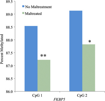
As illustrated in Figure 1, maltreated children had lower levels of FKBP5 methylation at CpG 1 and CpG 2 (t = 3.16, p = .002 and t = 2.29, p = .023, respectively).
(Color online) Association of child maltreatment with methylation of FKBP5. *p < .05, **p < .005.

With respect to other forms of adversity, there was a trend-level association of lifetime contextual stress and methylation of FKBP5 at CpG 1 (r = –.14, p = .064) but not CpG 2 (p = .54). In contrast, past-month contextual stress and the number of traumatic life events were not associated with FKBP5 methylation at either CpG site. The adversity composite was negatively associated with methylation of FKBP5 at CpG 1 (r = –.17, p = .026) but not CpG 2 (p = .36).
FKBP5 methylation and NR3C1 methylation
Methylation of FKBP5 at CpG 1 was negatively associated with mean methylation of NR3C1 at exon 1F (r = –.17, p = .023), but not exons 1D or 1H. This association remained after controlling for maltreatment (p = .04) or the adversity composite (p = .051). No significant associations were observed between methylation of FKBP5 at CpG 2 and methylation of NR3C1 at any of the alternate first exons. The associations of the adversity variables and FKBP5 methylation described above were consistent when methylation of NR3C1 at exons 1D, 1F, and 1H were covaried in the models.
Associations of FKBP5 genotype, adversity, and FKBP5 methylation
Seventy-seven children were homozygotes for the C allele, 68 were CT heterozygotes, and 11 were homozygotes for the T allele. The minor allele frequency (MAF) was .29 for the sample, and the distribution conformed to the Hardy–Weinberg equilibrium (p = .44). Our overall sample MAF was not statistically different from the National Center for Biotechnology Information SNP database reported MAF of 0.33 (p = .44). Sample age, sex, and race did not differ according to genotype. Calculated MAF for males and females were 0.34 and 0.27, respectively; the distribution conformed to Hardy–Weinberg equilibrium (p = .65 and .35, respectively). Calculated MAFs for Whites, Blacks, and Hispanics were 0.3, 0.29, and 0.28, respectively; the distribution conformed to the Hardy–Weinberg equilibrium (p = .36, .23, and .40, respectively). These did not significantly differ from the MAF reported in the National Center for Biotechnology Information SNP database for these populations of 0.21, 0.40, and 0.23, respectively (p = .36, .23, and .40, respectively).
There was no main effect of FKBP5 genotype on methylation of FKBP5. Child maltreatment status, past-month contextual stress, and the number of traumatic life events did not moderate links between FKBP5 genotype and methylation of FKBP5. There was a trend-level interaction of FKBP5 genotype and lifetime contextual stress; children with no lifetime contextual stress with the protective CC genotype had the highest levels of FKBP5 CpG 1 methylation, but this did not reach significance, F (1, 152) = 3.07, p = .082. The interaction of lifetime contextual stress and FKBP5 genotype was not associated with methylation of CpG 2 (p = .321).
Discussion
The central finding of this study is that childhood maltreatment was associated with lower levels of methylation of FKBP5 at two sites within a regulatory region in intron 7 of this gene. These sites were found by Klengel et al. (Reference Klengel, Mehta, Anacker, Rex-Haffner, Pruessner and Pariante2013) to exhibit lower levels of methylation in adults with a history of childhood adversity, with a moderating effect of rs1360870 variation. FKBP5 regulates GR sensitivity and function, so FKBP5 demethylation may be a mechanism of the effect of maltreatment on HPA axis dysfunction and associated risk for psychiatric and other stress-associated conditions.
FKBP5 plays a dynamic role in the regulation of the HPA axis. Prolonged glucocorticoid exposure increases FKBP5 levels, which in turn limit the activation and translocation of GRs, creating a negative feedback loop that ultimately downregulates FKBP5 activation (Klengel & Binder, Reference Klengel and Binder2015b; Klengel et al., Reference Klengel, Mehta, Anacker, Rex-Haffner, Pruessner and Pariante2013; Lee et al., Reference Lee, Tamashiro, Yang, Purcell, Huo and Rongione2011). Methylation of FKBP5 decreases the induction of FKBP5 that occurs following glucocorticoid binding to GR, and demethylation can be induced by prolonged glucocorticoid exposure (Klengel et al., Reference Klengel, Mehta, Anacker, Rex-Haffner, Pruessner and Pariante2013; Lee et al., Reference Lee, Tamashiro, Yang, Purcell, Huo and Rongione2011). Thus, demethylation at glucocorticoid response elements in FKBP5 allows for another level of feedback-loop control in response to glucocorticoids. Our finding in the present study that methylation of FKBP5 and NR3C1 1F were negatively correlated with each other and were associated with maltreatment in different directions suggests that both of these negative regulators of HPA axis activation are at work. These epigenetic changes may serve to establish a steady-state level and counteract elevated levels of glucocorticoids (Lee et al., Reference Lee, Tamashiro, Yang, Purcell, Huo and Rongione2011).
Converging lines of evidence indicate that methylation of glucocorticoid response elements in FKBP5 in DNA from the periphery may have functional effects in the body and may reflect changes to GR sensitivity in the brain. Treatment with glucocorticoids was associated with lower levels of methylation of a key regulatory locus in Fkbp5 in blood in mice, which in turn was associated with important physiological changes in glucocorticoid target tissues, including atrophy of the thymus, spleen, and the adrenal glands (Lee et al., Reference Lee, Tamashiro, Yang, Purcell, Huo and Rongione2011). Although epigenetic modifications to DNA are often tissue-specific, glucocorticoid-induced demethyation of regulatory regions of Fkbp5 in blood was associated with anxiety-like behavior (Lee et al., Reference Lee, Tamashiro, Yang, Purcell, Huo and Rongione2011), and levels of Fkbp5 methylation in blood were significantly correlated with both methylation and gene expression in the hippocampus (Ewald et al., Reference Ewald, Wand, Seifuddin, Yang, Tamashiro and Potash2014). Demethylation of the regulatory region examined in the present study was also shown in a human hippocampal progenitor cell line following glucocorticoid exposure (Klengel et al., Reference Klengel, Mehta, Anacker, Rex-Haffner, Pruessner and Pariante2013). Taken together, these findings suggest that studies using peripheral markers in humans may have direct implications for brain function. It is important to note that we utilized saliva DNA, which may originate from a combination of blood leukocytes (Endler, Greinix, Winkler, Mitterbauer, & Mannhalter, Reference Endler, Greinix, Winkler, Mitterbauer and Mannhalter1999; Thiede, Prange-Krex, Freiberg-Richter, Bornhauser, & Ehninger, Reference Thiede, Prange-Krex, Freiberg-Richter, Bornhauser and Ehninger2000) and buccal epithelial cells. Some evidence indicates that saliva DNA may actually more closely reflect methylation patterns in the brain than DNA from leukocytes (Smith et al., Reference Smith, Kilaru, Klengel, Mercer, Bradley and Conneely2015).
This system is complex, and the literature regarding the functional and dynamic effects on systemic cortisol levels and psychopathology remains at an early stage. There is evidence that lower levels of FKBP5 methylation are associated with higher FKBP5 expression (Lee et al., Reference Lee, Tamashiro, Yang, Purcell, Huo and Rongione2011; Sarapas et al., Reference Sarapas, Cai, Bierer, Golier, Galea and Ising2011), and reduced GR sensitivity (Klengel et al., Reference Klengel, Mehta, Anacker, Rex-Haffner, Pruessner and Pariante2013). FKBP5 expression is positively associated with cortisol levels (Lee et al., Reference Lee, Tamashiro, Yang, Purcell, Huo and Rongione2011; Sarapas et al., Reference Sarapas, Cai, Bierer, Golier, Galea and Ising2011). However, early stress and PTSD are often associated with low levels of cortisol (Morris, Compas, & Garber, Reference Morris, Compas and Garber2012; Tyrka et al., Reference Tyrka, Burgers, Philip, Price and Carpenter2013) and increased sensitivity of the GR has been shown in some studies (Rohleder, Joksimovic, Wolf, & Kirschbaum, Reference Rohleder, Joksimovic, Wolf and Kirschbaum2004; Daskalakis, Lehrner, & Yehuda, Reference Daskalakis, Lehrner and Yehuda2013). There is also evidence of lower FKBP5 expression in PTSD (Yehuda et al., Reference Yehuda, Cai, Golier, Sarapas, Galea and Ising2009). One report indicated that the FKBP5 risk allele that confers higher FKBP5 expression is linked to blunted cortisol response to dexamethasone challenge in adults over age 50, consistent with elevated GR sensitivity (Fujii et al., Reference Fujii, Hori, Ota, Hattori, Teraishi and Sasayama2014). Thus, it is not clear how findings of lower methylation and reduced GR sensitivity are related to some of these other findings, and more work is needed to clarify the role of FKBP5 in regulating glucocorticoid signaling in the wake of childhood maltreatment.
Most of the families in our sample were impoverished and exposed to a variety of contextual stressors, though we did not observe significant effects with our measure of socioeconomic adversity, and we only saw trend-level effects of contextual stressors. There may be effects of early life or in utero exposures that we did not measure. It is also possible that undocumented cases of maltreatment, such as neglect, may have occurred in the nonmaltreated group, and this would have diminished the magnitude of the effect. Moreover, maltreatment characteristics, such as type, timing of onset, and chronicity, may influence DNA methylation patterns and this should be examined in future studies.
In summary, this study shows that maltreatment in preschool-aged children is associated with lower levels of methylation of a functional glucocorticoid response element of FKBP5. Methylation of this region decreases the induction of FKBP5 that occurs when the GR is activated by glucocorticoids. Future work should examine associations of diurnal and provoked cortisol concentrations in relation to FKBP5 and NR3C1 methylation in maltreated children. Lower levels of methylation of FKBP5 and associated increases in FKBP5 induction by activated GRs may explain some of the neuroendocrine dysfunction and associated psychiatric and medical morbidity that result from childhood maltreatment.