Food-borne zoonotic diseases pose an immense threat to public health. Consumption of food contaminated with Listeria monocytogenes is the main route of human listeriosis infection. The Centers for Disease Control and Prevention (CDC, 2022) reports over 1,600 listeriosis cases every year, of which about 16% result in death. Overall, dairy products are associated with about 37% of L. monocytogenes illnesses (IFSAC, 2022). L. monocytogenes remains the most prevalent, however, other Listeria species are also of concern. Listeria ivanovii has been predominantly linked to animal disease including sheep and bovines (Orsi and Wiedmann, Reference Orsi and Wiedmann2016). According to Rossi et al. (Reference Rossi, Giaccone, Colavita, Amadoro, Pomilio and Catellani2022), a wide variety of food products can be a source of L. ivanovii, including dairy products. Listeria innocua is another established human pathogen where septicaemia and meningitis infections have been reported in humans and also in ruminants (Quereda et al., Reference Quereda, Moron-Garcia, Palacios-Gorba, Dessaux, Garcia-Del Portillo, Pucciarelli and Ortega2021).
L. monocytogenes is ubiquitous in nature and has been isolated from various food-processing environments, including dairy processing facilities (Melero et al., Reference Melero, Stessl, Manso, Wagner, Esteban-Carbonero, Hernandez, Rovira and Rodriguez-Lazaro2019; Kaszoni-Rückerl et al., Reference Kaszoni-Rückerl, Mustedanagic, Muri-Klinger, Brugger, Wagner, Wagner and Stessl2020; Nüesch-Inderbinen et al., Reference Nüesch-Inderbinen, Bloemberg, Müller, Stevens, Cernela, Kollöffel and Stephan2021). According to Kaszoni-Rückerl et al. (Reference Kaszoni-Rückerl, Mustedanagic, Muri-Klinger, Brugger, Wagner, Wagner and Stessl2020), the species L. innocua has also shown recurrent contamination of dairy processing surfaces. L. monocytogenes is well known as a producer of biofilms that can contaminate and persist in processing environments and the food supply chain for extended periods, representing a major challenge to ensure the microbial safety of food products (Osek et al., Reference Osek, Lachtara and Wieczorek2022). This is something that requires the implementation of rigorous strategic prevention, control and surveillance. The chemical preservatives and physical processes that are used in the food industry are not always effective and can alter the organoleptic characteristics of products (Mazaheri et al., Reference Mazaheri, Cervantes-Huamán, Bermúdez-Capdevila, Ripolles-Avila and Rodríguez-Jerez2021; Muthuvelu et al., Reference Muthuvelu, Ethiraj, Pramnik, Raj, Venkataraman, Rajendran, Bharathi, Palanisamy, Narayanan, Vaidyanathan and Muthusamy2023). For instance, L. monocytogenes exhibits a high tolerance profile to different biocides, especially quaternary ammonium compounds (Duze et al., Reference Duze, Marimani and Patel2021). In this context, alternative strategies, including bacteriocins, for Listeria control have been sought by the industry (Mazaheri et al., Reference Mazaheri, Cervantes-Huamán, Bermúdez-Capdevila, Ripolles-Avila and Rodríguez-Jerez2021).
Nisin is the bacteriocin most commonly used in food, with activity against Gram-positive bacteria, including L. monocytogenes, by forming pores in the membrane and inhibiting cell wall synthesis (Chen et al., Reference Chen, Skeens and Wiedmann2022). Although efficient, nisin can be degraded or have interactions with the food matrix with a consequent reduction in antimicrobial activity (Wu et al., Reference Wu, Dong, Ma, Yang, Aslam, Liu and Li2022a). In addition, studies have shown that strains sensitive to nisin may present resistance after consecutive exposures, therefore, it is necessary to isolate and study new bacteriocins effective against Listeria sp. (Chen et al., Reference Chen, Skeens, Orsi, Wiedmann and Guariglia-Oropeza2020, Reference Chen, Skeens and Wiedmann2022).
Bacteriocins produced by Enterococcus spp., named enterocins, have antimicrobial activity against bacteria from various phylogenetic groups and a strong antilistericidal effect (Darbandi et al., Reference Darbandi, Asadi, Mahdizade, Ohadi, Talebi, Halaj, Darb, Ghanavati and Kakanj2022). Enterocins can affect the cell membrane of both Gram-positive and Gram-negative bacteria by forming pores, leading to the dissipation of the proton motive force, or inhibiting cell wall synthesis (Masias et al., Reference Masias, Dupuy, da Silva Sanches, Farizano, Cilli, Bellomio and Minahk2017; Furlaneto-Maia et al., Reference Furlaneto-Maia, Ramalho, Rocha and Furlaneto2020). Although studies report the use of enterocin as a potential strategy to control Listeria sp., most are produced by Enterococcus faecalis and Enterococcus faecium (Darbandi et al., Reference Darbandi, Asadi, Mahdizade, Ohadi, Talebi, Halaj, Darb, Ghanavati and Kakanj2022), raising controversy regarding the virulence of some strains of these species (Sameli et al., Reference Sameli, Skandamis and Samelis2021). Moreover, the low levels of bacteriocins secreted from some strains and narrow antibacterial spectrum do not meet the requirements of the industrial scale and have prevented any meaningful application thus far. Thus, the search for non-virulent enterocin-producing isolates and high antimicrobial efficient has considerable importance.
An advantage in using enterocins is tolerance to different treatments applied during food processing, such as high temperatures, acidic environments, lyophilization, spray-drying and others. Enterocins are also active in low concentrations, which turn them into promising alternatives as natural and safe biopreservatives in the food industry (Ibarguren et al., Reference Ibarguren, Guitián, Lenz, Cecilia and Audisio2022). Enterocin from E. durans isolate MF5 has activity against foodborne pathogens, as demonstrated in previous studies (Tosoni et al., Reference Tosoni, Perini, Terra, Katsuda, Furlaneto and Furlaneto-Maia2019; Ramalho et al., Reference Ramalho, de Souza, Moreira, De Oliveira, Perini, Furlaneto, Leimann and Furlaneto-Maia2023). We reported a high antimicrobial activity of enterocin present in the cell-free supernatant of E. durans MF5 against Shiga-like toxin-producing Escherichia coli (Tosoni et al., Reference Tosoni, Perini, Terra, Katsuda, Furlaneto and Furlaneto-Maia2019). More recently, we showed that enterocin produced by this isolate when microencapsulated in whey protein was active against Listeria species and enterotoxigenic E. coli (Ramalho et al., Reference Ramalho, de Souza, Moreira, De Oliveira, Perini, Furlaneto, Leimann and Furlaneto-Maia2023). In the present study, we determined the antilisterial activity of E. durans MF5 enterocin (Ent-MF5). Our findings suggest that enterocin Ent-MF5 has potential application prospects in the food industry, as food safety is a global issue affecting health and the economy.
Materials and methods
Bacterial strains
Enterococcus durans MF5 isolated from whey (Tosoni et al., Reference Tosoni, Perini, Terra, Katsuda, Furlaneto and Furlaneto-Maia2019) was used for the production of enterocin MF5 (Ent-MF5). The indicator microorganisms for the antimicrobial tests were: Listeria monocytogenes CLIST 2048, Listeria monocytogenes CLIST 2042 3b, Listeria ivanovii CLIST 2056, Listeria innocua CLIST 2050 and Listeria seeligeri CLIST 2067. All strains were preserved in brain heart infusion (BHI-Acumedia) broth with 15% (v/v) glycerol (CAS 56-81-5, Sigma-Aldrich Brasil) at −80°C. Prior to the experiments, the frozen stock cultures were seeded in BHI broth, followed by incubation at 37°C for 20 to 24 h. All strains belong to the Laboratory of Basic and Applied Microbiology of the Federal University of Technology – Paraná (Londrina, PR, Brazil).
Detection of enterocin-encoding genes and prediction of the peptides three-dimensional structures
Total genomic DNA from the E. durans MF5 strain served as a template in the polymerase chain reaction (PCR) for detecting the presence of enterocin genes (A, B, P, and X). Gene amplification was conducted with an initial denaturation at 95°C for 5 min, followed by 30 cycles at 95°C for 30 s, annealing at 72°C for 30 s, and a final extension at 72°C for 5 min, as previously described (Furlaneto-Maia et al., Reference Furlaneto-Maia, Ramalho, Rocha and Furlaneto2020). The PCR products were analyzed on 2% agarose gel containing 10 μl of ethidium bromide (CAS 1239-45-8, Sigma-Aldrich, USA), electrophoresed at 110 V for 45 min and visualized under UV transilluminator and photographed (L-PIX ST, LOCCUS). The amplicons obtained were sequenced by ACTGene Molecular Analyses, Rio Grande do Sul, Brazil. The obtained sequences were aligned with reference sequences in NCBI's GenBank, translated into amino acid sequences, and compared with NCBI's Protein database (http://blast.ncbi.nlm.nih.gov/Blast.cgi). The prediction of the three-dimensional structure of the peptides was generated by the transform-restrained Rosetta (trRosetta) server that uses deep learning methods for highly accurate de novo protein modeling (Du et al., Reference Du, Su, Wang, Ye, Hong Wei, Peng, Anishchenko, Baker and Yang2021).
Antibiotic sensitivity testing and virulence traits
The antibiotic sensitivity of E. durans MF5 was determined by the Kirby-Bauer disk diffusion method (Clinical and Laboratory Standards Institute, CLSI-2020). Nine antibiotics disks (OXOID, USA) were examined: ampicillin 10 μg (AMP), chloramphenicol 30 μg (CHL), erythromycin 15 μg (ERY), gentamicin 120 μg (GEN), neomycin 30 μg (NEO), penicillin 10 U (PEN), streptomycin 300 μg (STR), tetracycline 30 μg (TET), and vancomycin 30 μg (VAN). E. durans MF5 was screened for the presence of virulence genes including cylA (cytolysin activator), gelE (gelatinase) and hylA (hyaluronidase) as described by Creti et al. (Reference Creti, Imperi, Bertuccini, Fabretti, Orefici, Di Rosa and Baldassarri2004).
Preparation of enterocin from E. durans MF5
Enterocin produced by E. durans MF5 (Ent-MF5) was obtained as previously described (Rocha et al., Reference Rocha, Perini, Souza, Schueler, Tosoni, Furlaneto and Furlaneto-Maia2019) with modifications. Briefly, cells of the MF5 isolate (106 CFU/ml) were inoculated into 100 ml of Man-Rogosa-Sharpe (MRS) broth (Neogen Culture Media, USA) at pH 6.2 followed by incubation at 37°C at 180 rpm for 24 h. The cell-free supernatant (CFS) was obtained by centrifugation at 12 000 × g for 15 min, neutralized with 1 m NaOH at pH 6.5, and filtered through a 0.22 μm filter. Enterocin present in CFS was subsequently precipitated by ammonium sulfate (CAS 7783-20-2, Sigma-Aldrich, Brasil) (70% saturation) with 12 h of shaking at 4°C. The precipitate was collected by centrifugation at 10 000 × g (4°C, 15 min) dissolved in deionized water and subsequently 15 volumes of a chloroform/methanol (J.T. Baker, USA) mixture (2/1, v/v) were added. After 1 h at 4°C, the precipitate formed in the organic phase was centrifuged at 10 000 × g (4°C, 15 min) and resuspended in 5 ml of ultrapure water. The Bradford assay was used to determine the protein concentration (Bradford, Reference Bradford1976).
Effect of hydrolytic enzymes, temperature, pH and surfactants on Ent-MF5 stability
To analyze enzyme sensitivity, Ent-MF5 samples (4.47 μg/ml) were treated with catalase (CAS 9001-05-2), trypsin (CAS 9002-07-7), chymotrypsin (CAS 9004-07-3), protease, and proteinase K (Sigma-Aldrich, Brasil) at a final concentration of 1.0 mg/ml and incubated at 37°C for 4 h. To evaluate the effect of protease inhibitor, Ent-MF5 samples (4.47 μg/ml) were treated with EDTA (CAS 6381-92-6, Thermo Fisher Scientific, USA) (37.22, 744.4, and 1.86 g/ml) and incubated at 37°C for 30 min. Thermal stability was assessed by exposing Ent-MF5 samples (4.47 μg/ml) to temperatures of 80 and 100°C for 30 min or to 121°C for 15 min and cooled to room temperature. To determine the effect of pH on the stability of Ent-MF5, the pH was adjusted to 2, 4, 6, and 8.0 with sterile 1 M sodium hydroxide (NaOH) or 1 M hydrochloric acid (HCl) solutions and incubated at 37°C for 2 h. For surfactant tests, Ent-MF5 samples (4.47 μg/ml) was added to 1% SDS (Sigma-Aldrich, Brasil), Triton X-100 (CAS 9036-19-5, Sigma-Aldrich, Brasil), Tween 20 (CAS 9005-64-5, Sigma-Aldrich, Brasil), and Tween 80 (CAS 9005-65-6, Sigma-Aldrich, Brasil) solution and incubated for 30 min. The residual antibacterial activity of treated Ent-MF5 was then measured against L. monocytogenes CLIST 2042 3b by the agar well diffusion method. Untreated Ent-MF5 was used for comparison.
Antimicrobial activity
The antimicrobial activity was determined by agar well diffusion assay with serial twofold dilutions of the Ent-MF5. Briefly, an aliquot of 100 μl of each bacterial suspension adjusted to 0.5 of the McFarland scale were spread evenly on the surface of Brain Heart Infusion agar (BHIA – Himedia) with a sterilized swab. Subsequently, 30 μl of non-diluted Ent-MF5 and 1:1 dilution (v/v) (ranging from 0.03 to 8.94 μg/ml) were added to 6-mm holes in agar obtained with the aid of a metal cork-borer and incubated at 37°C for 24 h. The minimum inhibitory concentration (MIC) was considered as the lowest concentration which inhibited the growth of the respective bacteria (Gonelimali et al., Reference Gonelimali, Lin, Miao, Xuan, Charles, Chen and Hatab2018). The broth microdilution method, according to the Clinical Laboratory Standard Institute (CLSI, 2020) document M11-A6, was used to determine the growth curve using Ent-MF5 (0.5× MIC, 1× MIC and 2× MIC) as treatments. Briefly, the bacterial suspension was standardized by the McFarland scale 0.5 and diluted 1:100 in Mueller–Hinton broth (KASVI, Brasil). A volume of 100 μl of bacterial suspension was mixed with 100 μl of Ent-MF5 at different concentrations (ranging from 0.008 to 4.47 μg/ml). The samples were incubated at 37°C for up to 20 h and bacterial growth was determined by optical density (OD600nm) for periods of 2 to 20 h. Untreated bacteria were tested as growth controls.
Time kill assay kinetics
To determine bactericidal activity, different concentrations of Ent-MF5 (ranging from 0.008 to 4.47 μg/ml) were tested against L. monocytogenes CLIST-2042 3b at different incubation times (2 to 20 h) according to the broth microdilution assay (CLSI, 2020). After each time point, bacterial suspensions were diluted and plated on nutrient agar and incubated at 37°C for 18 h. Colony forming units (CFU) were recorded and bacterial growth was determined by measuring optical density (OD600nm). Untreated bacteria were tested as growth control. Inhibition rates (%) were determined based on CFU and OD600nm compared to control growth (no treatment).
Elucidation of the mechanism of action of Ent-MF5 against L. monocytogenes
The mechanism of action of Ent-MF5 against L. monocytogenes was investigated by flow cytometric analysis. Cell fluorescence was determined using a flow cytometer (BD Accuri™ 6 Plus personal flow cytometer) operating at a rate of <1000 events per second. L. monocytogenes CLIST – 2042 3b cells grown overnight in BHI at 37°C and adjusted to an inoculum of 106 CFU/ml by a 0.5 McFarland standard were treated with different concentrations of Ent-MF5 (0.5× MIC, 1× MIC, 2× MIC) and then incubated for 18 h at 37°C. Propidium iodide (PI: ThermoFisher Scientific) was added to all samples at a final concentration of 10 μg/ml and Dibac4(3)(bis-1-3-dibutylbarbituric acid-thymine) (ThermoFisher Scientific) at a final concentration of 1 μg/ml and incubated for 10 min at room temperature in the dark to analyze membrane integrity and membrane potential, respectively. A sample without treatment was prepared as control (negative). At the end of the incubation, the bacterial cells were analyzed by flow cytometry.
Fluorescence microscopy analysis was done with PI (ThermoFisher Scientific). L. monocytogenes CLIST – 2042 3b cells were treated with different concentrations of Ent-MF5 (0.5× MIC, 1× MIC, 2× MIC) for 12 h at 37°C. Samples were washed twice with PBS and incubated for 15 min at room temperature in darkness with 30 μg/ml of PI in PBS, as described elsewhere (Johnson and Criss, Reference Johnson and Criss2013). After incubation, the solution was centrifuged, cells were washed with PBS and observed using an epifluorescence microscope (Zeiss, Germany). The cells with a reddish color were considered nonviable.
Statistical analysis
All statistical analyses were carried out using the Graph-Pad Prism 7.0 program. All data were obtained in triplicate in two independent repetitions and analyzed using one-way ANOVA and Student's T-test in order to make comparisons between means. Data with P-values < 0.05 were considered statistically significant.
Results and discussion
Characterization of the enterocin producer E. durans MF5
Enterococcus is the third major genus of lactic acid bacteria (LAB) that exhibits interesting functions, including the production of peptide bacteriocins with broad-spectrum antimicrobial activity against foodborne pathogens (Darbandi et al., Reference Darbandi, Asadi, Mahdizade, Ohadi, Talebi, Halaj, Darb, Ghanavati and Kakanj2022; Wu et al., Reference Wu, Pang, Wu, Liu and Zhang2022b).
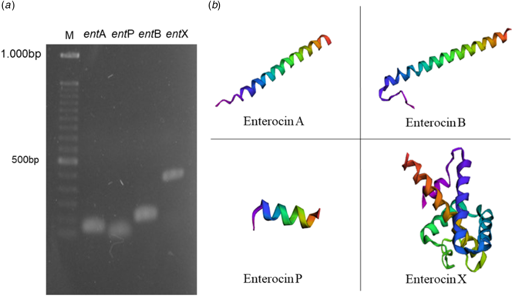
In the present study, we show that E. durans MF5 harbors all four tested enterocin-coding genes (entA, entB, entP, and entX: Fig. 1A), revealing that this strain can be considered a multiple enterocin producer. The nucleotide sequences of these genes analyzed by BLAST show high homology with the sequences corresponding to the consolidated bacteriocins of E. faecium, with up to 99% identity (Table 1). The predicted three-dimensional structure of the antimicrobial peptides corresponding to the genes present in isolate MF5 is illustrated in Fig. 1B, which predicts enterocins A, B, P, and X. One α-helix can be seen for enterocins A, B, and P, while enterocin X has five α-helices. These results indicate that E. durans isolate MF5 is a potential source of enterocins that resemble bacteriocins described by other Enterococcus species. Another interesting feature is that this isolate harbors entB and entX in addition to entA and entP, differentiating it from the literature that reports entA and entP genes as the most commonly prevalent together or individually in E. durans isolates (Sioziou et al., Reference Sioziou, Kakouri, Bosnea and Samelis2024). Bacterial strains that comprise genes encoding multiple enterocins may possess unique combinations of antimicrobial properties, as synergistic effects can be observed when more than one enterocin are used in combination (Hanchi et al., Reference Hanchi, Mottawea, Sebei and Hammami2018). For instance, according Sameli et al. (Reference Sameli, Skandamis and Samelis2021), strains of Enterococcus capable of producing multiple antilisterial enterocins are among the best LAB candidate species for use as bioprotective adjunct cultures against L. monocytogenes in traditional acid-curd cheese fermentations. In a recent study, Comerlato et al. (Reference Comerlato, Zhang, Walker, Mayne, Figeys and Brandelli2023) showed favorable effects of E. durans secretomes in improving gut microbial composition, showing its potential as a probiotic. In the dairy industry, bacteriocins and bacteriocin-producing LAB, including some Enterococcus species, can be applied to reduce the microbiological spoilage and improve safety of dairy products (Silva et al., Reference Silva, Silva and Ribeiro2018; Wu et al., Reference Wu, Pang, Wu, Liu and Zhang2022b).
Molecular characterization of the enterocins. (A) Agarose gel electrophoresis image of entA (enterocin A), entB (enterocin B), entP (enterocin P), and entX (enterocin X) amplicons obtained from E. durans MF5; (B) Predicted three-dimensional structure of the antimicrobial peptides corresponding to the genes present in isolate MF5 (Enterocin A, Enterocin B, Enterocin P, and Enterocin X) were established by the trRosetta v1.0 platform. The red and purple colors represent the C-terminal and N-terminal ends, respectively.

Enterococcus durans MF5 enterocin genes sequencing analysis and database search

Although there has been a lot of research on evaluating the use of bacteriocins, virulence factors and antibiotic resistance of the producing bacteria are important questions regarding their applications. Therefore, we tested the E. durans MF5 isolate for sensitivity to antibiotics and genotype in relation to some virulence genes. Testing revealed that E. durans MF5 is sensitive to eight clinically important antibiotics: ampicillin, chloramphenicol, erythromycin, gentamicin, penicillin, streptomycin, tetracycline, and vancomycin. Desirably, the presence of virulence genes, investigated by PCR, associated with cytolysin activator (cylA), and production of gelatinase (gelE) and hyaluronidase (hylA) was not detected (data not shown). Furthermore, E. durans MF5 does not produce hemolysin as shown in a previous study (Tosoni et al., Reference Tosoni, Perini, Terra, Katsuda, Furlaneto and Furlaneto-Maia2019), which are positive results regarding its safety.
Effect of enzymes, temperature, pH and surfactants on Ent-MF5 activity
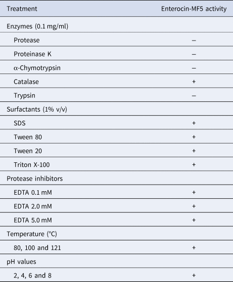
As shown in Table 2, the protein nature of Ent-MF5 was confirmed with loss of antibacterial activity after treatment with trypsin, chymotrypsin, protease and proteinase K. The effect of pH showed that Ent-MF5 remained active over a wide pH range from 2 to 8 as it showed no changes in the zones of inhibition compared to untreated Ent-MF5 (control: not shown), similar to enterocins that exhibit activities at low pH values of the gastrointestinal tract (Kang et al., Reference Kang, Seo, Lee, Kim, Kim, Han, Kang, Kim, Rhee, Chung and Park2009; Vijayendra et al., Reference Vijayendra, Rajashree and Halami2010). Furthermore, Ent-MF5 was heat stable during heat treatment (80, 100, and 121°C: Table 2), which makes it a promising candidate for food application. According to the literature, various enterocins obtained primarily from E. faecium and E. faecalis have high tolerance and resistance to temperature (Darbandi et al., Reference Darbandi, Asadi, Mahdizade, Ohadi, Talebi, Halaj, Darb, Ghanavati and Kakanj2022). For instance, Abanoz and Kunduhoglu (Reference Abanoz and Kunduhoglu2018) reported that bacteriocin KT11 from E. faecalis isolated from Kargı Tulum cheese remained stable after autoclaving at 121 ℃ for 30 min. On the other hand, Perumal and Venkatesan (Reference Perumal and Venkatesan2017) reported the loss of activity of bacteriocin CV7 from E. faecalis after treatment at 120°C for 15 min, reinforcing the importance of thermal stability for antimicrobial activity.
Factors affecting the antilisterial activity of enterocin produced by Enterococcus durans MF5 (Ent-MF5)

(+) activity; (−) absence of activity.
Ent-MF5 activity was unaffected by treatment with several surfactants, including 1% SDS, Triton X-100, Tween 20, and Tween 80, commonly used in the food industry (Table 2). Similar to our findings, it has been previously reported that enterocins remain stable after treatment with different surfactants (Perumal and Venkatesan, Reference Perumal and Venkatesan2017; Abanoz and Kunduhoglu, Reference Abanoz and Kunduhoglu2018). However, surfactants can inhibit bacteriocin activity, for example, Tween 20 reduced the AU/ml value of plantaricin by 4-fold (Daba et al., Reference Daba, Mostafa, Saleh, Elkhateeb, Awad, Nomiyama, Zendo and El-Dein2022).
Antimicrobial activity of Ent-MF5
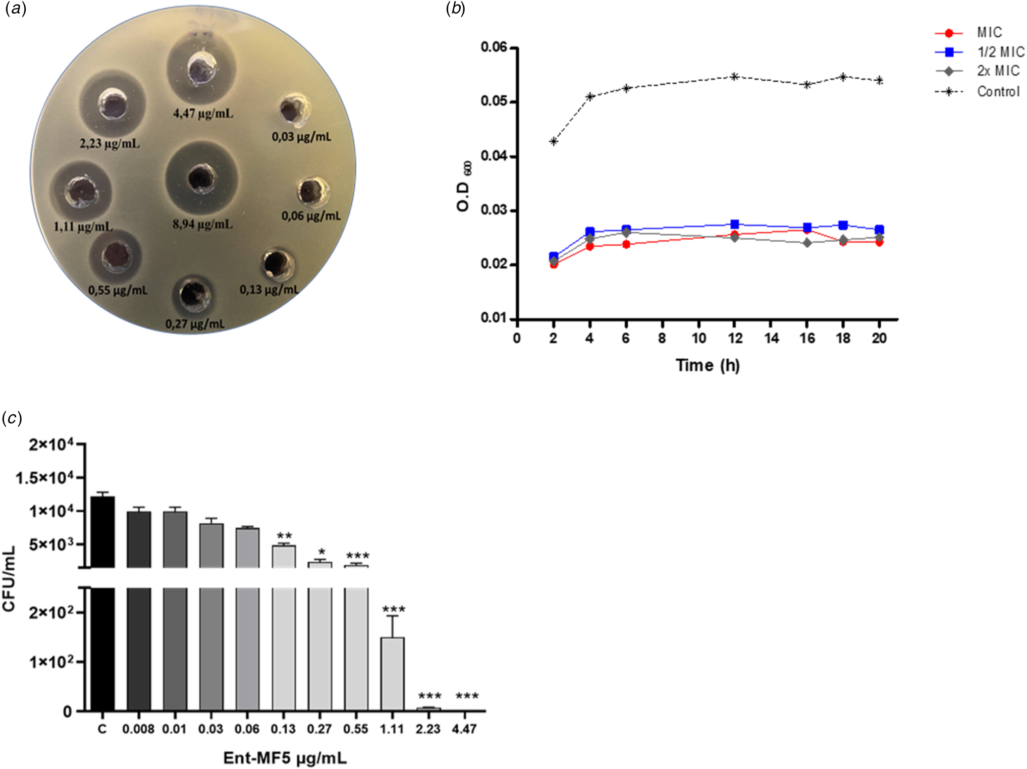
Ent-MF5 exhibited antimicrobial activity against all Listeria strains tested (L. monocytogenes CLIST 2048 and CLIST 2042 3b, L. ivanovii CLIST 2056, L. innocua CLIST 2050 and L. seeligeri CLIST 2067) with an MIC of 0.13 μg/ml. For L. monocytogenes 2042 3b, zones of inhibition as large as 17 mm were observed at the highest concentration of Ent-MF5 tested (8.94 μg/ml), as illustrated in Fig. 2A, and was the isolate selected for further experiments.
Antimicrobial activity of Ent-MF5 against L. monocytogenes 2042-3b. (A) Inhibition halos of L. monocytogenes post-treatment with Ent-MF5; (B) Growth curve in OD with the Ent-MF5 treatments (0.5× MIC, 1× MIC, and 2× MIC); (C) CFU of L. monocytogenes 2042-3b with different concentrations of enterocin-MF5 at 37°C for 18 h incubation. *P < 0.05; **P < 0.01; ***P < 0.001.

To further verify the antilisterial effect of Ent-MF5, the growth rate of L. monocytogenes was evaluated after treatment with Ent-MF5 (0.5× MIC, 1× MIC, and 2× MIC). A decrease in the OD of treated cells compared to the control (untreated bacteria) was observed during all incubation periods tested (2 to 20 h: Fig. 2B). Furthermore, our data revealed that Ent-MF5 has a bactericidal effect against L. monocytogenes with a significant reduction in surviving cells (CFU/ml) at concentrations equal to or greater than 0.13 μg/ml (0.13 to 8.94 μg/ml), as shown in Fig. 2C.
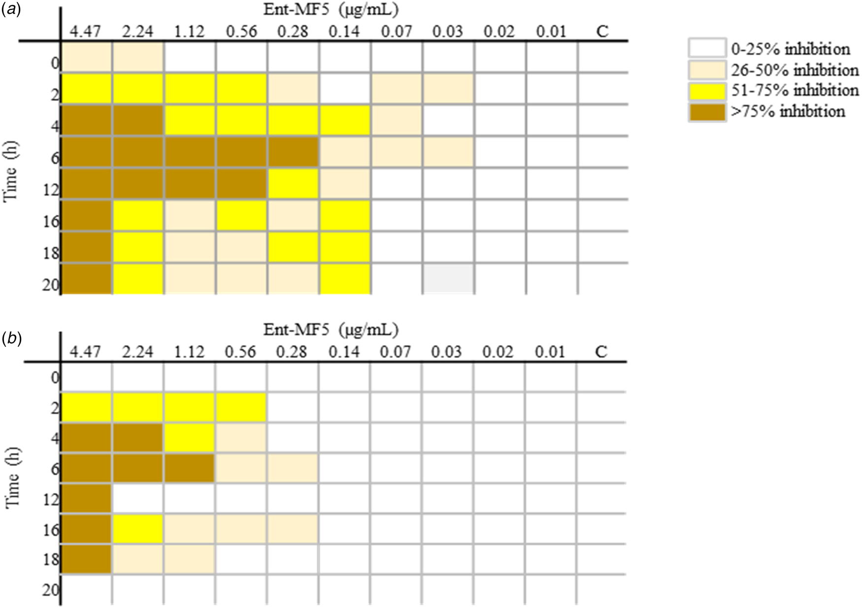
In Fig. 3, the percentage inhibition of L. monocytogenes growth by Ent-MF5 is presented in quartiles, where the higher the quartile, the higher the percentage inhibition. After exposure of L. monocytogenes to 4.47 μg/ml Ent-MF5 at time points starting at 2 h, a greater than 75% reduction in growth was observed, and a reduction of at least 26% after exposure to 0.14 μg/ml starting at 2 h of incubation (Fig. 3A). At the lowest concentrations of Ent-MF5 tested (0.03 μg/ml), growth rate was reduced, as was bactericidal activity at 0.02 μg/ml with up to 10% inhibition. Overall, the highest inhibitory activity was observed at concentrations of 4.47 and 2.24 μg/ml, with up to 100% inhibition. The lowest concentration of Ent-MF5 (0.01 μg/ml) had no inhibitory effect on L. monocytogenes. The most significant bactericidal effect determined by CFU counts was obtained after exposure to Ent-MF5 at 4.47 μg/ml at time points starting at 4 h. At shorter exposure times, even lower concentrations of Ent-MF5 (1.12 and 2.24 μg/ml) resulted in high rates of cell death (Fig. 3B). Our data show that Ent-MF5 presents effective antilisterial activity. Sioziou et al. (Reference Sioziou, Kakouri, Bosnea and Samelis2024) attributed the strong antilisterial capacity of an E. faecium strain to the fact that this strain possesses more than one enterocin-coding gene (entA/entB/entP). The low concentration values of Ent-MF5 with antilisterial activity are attractive for application in the control of foodborne pathogens. Previous studies in the literature reported MIC values of 5.26 μg/ml with enterocin TJUQ1 (Qiao et al., Reference Qiao, Du, Wang, Han and Zhou2020) and 50 μg/ml with enterocin AS-48 produced by E. faecium against L. monocytogenes (Gómez et al., Reference Gómez, Abriouel, Grande, Pulido and Gálvez2012). On the other hand, 0.3 μg/ml of enterocin CRL35 was effective against L. monocytogenes after 4 h of treatment (Minahk et al., Reference Minahk, Farías, Sesma and Morero2000).
Percentage inhibition of growth of L. monocytogenes CLIST 2042 3b after treatment with increasing concentrations of Ent-MF5, (A) OD600nm and (B) CFU, for a 20 h incubation period, corresponding to quartiles (Q1, Q2, Q3 and Q4). The colors mean the percentage inhibition from the lowest to the highest percentage, classified into quartiles, represented by white (Q1) and shades of yellow (Q2, Q3 and Q4), respectively. C: control (untreated bacteria).

Mode of action of Ent-MF5 against L. monocytogenes
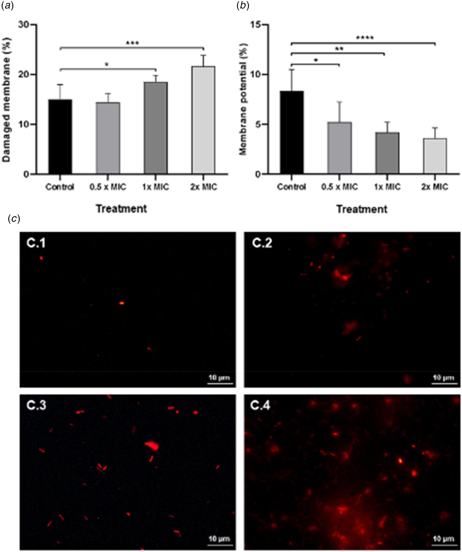
Bacteriocins primarily kill target cells by membrane permeabilization (Vasilchenko and Valyshev, Reference Vasilchenko and Valyshev2019). Thus, we examined whether Ent-MF5 induces permeation and disruption of the L. monocytogenes cell membrane. For this, PI and DiBAC4(3) uptake by flow cytometry were used to assess membrane damage, such as changes in membrane integrity and depolarization, respectively. As shown in Fig. 4, L. monocytogenes treatment with Ent-MF5 (1× MIC and 2× MIC) resulted in increased cell damage and decreased membrane potential, compared to the control (untreated cells: Fig. 4A, B) with a reduction of up to 3.16 ± 0.57% in membrane potential. Consistent increases in PI fluorescence intensity suggest alterations in membrane integrity and, as a consequence, disruption of the bacterial cell membrane. The membrane damage observed following exposure of cells to 2× MIC Ent-MF5 (Fig. 4A) was correlated with a significant reduction in cell viability (24 ± 2.19%, P < 0.05) as determined by CFU counts. The action of Ent-MF5 is illustrated using fluorescence microscopy, where red-labeled cells indicate reduced metabolic activity (Fig. 4 C.2–C.4). Sharma et al. (Reference Sharma, Kaur, Chadha, Kaur, Kaur and Kaur2021) identified a similar pattern of altered membrane permeability using an E. faecium enterocin 12a, indicating dissipation of the proton motive force by inducing membrane damage, which may lead to cell death.
Analysis of cell membrane damage by flow cytometry in L. monocytogenes 2042 3b, post-treated with Ent-MF5 for 18 h. (A) Cell membrane integrity was measured by flow cytometry in terms of % of cells stained with propidium iodide uptake-positive cells; (B) Cell membrane potential in terms of % DiBac uptake- positive cells; (C) fluorescence microscopy using propidium iodide of cells before treatment (C1) and after treatment with Enterocin MF5 at 0.5× MIC (C2), 1× MIC (C3), and 2× MIC (C4).

Bacteriocins from Enterococcus spp. have demonstrated activity against foodborne pathogens, however, there are limited reports on their characteristics and applications in the literature. This emphasizes the need to study and characterize new enterocin-producing strains and enterocins from diverse sources regarding their physicochemical properties, inhibitory spectrum and mode of action. To our knowledge, this is the first report to evaluate the antimicrobial activity of enterocin produced by E. durans from whey.
The antibacterial activity of bacteriocins in food depends on several factors, such as temperature, pH, proteases, salts, and metal ions (Yan et al., Reference Yan, Aizhan, Lu, Li, Wang, Yi, Shan, Liu, Zhou and Lü2021). Furthermore, commercial bacteriocins are often limited by their narrow antibacterial spectrum. From this perspective, Ent-MF5 showed effective activity against Listeria species that are associated with a wide variety of food sources, including dairy products. Furthermore, the activity of Ent-MF5 remained effective at different temperature and pH ranges, as well as in the presence of surfactants. Our results revealed that even low concentrations of Ent-MF5 (0.28 μg/ml) controlled Listeria growth at rates of more than 76–100%, making it highly promising for application in food matrices. Future research should focus on the application of Ent-MF5 in food matrices for the control of Listeria species, and on the expression of genes encoding enterocin from E. durans MF5, in vitro and in situ, aiming at its application in foods in addition to its enterocin (Ent-MF5).
In conclusion, the bacteria E. durans MF5 isolated from whey produces enterocin (Ent-MF5) active against pathogenic Listeria species. Ent-MF5 exhibited good thermal and pH stability, and was not affected by surfactants. Furthermore, Ent-MF5 showed efficient antilisterial and bactericidal activity at low levels in the first 4 h of exposure, due to changes in cell membrane integrity, which makes it an excellent candidate for application in the food preservation sector.
Acknowledgments
This work was supported by Fundação Araucária/SETI/Governo do Paraná–Brazil and PROPPG/UEL-Brazil. NAAS, LC and MHN were fellowship holders of Coordenação de Aperfeiçoamento de Pessoal de Nível Superior-Brasil (CAPES). MCF is grateful to CNPq for the PQ fellowship.