Introduction
The impact of major depressive disorder (MDD) on both the individual and society is considerable and it continues to be a major cause of morbidity in older people. Although some risk factors have been identified, our understanding of the causes of MDD remains poor at all ages. Whilst the precise neurobiological mechanisms underlying MDD are not fully understood, numerous co-morbidities have also been linked with MDD, including increased incidence of acute myocardial infarction and stroke (Plante, Reference Plante2005) and diabetes (Ali et al., Reference Ali, Stone, Peters, Davies and Khunti2006), The “vascular depression” model has been proposed to describe vascular damage to brain circuitry associated with affective regulation (Alexopoulos et al., Reference Alexopoulos, Meyers, Young, Kakuma, Silbersweig and Charlson1997; Baldwin and O'Brien, Reference Baldwin and O'Brien2002). Intriguingly, this has suggested a different series of pathological events that lead to MDD and possibly a different set of risk factors in MDD, depending on age.
Neural circuitry in MDD
Five parallel frontal-subcortical circuits have been proposed in primates, three of which are associated with cognitive and emotive function in humans: the dorsolateral prefrontal cortex (DLPFC), orbital frontal cortex (OFC) and anterior cingulate cortex (ACC) circuits (Alexander et al., Reference Alexander, DeLong and Strick1986). Components of these circuits are integral to mood regulation and have been implicated in underlying the symptomatic expression of MDD. The DLPFC circuit originates in Brodman's areas (BA) 9 and 46 of the prefrontal cortex, and is associated with executive functions such as problem solving, organization and working memory. Fibers extend from the DLPFC to the dorsolateral aspect of the caudate nucleus; projections from the rostral caudate then extend to the dorsomedial globus pallidus and the rostral substantia nigra, which in turn project fibers extending to the parvocellular portions of the of the ventral anterior and medial dorsal thalamus respectively. Projections then arise from the respective areas of the thalamus back to the DLPFC, thus completing the circuit (Bonelli and Cummings, Reference Bonelli and Cummings2007; Middleton and Strick, Reference Middleton and Strick2002). The OFC circuit, which is thought to facilitate in the integration of limbic and emotional information into behavioral responses, begins in BA 10, where projections are sent to the ventromedial head of the caudate nucleus; from here, projections are sent to the lateral dorsomedial globus pallidus interna, then rostromedial substantia nigra, before projecting to the ventral anterior, then the medial dorsal magnocellular regions of the thalamus. The loop is completed as thalamic projections are then sent back to the OFC (Bonelli and Cummings, Reference Bonelli and Cummings2007). In the ACC circuit, fibers from the areas within the ACC project to the ventral striatum, then to the ventral pallidum, rostrodorsal substantia nigra and rostrodorsal globus pallidus interna, before returning to the ACC via paramedial regions of the mediodorsal nucleus (Bonelli and Cummings, Reference Bonelli and Cummings2007). The ACC is a functionally diverse region, and two distinct functional subdivisions have been described: the dorsal cognitive division (areas 24b′–c′ and 32′), and rostral-ventral affective division (rostral areas 24a–c and 32, and ventral areas 25 and 33) (Vogt et al., Reference Vogt, Finch and Olson1992). Consistent with its functional complexity, the ACC possesses a high degree of cytoarchitectural heterogeneity across its anatomical subdivisions: the supragenual, subgenual and pregenual ACC (Gittins and Harrison, Reference Gittins and Harrison2004b; Palomero-Gallagher et al., Reference Palomero-Gallagher, Vogt, Schleicher, Mayberg and Zilles2008). Care must therefore be taken on the precise location when selecting ACC tissue for morphometric analysis.
Volumetric studies using magnetic resonance imaging have detected significant decreases in volume in several cortical and subcortical structures implicated in affective functioning in MDD patients (Ballmaier et al., Reference Ballmaier2004; Taylor et al., Reference Taylor2007; Drevets et al., Reference Drevets, Price and Furey2008; Kim et al., Reference Kim, Hamilton and Gotlib2008). Furthermore, structural changes, such as white matter hyperintensities (WMH), have been associated with late-life MDD in several key areas involved in affective circuitry (O'Brien et al., Reference O'Brien, Ames, Schweitzer, Colman, Desmond and Tress1996; Krishnan et al., Reference Krishnan2006; Sheline et al., Reference Sheline2008). WMH are more common in late-life MDD than in younger MDD patients (Teodorczuk et al., Reference Teodorczuk2007; Herrmann et al., Reference Herrmann, Masurier and Ebmeier2008) or control subjects (Herrmann et al., Reference Herrmann, Masurier and Ebmeier2008; Sheline et al., Reference Sheline2008). Large-scale cross-sectional studies have also reported that basal ganglia lesions (Steffens et al., Reference Steffens, Helms, Krishnan and Burke1999) and subcortical white matter changes (de Groot et al., Reference de Groot, de Leeuw, Oudkerk, Hofman, Jolles and Breteler2000) are associated with MDD, and especially in the elderly (de Groot et al., Reference de Groot, de Leeuw, Oudkerk, Hofman, Jolles and Breteler2000).
Methodological considerations in morphometric postmortem analysis
Whilst imaging technology has proved a valuable tool in detecting gross macrostructural changes in various brain areas, it is restricted by a limited spatial resolution and the inability to detect morphological changes at a cellular level. Conversely, morphometric analysis allows for the detection of neuronal and glial cell population changes at a high degree of resolution. The term “stereology” describes a methodology that estimates changes in three-dimensional (3-D) morphology based on observations in two-dimensional (2-D) sections. “Design-based” stereology is planned in a manner making it unnecessary to have prior knowledge of size, shape, spatial orientation or spatial distribution of the object examined, thus eliminating potential sources of systematic error. This is in contrast to “assumption-based” 2-D analysis that assumes a relationship between the number of profiles of an object seen on a section and the number of objects within the whole structure that is sectioned (West, Reference West1999).
Despite the merits of such analysis, inherent problems exist in the use of human postmortem tissue in such studies. Whilst the 3-D morphometric studies featured in this paper follow the principles behind stereology, few can be strictly labelled “stereological” studies and are often referred to as “morphometric” or “morphological” studies. This is partly due to the fact that the postmortem tissue is sectioned from pre-defined paraffin or celloidin-embedded blocks, making serial sectioning of whole structures unfeasible. It is also virtually impossible to isolate precisely continuous cortical structures and surrounding features (e.g. ventricles, gyri/sulci) often determine where sectioning occurs. Other possible methodological confounders that must be considered when using postmortem tissue include the interval between death and brain fixation, as well as the length of time in fixative before analysis takes place. Neuronal size and shape have also been positively correlated with brain pH (Gittins and Harrison, Reference Gittins and Harrison2004a), meaning care should be taken when studying postmortem tissue with acidosis. In addition, there is the difficulty and cost of obtaining such postmortem tissue from depressed subjects initially, as this requires substantial planning and infrastructure if subjects with a reliable MDD diagnosis are to be recruited. These factors explain why there are very few brain banks worldwide with sufficient numbers of depressed subjects to conduct such studies.
Morphometric studies in MDD
3-D morphometric studies examining cytoarchitecture in all age groups have generally focused on components of the DLPFC, OFC and ACC circuitry, based on the findings from imaging studies mentioned earlier. Most morphometric studies in MDD have been conducted in younger and mixed age adult populations, comprising younger, middle age (and occasionally elderly adults), with only a few recent studies (Bowley et al., Reference Bowley, Drevets, Ongur and Price2002; Rajkowska et al., Reference Rajkowska, Miguel-Hidalgo, Dubey, Stockmeier and Krishnan2005; Khundakar et al., Reference Khundakar, Morris, Oakley, McMeekin and Thomas2009) obtaining tissue from patients with the mean age in both groups exceeding 60 years. This review attempts to stratify the findings of the various studies on cell morphology in MDD according to age and will assess possible differences in neuronal and glial cell changes in the two age brackets.
Studies in younger age groups
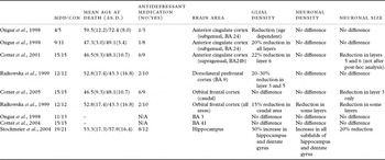
A consistent finding in studies of younger group patients has been that of reduced glial cell density in cortical areas in patients with MDD. These changes, however, have not been accompanied by consistent corresponding changes in neuronal density or volume, though some studies suggest some evidence of neuronal pathology in individual cortical layers (Table 1).
Summary of morphometric studies examining glial cell density and neurone density and volume in younger depressed patients

MDD = major depressive disorder; CON = control; BA = Brodmann area.
Two major studies have assessed morphological alterations within the subgenual (Ongur et al., Reference Ongur, Drevets and Price1998) and supragenual (Cotter et al., Reference Cotter, Pariante and Everall2001) subdivisions of the ACC. Ongur and colleagues obtained postmortem tissue from the subgenual ACC (BA 24) from two tissue banks and performed separate analyses on both sets. Five controls (one obtained from the Harvard Brain Tissue Resource Centre and four from Washington University Department of Pathology), and four MDD patients (all from Harvard) were used in a preliminary study. The study found a marked decrease in the density of glial cells across all layers. However, the mean age in the study was high and there was a substantial difference in mean age between the control and MDD group, with the control group averaging 15 years older. Consequently, the respective age of the individuals involved in the study may indicate an age-associated increase in glial cells in the control group.
In the subsequent study using 11 control and 10 MDD subjects (derived from the Stanley Foundation), the group found a significant reduction in mean glial cell number and density in MDD patients in the subgenual ACC across all layers (nine MDD vs. ten control) (Ongur et al., Reference Ongur, Drevets and Price1998). No changes were found in number or size of neurons in the subgenual ACC in either study.
Cotter et al. (Reference Cotter, Pariante and Everall2001) examined morphometric changes in the supragenual ACC, using tissue from the Stanley Foundation. They found lamina-specific changes, with a significant decrease in glial cell density detected in layer 6 in MDD patients (15 MDD vs. 15 control). They also detected a reduction in neuronal size in layers 5a and 6 of the ACC in MDD; however, this reduction was not significant after multiple comparison analysis adjustments.
Reductions in glial density have also been found in other cortical areas associated with the neurobiology of MDD. Rajkowska et al. (Reference Rajkowska1999) examined cytoarchitectural changes in frontal cortical areas in 12 mixed age MDD and 12 control subjects: the rostral OFC, the caudal OFC and the DLPFC. In accordance with the Stanley Foundation study in the ACC (Cotter et al., Reference Cotter, Pariante and Everall2001), the study found lamina-specific reductions in glial cells in the DLPFC and the caudal part of the OFC (Rajkowska et al., Reference Rajkowska1999). The reductions in glial cell density, however, varied according to brain area. In the DLPFC, substantial reductions were present in layers 3 and 5. In the OFC, reductions were limited to caudal layers 5 and 6. No significant reductions were found in the rostral region. The alterations in glial cell density in MDD patients were accompanied by a corresponding decrease in neuronal size in specific cortical layers. An increase in the density of the larger glial cells was also found in MDD subjects, and this was attributed to an enlargement of the remaining glial cells as a mechanistic response to the apparent neuronal pathology.
A subsequent study (Cotter et al., Reference Cotter, Hudson and Landau2005) conducted in Stanley Foundation subjects examined the six layers of the caudal OFC in 14 in mixed aged MDD patients and 14 control subjects. They found further evidence suggesting lamina-specific neuronal pathology in the caudal OFC through a reduction in neuronal size in layer 3. In contrast to the previous study, no changes were found in either glial cell density or neuronal density in any layer of caudal OFC.
Studies have been performed in other areas of the cortex not associated with MDD. As expected, no changes in neuronal or glial cell morphology were found between depressed and control groups in Heschl's gyrus (BA 41) (Cotter et al., Reference Cotter, Hudson and Landau2005) or the somatosensory cortex (BA 3b) (Ongur et al., Reference Ongur, Drevets and Price1998), suggesting localized cortical pathology in MDD (Table 1).
Few studies have examined subcortical areas and to our knowledge, only two areas, the hippocampus and thalamus have been examined in younger or mixed age patients with MDD (and only one, the amygdala (Bowley et al., Reference Bowley, Drevets, Ongur and Price2002), in patients with a mean age of over 60 years). The paucity of 3-D studies in these subcortical areas is particularly disappointing because volumetric changes have been reported in neurons of the hippocampal formation and thalamus in MDD patients (Stockmeier et al., Reference Stockmeier2004; Young et al., Reference Young, Holcomb, Yazdani, Hicks and German2004)
A study on the thalamus in MDD in patients with a younger mean age was limited to assessing possible changes in neuronal pathology (Young et al., Reference Young, Holcomb, Yazdani, Hicks and German2004). Glial pathology was not examined. Serial sections obtained from the entire mediodorsal and anteroventral/anteromedial nuclei of the thalamus were studied and the total number estimated in eleven controls and 13 MDD patients. There was a significant increase in neuronal number in both mediodorsal and anteroventral/anteromedial nuclei of the thalamus in MDD subjects. No changes were found in neuronal density or size. As no distinction was made between neurons in the thalamic nuclei, it is impossible to say whether the change occurred in interneurons or projection neurons that comprise the thalamus (Young et al., Reference Young, Holcomb, Yazdani, Hicks and German2004).
Stockmeier et al. (Reference Stockmeier2004) found a significantly increased density of granule cells and glial cells in the dentate gyrus and increased density of pyramidal neuron and glial cells in all subfields of the hippocampus, coupled with a reduction in neuronal size, in MDD subjects. However, these changes were present only after correction for differential shrinkage of the frozen sections between groups. Further studies are needed in subcortical areas in order to confirm these intriguing findings, which appear to differ from those in cortical areas.
The effect of pharmacological treatment (e.g. antidepressant medication, mood stabilizers, neuroleptics) was performed in all of the studies in younger age groups by covariate analysis. No significant effects were found in any study; however, one study (Ongur et al., Reference Ongur, Drevets and Price1998) was unable to eliminate the possibility of psychotropic effects on glial cell reduction due to low medication-free sample numbers in the MDD group.
Studies in older age groups
In contrast to studies examining younger and mixed age MDD patients, studies performed on patients with a mean age of over 60 years in both groups have reported no evidence of reduced glial density in cortical areas but one study did find a significant reduction in the amygdala (Bowley et al., Reference Bowley, Drevets, Ongur and Price2002) (Table 2). Rajkowska et al. (Reference Rajkowska, Miguel-Hidalgo, Dubey, Stockmeier and Krishnan2005) examined the glial and neuronal density in 15 subjects and 11 age-matched controls aged over 60 years (except one patient aged 58 years) in the six laminae from the rostral region of the OFC (BA 47) (Rajkowska et al., Reference Rajkowska, Miguel-Hidalgo, Dubey, Stockmeier and Krishnan2005). Furthermore, pyramidal and non-pyramidal neurons were distinguished on the basis of morphology. Prefrontal pyramidal neurons are mainly glutamatergic, and as well as projecting to other cortical areas, also project to the striatum as part of frontal-subcortical circuitry mentioned earlier. Interestingly, the pathological changes in the elderly MDD patients differed slightly from those in the younger patient groups from the previous study. Unlike the modest reduction seen in caudal areas of the OFC in younger patient groups, there was no difference in glial cell density between the MDD patients and controls. A significant reduction in pyramidal neuron density was found in MDD subjects in the OFC overall, largely due to significant reductions in layer 3 and 5 of the OFC. The study also showed a negative correlation between age and overall neuronal density in both MDD and control groups.
Summary of morphometric studies examining glial cell density and neurone density and volume in older depressed patients

*No SD figures available.
In accordance with the earlier study (Rajkowska et al., Reference Rajkowska, Miguel-Hidalgo, Dubey, Stockmeier and Krishnan2005), Khundakar et al. (Reference Khundakar, Morris, Oakley, McMeekin and Thomas2009) used a similar methodological approach in the DLPFC but also estimated neuronal volumes in 17 MDD patients and 10 matched controls all aged over 60 years of age in tissue obtained from the Newcastle Brain Tissue Resource. As in the previous study, no differences in glial cell density were found between groups. Furthermore, evidence of pyramidal cell pathology was found through a reduction in volume across all layers. Analysis of individual cortical layers revealed lamina-specific reductions in pyramidal neuron volume in layer 3, and more markedly, layer 5 of the DLPFC.
Components of the limbic system, the amygdala and the entorhinal cortex, have been examined in tissue blocks acquired from either left or right hemispheres of elderly MDD (seven in the amygdala, six in the entorhinal cortex) and control (ten) cases (though some younger patients were included in the control group) (Bowley et al., Reference Bowley, Drevets, Ongur and Price2002). Glial density was significantly reduced in the amygdala in MDD cases, mainly as a result of reductions in the sections obtained from the left hemisphere. Glial density was also reduced in the entorhinal cortex, though not significantly. No changes were found in neuronal density in either the amygdala or the entorhinal cortex and neuronal size was not analyzed. Further histopathological examination appeared to suggest that two of the subjects (one MDD and control) had indicators of preclinical Alzheimer's disease. Both of the subjects had higher glial densities on average in both diagnostic groups.
As with younger age groups, all of the three studies in older age groups factored the effect of pharmacological treatment into the analyses on cellular changes. Co significant treatment effect was found in any study, though one study (Bowley et al., Reference Bowley, Drevets, Ongur and Price2002) did find that the mood stabilizers valproate and lithium had an effect on modulating glial density reduction in bipolar disorder.
Patterns emerging from postmortem morphometric studies
Glial cells
Whilst it is difficult to establish any definitive patterns of cellular changes in the brains of MDD patients due to the limited number of these morphometric studies, certain trends exist in glial pathology among differing age groups in published studies. Thus, the glial cell reduction reported in cortical areas such as the DLPFC and OFC (as well as the subgenual and supragenual ACC) in studies in younger patient groups has not been found in either of the published studies in older adults; indeed, these studies did not even suggest a trend to such a reduction in glial density. The Nissl staining method used in 3-D stereological procedures does not allow for differentiation between astrocytes, oligodendrocytes and microglia. However, as astrocytes are the most abundant of cortical glial cells, one could speculate that they may be significantly affected in these studies. Furthermore, immunohistochemical analysis has shown an increase in the density and areal fraction for glial fibrillary acidic protein (GFAP) packing density in older MDD patients, than younger MDD patients and controls, suggesting a difference in astroglial pathology in MDD amongst different age groups (Miguel-Hidalgo et al., Reference Miguel-Hidalgo2000). A further GFAP study in elderly MDD patients (Davis et al., Reference Davis, Thomas, Perry, Oakley, Kalaria and O'Brien2002) showed increased GFAP immunoreactivity in a single layer (1) of the DLPFC compared with age-matched controls. Further immunohistochemical research is needed to establish the role of oligodendrocytes and microglia in MDD.
Taken together, data from the immunohistochemical and 3-D morphometric studies suggest that a biphasic glial response may occur in cortical areas in MDD, dependent on age. The significant reductions in glial numbers in cortical areas found in studies with younger patient groups have not occurred in elderly patient groups (though the ACC has not yet been examined), suggesting a different neurobiology may be associated with cortical glial differences in MDD at different ages. Enhanced glucocorticoid levels, as a result of hyperactivity of the hypothalamic-pituitary-adrenal (HPA) axis, have been implicated in the pathophysiology of MDD (McQuade and Young, Reference McQuade and Young2000). Chronic glucocorticoid administration has been shown to result in degeneration of hippocampal neurons (Sapolsky et al., Reference Sapolsky, Romero and Munck2000) and increased neuronal vulnerability to ischemic damage (Ahlbom et al., Reference Ahlbom, Gogvadze, Chen, Celsi and Ceccatelli2000). Glucocorticoid receptors are also present on glial cells (Cadepond et al., Reference Cadepond2002), and corticosteroids have been shown to reduce astrocytic activation (Laping et al., Reference Laping, Teter, Nichols, Rozovsky and Finch1994) and inhibit glucose uptake (Virgin et al., Reference Virgin1991), leading to impaired glial cell function and possible glial cell loss. Therefore, a prolonged elevation of corticosteroid levels, as found in MDD patients, may have a detrimental effect on glial cell function and may exacerbate pre-existing glucocorticoid-influenced neuronal dysfunction. As astrocytic glial cells also possess 5-hydroxytryptamine (5-HT), noradrenaline and dopamine transporters (Inazu et al., Reference Inazu, Takeda, Ikoshi, Sugisawa, Uchida and Matsumiya2001), monoaminergic transmission may be affected by, or indeed result in, glial cell loss. Glial cells also possess glutamate transporters, and glial glutamate uptake is the primary mechanism for glutamate removal from the synapse (Krystal et al., Reference Krystal2002). Reductions in glial cell numbers could therefore result in high levels of extracellular glutamate and consequent excitotoxic damage.
Conversely, as no changes have been found in glial cell density in cortical areas in studies with older patients, one could speculate that either glial cell density is not significantly affected by MDD in elderly patients in cortical areas, or that gliosis may partially counteract a reduction in earlier life. Gliosis is a hallmark of the brain's response to a range of insults, such as neurotrauma, brain ischemia and neurodegenerative diseases. As patients who exhibited such disease and damage were excluded from all three elderly MDD morphometric studies, one may assume that any gliosis (balancing out earlier life glial reduction) in elderly patients may be as a result of a more subtle damage, such as an ischemic/hypoxic process as proposed in the “vascular depression” hypothesis (Alexopoulos et al., Reference Alexopoulos, Meyers, Young, Kakuma, Silbersweig and Charlson1997).
Neurons
In contrast to the changes found in glial cells, the morphological changes in neurons between younger and older MDD patient groups appear less consistent and more subtle. Significant reductions in either neuronal cell density or volume have only been found in prefrontal cortical areas (DLPFC and OFC) when either specific cell type or individual cortical layers are analyzed. Interestingly, in studies in cortical areas in elderly patients that classified cell morphology (Rajkowska et al., Reference Rajkowska, Miguel-Hidalgo, Dubey, Stockmeier and Krishnan2005; Khundakar et al., Reference Khundakar, Morris, Oakley, McMeekin and Thomas2009) pyramidal neurons appeared to be selectively affected, especially in layers 3 and 5. Glutamatergic pyramidal projection neurons extend from layers 3 and 5 (especially layer 5) of the DLPFC and OFC to the striatum, where they synapse with the γ-aminobutyric acid (GABA)ergic neurons of the dorsolateral and ventromedial caudate nucleus respectively (Bonelli and Cummings, Reference Bonelli and Cummings2007) as part of the fronto-cortical circuitry implicated in mood regulation mentioned earlier. Abnormalities in the glutamatergic circuitry have long been implicated in the pathophysiology of MDD, and reductions in size and/or density could be associated with a reduced activity in circuitry associated with MDD. In order to reach subcortical areas pyramidal neurons traverse through white matter areas thus leaving them vulnerable to damage, such as from WMH found in neuroimaging studies. Our earlier report has found such white matter damage to be ischemic in origin, especially in depressed subjects (Thomas et al., Reference Thomas2002). Immunohistochemical examination has shown an upregulation in inflammatory markers, intercellular adhesion molecule (ICAM)-1 (Thomas et al., Reference Thomas2000) and interleukin (IL)-1β (Thomas et al., Reference Thomas, Davis, Morris, Jackson, Harrison and O'Brien2005) in cortical areas of elderly MDD patients, which may be due to inflammatory changes caused by alterations in vascular function (Thomas et al., Reference Thomas2002). Therefore, WMH and inflammatory processes may be associated with demyelination and dysfunction of the axons of the neurons resulting in grey matter volumetric and densitometric changes, as seen in studies on elderly MDD patients (Rajkowska et al., Reference Rajkowska, Miguel-Hidalgo, Dubey, Stockmeier and Krishnan2005; Khundakar et al., Reference Khundakar, Morris, Oakley, McMeekin and Thomas2009). Such findings would be consistent with the “vascular depression” model (Alexopoulos et al., Reference Alexopoulos, Meyers, Young, Kakuma, Silbersweig and Charlson1997) (Figure 1).
Morphological changes in pyramidal neurones in layers 3 and 5 in depression. The figure shows a simplified hypothetical scheme, whereby ischemic/hypoxic damage causes white matter hyperintensities and periventricular damage and subsequent demyelination and dysfunction of the axons of neurones, resulting in grey matter volumetric and densitometric changes in late-life depression.

Glutamate is also the major key element in the regulation of brain-derived neurotrophic factor (BDNF) (Zafra et al., Reference Zafra, Hengerer, Leibrock, Thoenen and Lindholm1990; Khundakar and Zetterstrom, Reference Khundakar and Zetterstrom2006). BDNF and its receptor, tropomyosin-related kinase (trk) B are found pre- and post-synaptically on glutamategic neurons, and are modulators of dendritic growth and morphology; BDNF, in turn, regulates glutamatergic function. Dysfunction of this bidirectional relationship may disrupt the modulation of synaptic transmission and plasticity in affective circuitry. One may therefore speculate that this may result in, or be a symptom of, the morphological pyramidal neuron changes found in these studies.
Conclusion
Whilst establishing any firm conclusions from the limited data available from postmortem studies in MDD patients would be highly speculative, certain trends do exist in the morphological changes found in MDD between studies with younger patients and those with older adults. Reductions in glial cells found in cortical areas of younger subject groups with MDD against age matched controls have not been found in older patient groups. Changes in neuron pathology in cortical areas appear to be layer-specific and dependent on cell type, with pyramidal neurons predominantly affected. Reductions in either neuronal density or volume have been found among elderly patient groups. The apparent differences in the morphological changes between younger and elderly patients perhaps suggest a differing pathological basis in MDD, dependent on age, in accordance with the “vascular depression” hypothesis.
Conflict of interest
None.
Description of authors' roles
AK and AT were equally involved in the research, analysis and writing of this manuscript.