Introduction
Hypospadias is the abnormal placement of the urethral opening, resulting from disrupted urethral closure during development. It affects 1 in 125 live male births in developed countries, and its incidence doubled in the USA between 1970 and 1993,Reference Paulozzi1 with a similar prevalence and increase in incidence reported in Australia between 1980 and 2000.Reference Nassar, Bower and Barker2 This increase is too rapid to be accounted for by genetic mutations and is not due to increased reporting.Reference Paulozzi1 Males clinically diagnosed with hypospadias often exhibit reduced fertility and chordee (curvature of the penis), which can have profound medical and psychological consequences for the individual and family, as well as placing a significant burden on the health care budgets.Reference Bouty, Ayers, Pask, Helourry and Sinclair3
Correct urethral closure is dependent on androgens and blocked by exogenous oestrogens.Reference Govers, Phillips and Mattiske4 Environmental toxicants, namely, endocrine disrupting chemicals (EDCs), can interfere with both androgen and oestrogen signalling pathways.Reference Gore, Chappell and Fenton5 EDCs are increasing exponentially in their abundance and are highly pervasive in our environment, due to their beneficial properties, such as being effective plasticisers, preservatives or pesticides, and their use in numerous industrial and manufacturing processes.Reference Gore, Chappell and Fenton5 Hence, it is postulated that exposure to EDCs may explain the increasing incidence of hypospadias. This statement is supported by a growing body of evidence from human and animal studies, most notably the increased incidence of hypospadias and other male reproductive tract abnormalities (i.e. cryptorchidism) in the sons of the >10 million women prescribed the synthetic oestrogen diethylstilbestrol, to reduce the risk of miscarriage, as well as females and their offspring exposed to phytoestrogens, polychlorinated biphenyls, perflurooctanoic acid, and especially, organochloride pesticides used in agriculture (e.g. dichlorodiphenyltrichloroethane, hexachlorobenezene and methoxychlor) (see reviewsReference Gore, Chappell and Fenton5–Reference Bergman, Heindel, Jobling, Kidd and Zoeller8).
Atrazine (2-chloro-4-ethylamino-6-isopropylamino-1,3,5-triazine, ATR) is one of the most commonly used herbicides in agriculture in the USAReference Farruggia, Rossmeisl, Hetrick, Biscoe and Branch9 and Australia.Reference Mnif, Hassine, Bouaziz, Bartegi, Thomas and Roig10 ATR is known to have endocrine disrupting effects in amphibians, fish, reptiles and rodents.Reference Gore, Chappell and Fenton5 ATR can increase the expression of the gene cytochrome P450 family 19 subfamily A member 1 (Cyp19a1) in vivo in a wide spectrum of animals ranging from fish to rodents,Reference Jin, Wang and Fu11 leading to an increase in the enzyme aromatase, that is responsible for converting testosterone to oestradiol.Reference Holloway, Anger, Crankshaw, Wu and Foster12 In addition, ATR has anti-androgenic properties, as it causes a decrease in the conversion of testosterone to the more potent dihydrotestosterone, via suppression of the enzyme 5α-reductase.Reference Kniewald, Osredecki, Gojmerac, Zechner and Kniewald13 Thus, ATR can have both oestrogenic and anti-androgenic actions, which can markedly affect male reproductive development. Here, we examined the effects of ATR exposure, specifically on penis development which has not yet been investigated.
Materials and methods
Animal treatments
Pregnant C57BL/6J mice were treated with atrazine (ATR, 5 mg/kg bw/day; Sigma-Aldrich, NSW, Australia, n = 3) in distilled water or vehicle control (0.5% dimethyl sulfoxide, Control, n = 3) in distilled water from embryonic day 9.5 through to weaning. Male mice were housed individually and maintained on ATR water (n = 7) or control (n = 8) water treatment from weaning (3 weeks of age) until sexual maturity (26 weeks post-partum). All mice were maintained under a 12 h light:12 h dark lighting regimen and fed ad libitum a soy-free diet (Specialty Feeds, Perth, WA, Australia) throughout their gestation, lactation and post-weaning to avoid the effects of phytoestrogens. Water intake and food consumption were monitored for the duration of the study. Body weights were recorded weekly.
Analyses of body and testis weight, and examination of external genitalia
At 26 weeks of age, mice were anesthetised with isoflurane (Forane; Abbott, NSW, Australia) and euthanised via cervical dislocation. At post-mortem, body weight was recorded, testes dissected and weighed, while the penis was dissected and imaged for gross morphology (n = 7 ATR and n = 8 control males). Comprehensive assessment of the morphological distinctions between the mouse and human penis, including similarities in their embryological origin, allows for the accurate comparison of analogous structures and hypospadias phenotypes.Reference Blaschko, Mahawong and Ferretti14,Reference Phillips, Wright, Gradie, Johnston and Pask15 Mouse penis development differs from that of humans in that they have an os penis bone and male urogenital mating protuberance (MUMP). MUMP development is highly androgen-dependent and forms a cartilaginous distal projection from the os penis bone that ensures the correct placement of the penis in the female urogenital tract for effective fertilisation.Reference Phillips, Wright, Gradie, Johnston and Pask15 Thus, measurement of the MUMP provides a reliable biological proxy for how endocrine factors can affect the penis. The MUMP was imaged using a Nikon Digital Sight DS-U3 (Nikon Australia, VIC, Australia), and all images were analysed and MUMP height measured with NIS Elements Analysis D 4.300.00 64-bit software.
The penis was then fixed in 4% paraformaldehyde and processed for histology. The tissue was serially sectioned at 7 μm and stained with Hematoxylin and Eosin Y (H & E; Sigma-Aldrich, NSW, Australia) following standard protocols.Reference Fischer, Jacobson, Rose and Zeller16 To determine the presence of hypospadias, we examined how proximal the urethral opening persisted in the penis using the os penis bone as a distal landmark. This is a well-established standard methodology for determining hypospadias in the mouse penis.Reference Govers, Phillips and Mattiske4,Reference Mahawong, Sinclair and Li17
Statistical analyses
All data were tested for normality using the Shapiro–Wilk test. Data subjected to repeated measures analysis were tested for sphericity using the Mauchly’s sphericity test. Body weight, cumulative weight gain, as well as food and water intake were analysed using a repeated measures ANOVA, with treatment as a fixed factor, using SAS version 9.2 (SAS Institute). Birth weight and relative testis weight at post-mortem were analysed using a one-way ANOVA. Non-normally distributed data (survival to weaning and MUMP height) were analysed by a Mann–Whitney test using R studio version 1.0.143. Sex ratio was compared with an expected 50:50 ratio as well as between groups by a corrected χ 2 procedure and was double-checked by binominal analysis. Results were considered significant when P ≤ 0.05, and all data are expressed as mean ± SEM, unless otherwise stated.
Results
Litter sex ratio, birth weight and survivability
ATR treatment had no effect on the sex ratio of pups when compared with controls (proportion male; control 0.44 (n = 8 male, n = 10 female), ATR 0.41 (n = 7 male, n = 10 female; P > 0.1)). Also, ATR treatment had no effect compared with controls on average pup birth weight (control 4.1 ± 0.2 g, n = 18; ATR 3.9 ± 0.4 g, n =17; P > 0.1) or survival to weaning (control n = 18/18 (100%); ATR n = 17/17 (100%; P > 0.1)).
Food and water intake, growth rate, body and relative testes weights at post-mortem
Food and water intake did not differ between control and ATR-exposed males (P > 0.1). Average water intake for control and ATR-exposed males was 25.9 ± 0.6 ml/week and 25.7 ± 0.8 ml/week, respectively, whilst average food intake was 27.6 ± 0.9 g/week and 27.9 ± 0.9 g/week, respectively. Equally, cumulative weight gain (growth rate from weaning to post-mortem; data not shown), body weight (control 37.2 ± 0.8 g, ATR 36.7 ± 1.1 g) and relative testes weight (control 0.0058 ± 0.0003 g/g bw, ATR 0.0063 ± 0.0004 g/g bw) at post-mortem were not different between control and ATR-exposed males (P > 0.1).
ATR alters MUMP development
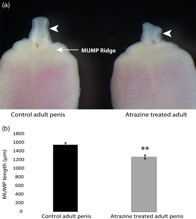
Males exposed to ATR displayed abnormal MUMP morphology (Fig. 1a, white arrowhead indicating the MUMP), being consistently asymmetric in shape. This phenotype was observed in all seven ATR-exposed males examined. In addition, the MUMP was also significantly shorter in ATR-exposed males when compared to controls (P = 0.0014, Fig. 1b). Again, this was consistent across all ATR-exposed males.
Effects of atrazine (ATR) on penis development in a mouse. (a) Gross morphology of the MUMP (indicated by white arrow heads) and MUMP ridge of the glans penis in the control (left) and the atrazine-treated male (right) at 26 weeks of age. (b) The mean (±SEM) length of the MUMP measure from the distal tip of the MUMP till the proximal base of the MUMP located in the MUMP ridge of the control (n = 8) and atrazine-treated (n = 7) adult mouse penis (** indicates P = 0.0014).

ATR-induced hypospadias
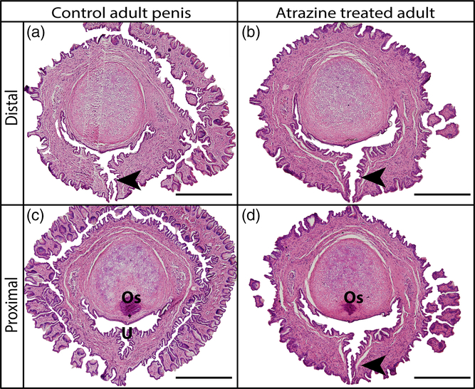
Analyses of the urethral opening showed that it was distal to the tip of the os penis bone, in its normal (wild type) location for control and the majority of ATR-exposed males (Fig. 2a and b). However, hypospadias (the proximal placement of the urethral opening) was observed in one of the seven ATR males examined (14.3% incidence), as indicated by the persistent urethral opening (ventral cleft) (Fig. 2d, black arrowhead) in proximal sections of the penis where the os penis is visible (Fig. 2d; os – dark pink condensed cells). This phenotype is very rare in wild-type mice and was not observed in any vehicle control treated males in this cohort (n = 8, Fig. 2c).
Comparison of distal transverse section histology of the adult control mouse penis (a), and the adult atrazine-treated mouse penis (b), the ventral cleft (urethra opening) indicated by the black arrowhead. Comparison of proximal transverse section histology of the adult control mouse penis (c) where the ventral cleft is no longer present, and the adult atrazine-treated mouse penis with a persisting ventral cleft (d). The ventral cleft indicated by the black arrowhead, U = urethra, Os = os penis bone. Scale bars = 500 µm.

Discussion
Although ATR was banned in the European Union in 2003,18 due to its highly persistent levels in ground water and risks to human health, it continues to be used at very high rates especially across the USA and in Australia.Reference Gore, Chappell and Fenton5 ATR is extremely stable in the environment with a half-life of more than 100 days in water19 and 240 days in soil.20 The dose given in this study, although supra-environmental, was based on previous papers that showed impacts on male reproductive development in rodents. These included decreased testis weight, altered testis morphology and steroidogenic enzyme expression, as well as reduced testosterone concentrations, the number of epididymal spermatozoa and delayed meiosis of spermatozoa.Reference Jin, Wang and Fu11,Reference Victor-Costa, Bandeira, Oliveira, Mahecha and Oliveira21,Reference Gely-Pernot, Hao and Becker22 However, no previous study had examined the potential impacts of ATR on penis development.
Development of the penis, including the processes of urethral closure and differentiation of the MUMP, is all highly androgen-dependent processes.Reference Mahawong, Sinclair and Li17 Furthermore, exposure of the developing penis to exogenous oestrogen is known to prevent urethral closure and demasculinise the developing genitalia leading to a smaller overall penis size and reduction of structures including the MUMP.Reference Govers, Phillips and Mattiske4,Reference Chen, Yu and Pask23,Reference Sinclair, Cao, Baskin and Cunha24 Therefore, the stunted growth and altered morphology of the MUMP, evident in all the ATR-exposed mice, are consistent with an endocrine disruption caused by the exposure. This is further validated by the incidence of hypospadias observed in one ATR-exposed mouse (14.3% incidence in the ATR cohort). The ATR hypospadias phenotype was similar to that of mice exposed to the potent oestrogen diethylstilbestrol.Reference Govers, Phillips and Mattiske4,Reference Mahawong, Sinclair and Li17 Collectively, the stunted growth and disrupted morphology of the MUMP, as well as hypospadias phenotype of ATR-exposed mice, are consistent with ATR eliciting an anti-androgenic and pro-oestrogenic response during penis development. Given the association between EDCs and the increasing incidence of hypospadias in humans, these data implicate ATR to be one of the many potential causative agents in the etiology of this common disease.
Further rodent studies are required using lower, environmentally relevant doses and greater sample number, to determine if they also cause hypospadias. This is supported by the finding that men with higher urinary atrazine concentrations had poor semen quality.Reference Swan, Kruse and Liu25 Given the rapidly increasing incidence of hypospadias and our demonstrated effect of ATR exposure on urethral closure and penis development, the association between human exposures to ATR and the incidence of hypospadias warrants further investigations.
Acknowledgements
The authors thank Tania Long and Darren Cipolla for their technical contributions and help with animal husbandry.
Financial Support
This work was supported by University of Melbourne internal funds [R06000010 to M.P.G.], as well as NIH R01DK096263 and NHMRC APP1098480 to A.J.P.
Conflicts of Interest
The authors have no conflicts of interest to declare.
Ethical Standards
Animal experiments conform to internationally accepted standards and were approved by the University of Melbourne Animal and Ethics committee (AEC 1513481.5) in accordance with the Australian National Health and Medical Research Council guidelines.
Author Contributions
L.C.G. and D.M.M. undertook the histological studies, data analyses, drafted the manuscript and prepared the figures. A.P.H. and B.J.F. conducted the animal study and collected the tissues. M.P.G. and A.J.P. were responsible for the experimental design and supervised the study. All authors reviewed and edited the final manuscript.