INTRODUCTION
The ectoparasite Sarcoptes scabiei (Arthropoda: Arachnida: Acari) is a disease-causing mite of humans and animals that is prevalent worldwide. The mite burrows into the stratum corneum of the skin, a process that inflicts mechanical injury and causes cytological changes, which in combination with a mounting immune response leads to a dermal disease (Burgess, 1994). The most prominent symptom associated with the human disease scabies, is an intense itch, followed by a range of other symptoms including consistent cutaneous eruption and the formation of lesions (Cabrera et al. 1993; Chouela et al. 2002). Carriers of S. scabiei may remain asymptomatic for long periods of time, but the symptoms of scabies will eventually be manifested as an allergic type skin reaction, also known as a delayed hypersensitivity reaction. The most serious form of the illness is crusted scabies, a condition commonly confined to predisposed individuals with an impaired immune system (Donabedian and Khazan, 1992; Schlesinger et al. 1994). This condition, if left untreated, may have a fatal outcome. Although effective treatments are available (Buffet and Dupin, 2003), it has been estimated that the prevalence of scabies is about 300 million infected humans, the majority of them in developing countries (Taplin et al. 1990).
S. scabiei is also of great veterinary importance, as it occurs in a wide range of wild and domestic animals (Burgess, 1994; Walton et al. 2004b). In particular, canines are susceptible to infection, and sarcoptic mange is a highly contagious disease among dogs. S. scabiei also constitutes the most important ectoparasitic threat in pigs (Davies, 1995), and within pig herds a prevalence between 20 and 86% has been recorded (Löwenstein et al. 2004). Affected pigs scratch continuously, and as the disease progresses cuticular lesions form that are accompanied by strong pruritus, and the animal's health may become impaired. The animal welfare aspects are important, but the pig industry also has to take preventive measures in order to limit the economic losses caused by mange (Firkins et al. 2001). Zoonotic scabies is usually contracted from dogs. However, in humans the infection is normally less severe when of animal origin compared with human-to-human transfer, and it disappears in a few weeks (Burgess, 1994). Indeed, molecular data suggest that control programmes for human scabies in endemic regions should focus on human-to-human transmission even when the disease is common in companion dogs as well (Walton et al. 2004a).
The importance of S. scabiei as a pathogen in humans and animals is well documented, but this has not been reflected in the scientific literature, as very few basic studies employing molecular tools have been reported (Kemp et al. 2002). Indeed, scabies has historically been an ignored disease, as a recent review points out (Walton et al. 2004b). However, as indicated in the review, much effort has been made lately and more efforts are in progress to increase our understanding of the parasite and the disease. The lack of parasite material and the absence of an in vitro propagation system for S. scabiei make this parasite an excellent candidate for a molecular approach. In recent years, we have reported the construction of an S. scabiei cDNA library and the identification of several antigens generated from mites isolated from the red fox (Vulpes vulpes) (Mattsson, 2001; Mattsson et al. 2001), followed by an analysis of over 1000 expressed sequence tags (ESTs) (Ljunggren et al. 2003). Additionally, several S. scabiei cDNA libraries generated with mites isolated from humans have been reported (Fischer et al. 2003a,b; Harumal et al. 2003). A pilot-scale EST project with 156 cDNA clones of the S. scabiei var. hominis cDNA library has been reported (Fischer et al. 2003a). An extensive sequencing of cDNA clones by the Australian Genome Research Facility has followed this small-scale EST project, although the analysis of these sequences is still underway (Walton et al. 2004b). In the meantime, only a limited amount of molecular data are available in the public databases, which can provide us with clues to the infection process or the molecular targets for the host's immune response to the parasite.
Here we report the isolation of an immunodominant antigen that was identified with sera from S. scabiei infected dogs and that was found again in our EST data. The full-length open reading frame has been sequenced and a fusion protein has been expressed. We have looked at the antibody response against this protein along with a previously reported immunodominant antigen, MSA1 (Mattsson et al. 1999). We have also determined the actual localization of the proteins in S. scabiei mites.
MATERIALS AND METHODS
The S. scabiei cDNA library
A detailed description of the construction of the S. scabiei cDNA library has previously been published (Mattsson et al. 2001). Briefly, mites of both sexes and of different developmental stages were isolated from the skin of red foxes (Vulpes vulpes) as described by (Bornstein and Zakrisson, 1993). Total RNA was isolated from the mites and mRNA was isolated by oligo(dT) cellulose chromatography and transcribed into cDNA. After size-fractionation the cDNA was ligated into the EcoRI-XhoI sites of the UNI-ZAP XR vector (Stratagene, La Jolla, CA).
Immunoscreening and PCR amplification of cDNA inserts
The S. scabiei cDNA library was diluted in LB-media in order to avoid overlapping plaque formation on the agar plates. Escherichia coli strain XL-1 Blue MRF′ (Stratagene) was infected with the diluted phage cDNA library, followed by an overnight incubation at 42 °C.
The library was screened with 1[ratio ]100 dilutions of sera from 5 S. scabiei infected dogs (14336, 14401, 14404, 14408 and 14533) as previously described (Mattsson et al. 2001). Positive plaques were re-screened at a lower density, and this process was repeated until populations of only positive plaques were obtained. Single plaques containing recombinant clones were picked, and transferred to sterile tubes with 100 μl of SM buffer (100 mM NaCl, 50 mM Tris-HCl (pH 7·5); 10 mM MgSO4). The agar plugs were incubated in SM buffer for 15–60 min before samples were used as templates in the individual PCR reactions. The cDNA inserts were amplified by PCR in 40 μl reactions using the vector-specific primers T3 and T7, as previously described (Ljunggren et al. 2003). All PCR reactions were purified using the QIAquick PCR purification kit (Qiagen, Hilden, Germany) and were eluted with 30 μl of distilled H2O. To analyse the amplification results, 2 μl of each purified PCR sample was loaded onto 1% agarose gels containing ethidium bromide. After the PCR, chloroform was added to SM buffer for long-time storage of immunopositive clones.
DNA sequencing and sequence similarity searches
Purified templates were initially sequenced in single-pass reactions with the T3 primer. Primer walking was then used to generate overlapping sequence data in order to provide full sequence information over the cDNA inserts. Each 20 μl reaction contained 2 μl of Big Dye solution (Applied Biosystems), 5 pmol of primer, 6 μl of dilution buffer (200 mM Tris-HCl, pH 9·0 and 10 mM MgCl2), distilled H2O and 10–20 ng purified PCR-product. Sequence reactions were carried out with the following programme: 25×(96 °C for 10 sec, 45 °C for 5 sec and 60 °C for 4 min). The reaction products were purified and concentrated through ethanol and sodium acetate precipitation, and the resulting pellets were resuspended in 11 μl of formamide before product separation was performed with an ABI PRISM 3100 Genetic Analyzer (Applied Biosystems). The sequences were processed using the Vector NTI program suite 9 (Informax Inc., Oxford, UK). Protein sizes were calculated using the Compute pI/Mw program available through the ExPASy server (http://www.expasy.org).
Edited sequences were compared with public databases (GenBank non-redundant nucleotide and protein databases) using the BLAST family of algorithms with default parameters (BLOSUM62, gap existence and extension penalties 11 and 1, E=10 and wordsize 3 without complexity filtering) (Altschul et al. 1990; Gish and States, 1993). The conserved-domain searches were performed using NCBI conserved domain search (Marchler-Bauer et al. 2003, 2005). Any significant database matches that were detected were followed up by down-loading the relevant family alignment for the conserved domain, and then individually comparing the query sequence to each of the database sequences in terms of its genetic distance using the SplitsTree program (http://www-ab.informatik.uni-tuebingen.de/software/jsplits/welcome.html). Only matches that clearly fitted within the range of all of the domain sequences were accepted as putative homologues. Then, the sequences were examined for the number and length of matching motifs, along with conserved alignment positions, as further evidence of homology.
Subcloning of cDNA fragments
The full-length open reading frame (ORF) of S. scabiei atypical Sarcoptes antigen 1 (ASA1) cDNA was amplified by PCR from isolated immunopositive plaques using Pfu Turbo DNA polymerase (Stratagene), the forward primer OP236 (5′-CGGGATCCATGCAT CGCTTGTCTTTCG-3′) and the reverse primer OP241 (5′-AACTGCAGCTGTTTTGATGAGTCATT-3′). The underlined nucleotides form linkers that contain a BamHI site and a PstI site, respectively. The cycle conditions were: 95 °C for 5 min (95 °C for 15 sec, 50 °C for 30 sec, 72 °C for 1 min)×45 cycles, 72 °C for 10 min. The resulting 2·2 kb PCR product was purified as above and eluted with 30 μl of distilled H2O. The purified amplicon was digested with BamHI and PstI and cloned into pPU16, an MBP fusion expression vector, that allows for a C-terminal fusion to a hexahistidine tag (Mattsson et al. 2001). The resulting plasmid was denoted pPU97 (aa 1-719). Three deletion derivatives of ASA1 were also cloned into pPU16, employing the same strategy as above using the Pfu Turbo DNA polymerase (Stratagene). An 819 bp fragment corresponding to the N-terminal part of ASA1 (aa 1-273) was amplified with forward primer OP236 and reverse primer OP237 (5′-AACTGCAG TGGTTCTATCTTGATTCGACG-3′). The resulting plasmid was denoted pPU94. Forward primer OP238 (5′-CGGATCCGTACCAGTTGCTGGTCTT-3′) and reverse primer OP239 (5′-AACTGCAGATCCTCGAATTCAAG AATGG-3′) was used to amplify an 843 bp fragment corresponding to the central part of ASA1 (aa 240-521). The resulting plasmid was denoted pPU95. A 717 bp fragment equivalent to the C-terminal part of ASA1 (aa 480-719) was amplified by PCR with forward primer OP240 (5′-CGGGATCCAATTTATCTCAGGTCTCT-3′) and reverse primer OP241. The resulting plasmid was termed pPU96.
Expression and purification of recombinant proteins
For high-level expression, plasmids were freshly transformed into E. coli strain BL21(DE3). A single colony was inoculated into 20 ml of LB medium with ampicillin (50 μg/ml) and incubated on a shaker at 37 °C overnight. On day 2, 10 ml of the overnight culture was inoculated in 1 l of fresh minimal medium with casamino acids (MM/CA) (Pryor and Leiting, 1997) supplemented with 50 μg/ml carbenicillin. The MM/CA cultures were grown on a shaker at 37 °C until the OD600 reached ~0·9. Then the cells were cooled for 5 min in a water bath to a temperature of 20 °C. The recombinant gene expression was induced by the addition of isopropylthiogalactoside (IPTG) to a final concentration of 0·5 mM. Induced cells were transferred to a shaker and incubated at 18 °C overnight. After incubation, cells were collected by centrifugation (4000 g, 4 °C, 10 min). About half of the cells were resuspended in HiTrap Start buffer (1× phosphate buffer, pH 7·4 (20 mM sodium phospate buffer and 500 mM NaCl) with 10 mM imidazole) and the rest in PBS, pH 7·3. To prevent protein degradation, both buffers were supplemented with Complete Protease Inhibitor (Roche Molecular, Basel, Switzerland) according to the manufacturer's instructions. Harvested cells from the high-level expression were lysed by sonication. Cell debris was collected by centrifugation (9000 g, 4 °C, 30 min) and the resulting supernatant was filtered through a 0·45 μm filter.
All recombinant versions of ASA1 were purified by affinity chromatography under native conditions (1 ml HiTrap chelating HP columns, Amersham Biosciences, Uppsala, Sweden) on an ÄKTA-FPLC system (Amersham Biosciences) or using a peristaltic pump. The recombinant proteins were eluted in 1× phosphate buffer, pH 7·4 with 500 mM imidazole (Sigma-Aldrich, St Louis, MO). As a control, recombinant MBP with the histidine tag was expressed and purified as above. All protein expressions and purification steps were checked by 10% or 12% sodium dodecyl sulfate-polyacrylamide gel electrophoresis (SDS-PAGE) and stained in 0·1% Coomassie Brilliant Blue solution. Final protein concentrations were estimated by a spectrophotometer (Ultrospec 1000, Pharmacia Biotech) analysis.
Generation of hyper-immune sera from rabbits
Recombinant MSA1 expressed from pPU17 and a deletion derivative of ASA1, denoted ASA1a, expressed from pPU94 (Fig. 1) were purified and used to immunize 1 rabbit each, using standard protocol procedures (MedProbe, Oslo, Norway). The animals were immunized according to European Guidelines for Animal Health under ISO 9001[ratio ]2000 standards; for production details see http://www.medprobe.com.

Fig. 1. Schematic overview of the cDNA containing the ASA1 ORF. Indicated in the figure are the parts used for the construction of 4 different expression vectors, pPU94-pPU97. ASA1-ASA1c denote the names given to the recombinant expressed fusion proteins. See the Material and Methods section for more information.
SDS-PAGE and Western blot analysis
Separations of E. coli lysates and HiTrap purified proteins were done by SDS-PAGE in mini gels (10 or 12%) according to standard procedures. A total of 100 μg of S. scabiei antigen extract or 20 μg of recombinant antigen were loaded onto the gel. Separated proteins were blotted onto a nitrocellulose filter with an electrophoretic transfer cell (Bio-Rad). To assess the quality of the protein transfer, the membranes were briefly incubated in a 0·2% Ponceau-S solution and then in distilled water. All membranes were blocked with Tris-buffered saline with Tween 20 (T-TBS) containing 5% non-fat dry milk for 1 h. Following the blocking, the membranes were rinsed with T-TBS and then incubated for 1 h with different sera diluted in T-TBS containing 1% non-fat dry milk. Hyperimmune sera from immunized rabbits were used as positive controls in 1[ratio ]2000 dilutions. Sera from naturally infected dogs that had been confirmed by ELISA (Bornstein et al. 1996) to be S. scabiei positive were diluted 1[ratio ]100. The membranes were rinsed and washed once for 15 min and twice for 5 min with T-TBS, before they were incubated for 1 h with the appropriate anti-IgG peroxidase conjugate. The membranes were washed for 1×15 min and 3×5 min with T-TBS, before bound antibodies were visualized by chemiluminescence detection by exposure to film using the ECL-system (Amersham Biosciences). A subset of the dog sera was also analysed for the presence of reactive IgE-antibodies using a goat anti-dog-IgE peroxidase conjugate (ICL, Newberg, OR) diluted 1[ratio ]1000. As negative controls we used sera from dogs that were negative in the ELISA as well as in Western blots using S. scabiei protein extracts.
Immunolocalization
Paraffin-embedded and formalin-fixed skin biopsies, 5–6 μm in thickness, from an S. scabiei-infected fox were placed on Superfrost Polysine slides (Histolab Products AB). The biopsies were deparaffinized, rehydrated and rinsed in 50 mM Tris-HCl, pH 7·6 with 9% NaCl. The material was then treated against endogenic peroxidases by impregnation with 1·5 ml of 30% H2O2/50 ml of 0·05 M Tris-HCl for 20 min in a darkroom. All biopsies were pre-treated in a microwave (750 W) for 2×5 min in 10 mM Tris and 1 mM EDTA, pH 9·0, and thereafter for 15 min at RT in fresh 10 mM Tris and 1 mM EDTA, pH 9·0. The ASA1 and MSA1-specific polyclonal immune sera were diluted 1[ratio ]2000 and 1[ratio ]800, respectively, in 0·05 M Tris-HCl, pH 7·6. As negative controls we used pre-immune sera. For detection, a streptavidin-biotin system, DAKO LSAB+ (DAKOCytomation, Glostrup, Denmark), was used according to Dakopatt's instructions. As chromogenic substrate, the AEC Substrate kit for peroxidase (Vector Laboratories, Burlingame, CA) or 3,3′-diaminobenzidine (DAB) was used, and Mayer's haematoxylin was used as the counter-stain. The cover-slips were mounted with an aqueous mountant, Aqueos (DAKOcytomation).
RESULTS
Sequence analysis of ASA1
The initial screening of the cDNA library with sera from 5 S. scabiei-positive dogs yielded 2 positive clones, both identified with serum 14401. After re-screening to single plaque clones, 1 of these clones was excluded from further analysis as it turned out to be a false positive. The protein product of the remaining clone was later named atypical Sarcoptes antigen 1 (ASA1). The plaque was used as template in a PCR with vector-specific T3 and T7 primers. After sequence determination, the insert length was established to be 2847 bp, and the insert contained a 5′-UTR, a 3′-UTR and a 2157 bp long ORF corresponding to a protein product of 719 amino acids (Fig. 1), with a predicted molecular weight of 79 kDa. The sequence has been deposited in GenBank (Benson et al. 2004) under the Accession no. AY895012.
One of our EST clones, available in dbEST, matches the ASA1 clone (Identity=98%, E=0·0, accession no. BG817872) (Ljunggren et al. 2003), whereas we did not find any other significant matches in dbEST. The ASA1 sequence was then compared with sequences in the GenBank non-redundant databases. A BLAST×comparison revealed a 26% identity with a protein named GA10629-PA of unknown function (E=1×e−6, scorebit 57·8, accession no. EAL25440) sequenced from Drosophila pseudoobscura. A BLASTp comparison combined with conserved domain (CD) search (Marchler-Bauer et al. 2003) using the translated ASA1 ORF identified the presence of a MADF domain (E=4×e−14) (Fig. 2), which has been implicated to be involved in transcription events in Drosophila spp. (England et al. 1990, 1992). To help detect whether this is a fortuitous match to the consensus or is evidence of putative homology with all of the sequences, we down-loaded the MADF family alignment from the Simple Modular Architecture Research Tool database (SMART; http://smart.embl-heidelberg.de). We then compared the ASA1 sequence to each of the 55 aligned sequences using maximum-likelihood estimation of the genetic distance under the Jones-Taylor-Thornton amino-acid substitution model. The range of genetic distances from ASA1 to the other sequences (1·79–3·44, median 2·32) is well within the range for the sequences currently recognized as containing the domain (0·78–4·56, median 2·48), indicating that our sequence has as much global sequence similarity to each of the current domain members as they do to each other. We interpret this as evidence of likely homology between the ASA1 sequence and the MADF domain.

Fig. 2. CD-alignment showing amino acid sequence similarity between the MADF found in ASA1 (ASA1) and the consensus sequence (Cons.) in the conserved domain database.
The family members of the MADF domain have very few motifs in common when examined pairwise, the longest consisting of 7 contiguous identical amino acids (out of the 78–100 amino acids in the domain), and some members do not share motifs of even 3 contiguous amino acids. However, the ASA1 sequence shares a motif of 5 amino acids with one MADF family member, and motifs of 4 amino acids are shared with 11 other members, 1 of which shares two 4-aa motifs. There are no universally conserved alignment positions among the MADF members, although these are usually considered to represent functional sites, the best conservation being 50/55 members. Nevertheless, the ASA1 sequence shows conservation at 8 of the 9 positions that are conserved in >70% of the members. Thus, our sequence shares several types of detailed similarity with the MADF members, as well as global similarity. We interpret this as further evidence of likely homology.
Subcloning and expression of ASA1
Although ASA1 was identified with serum from an S. scabiei infected dog by immunoscreening, none of our BLAST searches with the ASA1 sequence found any significant matches with previously known antigens. The apparent species specificity of ASA1 and the presence of a MADF domain, implicated in sequence specific DNA binding, merited the protein for further characterization. We also included the major Sarcoptes antigen 1 (MSA1) (Mattsson et al. 1999), in this study, in part to serve as a reference antigen. The latter protein is homologous with antigens in other closely related mites (Aki et al. 1994; Fujikawa et al. 1996), and has also been denoted as S. scabiei antigen 1, Ssag1, by Kemp and colleagues (Harumal et al. 2003). Furthermore, the MSA1 sequence has been deposited in GenBank (Benson et al. 2004) under Accession no. DQ109676.
The ASA1 ORF was used in 4 different constructs to prepare recombinant proteins. Firstly the full-length ORF was expressed in fusion with the maltose binding protein (MBP) and with a C-terminal hexahistidine-tag. A product corresponding to the predicted size of 123 kDa for the MBP-ASA1 protein was expressed at a low level. The other 3 deletion derivatives containing the N-terminal (ASA1a, aa 1-273), central (ASA1b, aa 240-521) and C-terminal (ASA1c, aa 480-719) parts of ASA1, respectively, were expressed in the same manner as described above. All constructs containing derivatives of the ASA1 ORF were expressed to very high levels. The predicted molecular masses for the recombinant proteins generated from pPU94, pPU95 and pPU96 were 75 kDa, 74 kDa and 71 kDa, respectively. The sizes of the recombinant proteins were confirmed by Western blot analysis using a hyperimmune rabbit serum raised against native S. scabiei antigens (Mattsson et al. 2001). The Western blot data indicated a rather weak response towards the full-length MBP-ASA1, and not the shorter protein bands corresponding to the C-terminal parts of ASA1. Both recombinant proteins ASA1a and ASA1b were easily expressed, whereas the recombinant ASA1c protein, corresponding to aa 480-719, also displayed a pattern characteristic of truncated peptides. The 4 different protein preparations were analysed in a Western blot with the serum from dog 14401. This experiment verified the identity of ASA1 as the protein identified in our immunoscreening, and it also showed that ASA1a was the only peptide readily detected in the Western blot without any signs of minor proteins suggestive of translation problems (data not shown). Hence, ASA1a was used in parallel with a recombinant derivative of MSA1 in further studies.
ASA1 and MSA1 in S. scabiei mites
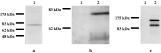
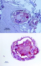
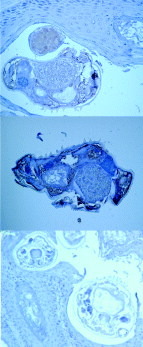
In Western blot analysis of S. scabiei protein extracts, the anti-ASA1a sera reacted with a band of the expected size as predicted from the sequence data for native S. scabiei ASA1 and the reaction with serum 14401 (Fig. 3A,B). No reaction with any of the pre-immune sera could be observed. In keeping with other studies, the anti-MSA1 sera reacted with a protein with a molecular mass of 164 kDa as well as 123 kDa (Fig. 3C). It is not clear if these are 2 independent proteins sharing the same epitopes or a singular protein that is differentially processed. In the next step we looked at the localization of the two proteins in mites within tissue samples. The staining of sections through red fox skin biopsies infected with S. scabiei showed that anti-ASA1a and MSA1 antibodies bound strongly to the mites. The polyclonal antisera stained numerous vital organs and tissues in the mite's body cavity. The only part of S. scabiei that omitted staining was the thin cuticle, including legs and mouthparts. This was also true for any of the visible surface structures such as stout setae (Figs 4 and 5). The anti-ASA1 serum also stains what appears to be the inside of the burrow wall, suggesting that the mite somehow deposits the protein there. No staining of any host tissue beyond the burrow could be detected (Figs 4 and 5). It appears that deposited eggs were stained by anti-MSA1 serum, in contrast to eggs in utero. Control sera from naïve rabbits did not stain the mites or red fox tissue.

Fig. 3. Western blots against (A) whole Sarcoptes scabiei antigen extract with serum 14401 from a S. scabiei-infected dog, (B) against whole S. scabiei antigen extract with rabbit pre-immune serum (lane 1) and rabbit anti-ASA1a serum, showing the immunoreaction towards an 79 kDa protein corresponding to the theoretical size of the full-length ASA1 protein (lane 2), and (C) against whole S. scabiei antigen extract with rabbit pre-immune serum (lane 1) and rabbit anti-MSA1 serum, showing the immunoreaction towards a 164 kDa and 123 kDa protein (lane 2). Size standards are the broad range pre-stained protein marker from New England Biolabs.

Fig. 4. Immunoperoxidase red staining of Vulpes vulpes skin section (top) and single mite with anti-ASA1a rabbit serum (bottom).

Fig. 5. Immunoperoxidase brown staining of Vulpes vulpes skin section (top) and single mite with anti-MSA1 rabbit serum (middle) and section stained with pre-immune serum (bottom).
The immune response against ASA1 and MSA1
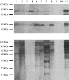
To analyse the immune response against recombinant ASA1a and recombinant MSA1 we used a panel of 107 sera from dogs. The sera had initially been tested with an ELISA based on a protein extract from S. scabiei mites (Bornstein et al. 1996) and with Western blots against an S. scabiei protein extract. Of these 107 sera 62 had IgG antibodies positive in the ELISA and the Western blot. A total of 15 of sera reacted with the recombinant version of ASA1a whereas the remaining 92 sera were negative. In contrast, 53 of the samples were positive for MSA1 and the remaining 54 were negative. In Fig. 6 the banding pattern for 11 sera against the 3 antigen preparations are shown. As a positive control we used a serum from a rabbit immunized with native S. scabiei antigen. All sera from the full panel that tested negative against the native antigen were also negative against ASA1a and MSA1, with 1 exception (Fig. 6A, lane 10). This single serum reacted with ASA1a and not with the S. scabiei protein or MSA1. We also observed that both ASA1a and MSA1a appeared as doublet bands in the Western blots, probably as a consequence of the expression system used. None of the 11 sera reacted with the MBP (data not shown).

Fig. 6. SDS-PAGE/immunoblot of IgG binding to the ASA1a (A), MSA1 (B) fusion proteins and Sarcoptes scabiei whole antigen extracts (C) by serum antibodies from S. scabiei- positive and negative dogs. Positive dogs in lane 1: 14401, lane 2: 5083, lane 3: 5312, lane 4: 5326, lane 5: 4340, lane 6: 5081, lane 7: 5343 and lane 8: 4681. Negative dogs in lane 9: 9922 and lane 10: 9832. The positive control serum p 21 from a hyperimmunized rabbit is in lane 11.
These 11 sera were also tested for IgE-reactive antibodies against the 3 antigen preparations. In the IgE analysis, none of the dogs reacted with MSA1, and only weak reactions were observed in 1, possibly 2, of the dogs tested against the protein extract from S. scabiei mites. In contrast, 1 dog showed a comparatively strong IgE reaction against ASA1a (data not shown).
DISCUSSION
We have screened an S. scabiei cDNA library with sera from dogs naturally infected with S. scabiei. In this process we isolated a cDNA corresponding to a 79 kDa large protein. In Western blot experiments 15 out of 62 infected dogs reacted with a recombinant version of the protein. This is in contrast with the immunodominant antigen MSA1. The putative antigen also contained a protein domain not normally associated with antigens (Doerks et al. 2002). Consequently, we have given this new antigen the name atypical Sarcoptes antigen 1 (ASA1).
The most prominent feature of ASA1 is the N-terminus, corresponding to aa 11-110, which shares similarity and probable homology to the MADF domain (Doerks et al. 2002). The domain is predominantly found in Arthropoda and Nematoda, as is evident from searches in the Simple Modular Architecture Research Tool database (SMART; http://smart.embl-heidelberg.de). The MADF domain contains a helix-turn-helix DNA-binding domain, and to date it has not been found in mammals. Outside of the MADF domain, ASA1 has a low compositional complexity with several blocks of triplets and quadruplets of polar amino acids (Asn, Gln and Ser). In fact, these 3 amino acids represent 39·7% of all amino acids down-stream of the MADF domain. As a result, no significant similarities were found besides the MADF domain. Our experimental data suggested that it was the N-terminal part of ASA1, including the MADF domain that were recognized by sera from S. scabiei-infected dogs.
The first protein described with a MADF domain, the Adh distal factor-1 (Adf-1) from D melanogaster is a transcription factor that binds the promoters of a diverse group of genes (England et al. 1990, 1992). The domain is a distantly related member of the Myb helix-turn-helix family of DNA-binding proteins (Cutler et al. 1998). Although the list of MADF-containing proteins has expanded since then, very few of them have been experimentally characterized, as the vast majority originate from the ORF predictions of major genome sequencing projects. However, the few examples characterized are all transcriptional regulators from D. melanogaster (Clark and McKearin, 1996; Bhaskar et al. 2000; Barbash et al. 2003). It is therefore rather surprising that ASA1 is recognized as an antigen by IgG and IgE from dogs infected with S. scabiei.
In a recent study of the IgG and IgE antibody response against S. scabiei during experimental infections in dogs (Arlian and Morgan, 2000) showed that a set of proteins with the sizes 170, 155 and 142/133 kDa are the most likely candidates for the development of a diagnostic test monitoring the IgG response against S. scabiei. In their study, Arlian and Morgan also observed a band corresponding to an 81 kDa large protein that was recognized by circulating IgE antibodies in 4 of the 15 dogs. In keeping with our observations, we believe that the 170 and 142/133 kDa antigens correspond to MSA1, since polyclonal sera against recombinant MSA1 recognize 2 bands with those approximate sizes in Western blots against native S. scabiei antigens. We have also seen this banding pattern with anti S. scabiei sera affinity purified against recombinant MSA1 (unpublished data). We also believe that the 81 kDa antigen might correspond to ASA1, because of both the similarities in size and the comparable immune response in dogs.
The size of MBP is a disadvantage when it is used as a fusion partner to produce recombinant proteins. However, it has been shown MBP stabilizes the recombinant protein and increases the expression yield (Pryor and Leiting, 1997). This has in part been attributed to the function of MBP as a molecular chaperone that promotes the solubility and stability of proteins that are fused to (Kapust and Waugh, 1999). This is also in keeping with our observations where we have been successful in using MBP but not other common fusion partners to express recombinant antigens. In ELISA screening experiments with MBP as the sole antigen we have not observed signals above the background levels when using dog sera, suggesting that it is not an important natural immunogen.
Not only is the recombinant version ASA1 an immunogenic protein, recognized by IgG/IgE from S. scabiei-infected dogs, but we could also find evidence for a deposition of ASA1 outside the mite. In our immunohistochemistry staining we observed that antibodies against ASA1 bound to the inside wall of the mite's burrow. We cannot rule out the possibility that the staining we observed was a consequence of our experimental set-up, although this is less likely. Nevertheless, we did not find any evidence for any secretion or targeting signals, including any classical nuclear localization signal in the ASA1 sequence.
In conclusion, the protein ASA1 is somewhat of a puzzle. It is clear that the protein is actively expressed throughout the parasite and also exposed to the immune system of the host. At the same time, the most dominant feature is the presence of a MADF domain, normally reserved for proteins linked to transcriptional control. Whether this indicates that ASA1 is indeed involved in transcriptional regulation in S. scabiei remains to be shown. Alternatively, ASA1 might be the result of a gene that evolved from a recombination of pre-existing domains (Copley et al. 2002). If this scenario bears some relevance, the MADF domain might have a completely different function in ASA1. Clearly, more work is needed to understand the function of this intriguing antigen, and its role in S. scabiei and the parasite-host interaction.
This work was funded by the Swedish Research Council for Environment, Agricultural Sciences and Spatial Planning (FORMAS). We thank Ewa Westergren at the Department of Pathology, National Veterinary Institute, for help with the immunolocalization analysis and Eva Pettersson for technical assistance.