Implications
In dairy farming practices, rumen development and its functional improvement in calves, play an important role in nutrient delivery, absorption and metabolism, and have an important impact on calf health and future production performance. Dietary isovalerate could promote rumen development by the improving morphology and function of rumen mucosa. It is suggested that dietary isovalerate supplements have the potential ability to promote the growth of calves.
Introduction
In dairy farming practices, rumen development is crucial for calves due to its important role in nutrient digestion, delivery, absorption and metabolism as well as the future performance and health of animals (Liu et al., Reference Liu, Wang, Zhang, Pei, Zhang, Wang, Zhang, Yang, Wang, Guo and Huo2016a). Most of studies reported that the continuous presence and absorption of volatile fatty acids (VFA) improved the development and metabolic function of rumen epithelium (Sakata and Tamate, Reference Sakata and Tamate1978; Lane and Jesse, Reference Lane, Baldwin and Jesse1997; Baldwin and McLeod, Reference Baldwin and McLeod2000), but some studies found that rumen metabolic function was independent of VFA production (Lane et al., Reference Lane and Jesse2002). The inconsistent results were attributed to different rumen VFA concentrations especially butyrate and the exposure period of epithelial tissue to VFA (Lane et al., Reference Lane, Baldwin and Jesse2000). In addition, solid feed intake can stimulate rumen muscularization and increase rumen volume by promoting rumen motility (Zitnan et al., Reference Zitnan, Kuhla, Sanftleben, Bliska, Schneider, Zupcanova and Voigt1998; Baldwin and McLeod, Reference Baldwin and McLeod2000; Baldwin et al., Reference Baldwin, McLeod, Klotz and Heitmann2004). Therefore, additives that improve dry matter (DM) intake and rumen fermentation can be used to promote the development of morphology and function of calves. Branched-chain VFA supplements, including isobutyrate, isovalerate and 2-methylbutyrate, have a positive impact on rumen fermentation, cellulolytic bacteria population, rumen enzyme activity, nutrient digestion and productive performance in steers (Misra and Thakur, Reference Misra and Thakur2001; Liu et al., Reference Liu, Wang, Huang, Dong, Yang, Zhang and Wang2009 and Reference Liu, Wang, Pei, Li, Wang, Zhang, Zhang, He, Wang, Yang, Bai, Shi and Liu2014).
In our previous study, the increase in DM intake, rumen total VFA concentration and butyrate production was observed with isovalerate supplements in calves (Liu et al., Reference Liu, Wang, Zhang, Pei, Zhang, Li, Guo, Huo, Yang and Wang2016b), and rumen development was improved by 2-methyl butyrate supplements (Liu et al., Reference Liu, Wang, Zhang, Pei, Zhang, Wang, Zhang, Yang, Wang, Guo and Huo2016a). Furthermore, there is no literature about the effects of isovalerate supplements on rumen development in calves. Based on the above considerations, the present study was designed to evaluate the effects of isovalerate supplements on the development and metabolic function of rumen epithelium in pre- and post-weaning calves.
Material and methods
Animals and experimental design
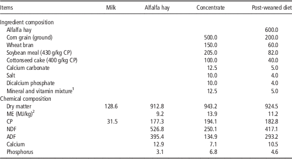
The experimental protocol was approved by the Animal Care and Use Committee of Shanxi Agriculture University. In total, 48 Chinese Holstein male calves of 15 days of age and 45.1±0.36 kg of BW were randomly assigned to four groups by using random numbers, 12 calves per group. The treatments were: control (without isovalerate), low-isovalerate (LIV), moderate-isovalerate (MIV) and high-isovalerate (HIV) with 0, 3, 6 and 9 g isovalerate per calf per day, respectively. The isovalerate supplements (98.5% of isovalerate) were purchased commercially (Shanghai Aladdin biological technology co., Ltd, Shanghai, China) and were mixed manually into milk for pre-weaning calves and the concentrate portion of post-weaning calves. The study lasted 75 days, including a 15-day-adaptation period and a 60-day-sampling period. Calves were weaned at 60 days of age. During the pre-weaned period, calves were offered whole milk (10% of BW) twice daily via nipple at 0800 and 1500 h for 25 days. From 26 to 30 days, the daily offer of milk was decreased to half and calves were fed a commercial concentrate ad libitum. Post-weaned diets consisted of 60% alfalfa hay and 40% commercial concentrate (DM basis), and was offered ad libitum. Chemical composition of the feeds is shown in Table 1. Calves were housed in individual pens (2.5×3 m) and fresh water was available throughout the experimental period.
Ingredient and chemical composition of the food and diet (in g/kg dry matter)

1 Contained 8 mg Co, 700 mg Cu, 4000 mg Fe, 2400 mg Mn, 2400 mg Zn, 240 mg I, 120 mg Se, 600 000 IU vitamin A, 100 000 IU vitamin D and 3000 mg vitamin E per kilogram premix.
2 ME=metabolic energy, calculated from National Research Council (2001).
Data collection and sampling procedures
All calves were weighed at 15, 30, 60 and 90 days of age. Milk, feed offered and refusals were recorded daily to calculate DM intake. Milk, feed and refusals were sampled once weekly for DM determination. Milk samples were preserved with 2-bromo-2-nitropropane-1, 3-diol and pooled for each group per period until chemical analysis. Feeds and refusals were dried in an oven at 55°C for 48 h, and ground to pass a 1-mm screen with a mill (FZ102; Shanghai Hong Ji instrument Co., Ltd, Shanghai, China) for later chemical analysis. At 28, 29, 58, 59, 88 and 89 days of age, blood samples were obtained at 2 h after morning feeding, via the coccygeal vein by 10 ml vacuum tubes containing sodium heparin for analyses of β-hydroxybutyrate (BHBA), and 20 ml vacuum tubes for analyses of growth hormone (GH) and IGF-1. Blood samples were immediately placed on ice and transported to the laboratory to separate plasma and serum by centrifuging at 2000×g for 15 min at 4°C. All plasma and serum samples were stored at −20°C for later analyses. At 30, 60 and 90 days of age, three calves per treatment were chosen from each treatment at random and slaughtered. The entire digestive tract was removed and placed on ice. Rumens, reticulums, omasums and abomasums were separated, emptied, washed clean with normal saline and weighed. Ruminal tissue samples (8×8 cm) were obtained within 20 min of death and collected from the dorsal, anterior dorsal, anterior ventral, posterior dorsal and posterior ventral blind sacs. For morphometry, a 1×1 cm area of the rumen wall was retrieved. Three 1×1 cm samples were collected and stored at −80°C for RNA isolation and analysis.
Chemical analysis
Milk samples were analyzed for fat, true protein and lactose using IR spectrophotometry (Foss 120 Milko-Scan; Foss Electric, Hillerød, Denmark) according to Association of Official Analytical Chemists (AOAC) (1997; method 975.16) procedures. Analytical DM content of oven-dried samples was determined by drying at 135°C for 3 h (AOAC, 1997; method 930.15). Organic matter content was calculated as the difference between DM and ash contents, with ash determined by combustion at 550°C for 5 h. Content of NDF and ADF were determined using the methods described by Van Soest et al. (Reference Van Soest, Robertson and Lewis1991) with heat stable α amylase and sodium sulfite used in the aNDF procedure, and expressed inclusive of residual ash. Content of N in the samples was determined by the Kjeldahl method (AOAC, 1997; method 976.05). Serum BHBA content was determined (Sigma Kit 310-A; Sigma Diagnostics, St. Louis, MO, USA) following the method of Williamson et al. (Reference Williamson, Mellanby and Krebs1962). Serum GH content was determined using an radioimmunoassay (RIA) (GH Assay Kit; Nanjing Jian Cheng Institute of Bio-Engineering, Nanjing, China) based on the method of Kasuya et al. (Reference Kasuya, Hodate, Matsumoto, Sakaguchi, Hashizume and Kanematsu1996). Serum IGF-1 content was determined using an RIA (IGF-1 Assay Kit; Nanjing Jian Cheng Institute of Bio-Engineering) after acid–ethanol extraction of the plasma based on the method of Kasuya et al. (Reference Kasuya, Hodate, Matsumoto, Sakaguchi, Hashizume and Kanematsu1996). The intra- and inter-assay CV of IGF-1 RIA were 5% and 10%, respectively.
Light microscopy and morphometry
Ruminal samples (1×1 cm surface) were fixed in 4% formalin solution. After rinsing with water, the rumen wall tissue was dehydrated in a graded series of ethanol (70%, 80%, 85%, 90%, 95% and absolute ethanol), cleared with xylene twice, saturated and embedded in paraffin. Serial sections were then cut at 4-μm thicknesses, 10 slices of each sample, deparaffinized in xylene, dehydrated and were stained with haematoxylin and eosin, and observed under a light microscope. The thickness of rumen wall and keratinized layer, and the length and width of rumen papillae were determined by the computer operated Image C picture analysis system (Intronic GmbH, Berlin, Germany) and the IMES analysis program, using a color video camera (Sony 3 CCD, CCD; Sony Electronics Inc., Tokyo, Japan) and a light microscope (Axiolab; Carl Zeiss, Jena, Germany).
Extraction of RNA and quantitative real-time PCR (qRT-PCR)
Total RNA was extracted from the rumen biopsies by using a Total RNA isolation kit (Invitrogen, Carlsbad, CA, USA) according to the manufacturer’s protocol. The quality and concentration of the isolated RNA were determined by using a NanoDrop ND-1000 Spectrophotometer (NanoDrop Technologies, Rockland, DE, USA). The ratios of absorbance at 260 and 280 nm of all preparations were ~2.0. The integrity of RNA was checked by denaturing agarose gel electrophoresis and ethidium bromide staining. The iScriptTM cDNA Synthesis Kit (Bio-Rad Laboratories GmbH, Munich, Germany) was used to synthesize complementary DNA (cDNA) from 500 ng total RNA from each sample per 10 μl sample reaction according to the manufacturer’s instructions. Reaction conditions were 15 min at 37°C and 5 min at 85°C. Negative control reactions without reverse transcriptase, were performed on each sample to detect possible contamination of genomic DNA or environmental DNA (Kuzinski et al., Reference Kuzinski, Zitnan, Viergutz, Legath and Schweige2011).
The abundance of messenger RNA (mRNA) for the growth hormone receptor (GHR), IGF-1 receptor (IGF-1R) and 3-hydroxy-3-methylglutaryl-CoA synthase 1 (HMGCS1) was quantified by qRT-PCR using the iCycler and the iQ-SYBR green detection (Bio-Rad Laboratories, Hercules, CA, USA). The primer sets used for RT-PCR were forward primer (5′ to 3′) CAG ACA AAT CAC TCC ACC AA, reverse primer (5′ to 3′) GAA GGG CAC CAC CAG GAG T for 18S ribosomal RNA (rRNA) as reference gene, forward primer (5′ to 3′) AAA TTC ACC AAG TGC CGT TC, reverse primer (5′ to 3′) GGG GCA TTC TTT CCA TTC TT for GHR (GenBank No. NM176608), forward primer (5′ to 3′) CCT CAT TAT TCC TGC TAA CCA A, reverse primer (5′ to 3′) AGA TAG AAG AGA TGC GAG GAG GAT for IGF-1R (GenBank No. NM001244612), forward primer (5′ to 3′) CTC ACG CAG TTC TTT CAC CA, reverse primer (5′ to 3′) GGC AAC AAT TCC CAC ATC TT for HMGCS1 (GenBank No. BC102850). Subsequent qPCR was performed on a MxPro – Mx3000P multiplex qPCR systems (Stratagene, La Jolla, CA, USA) at a minimum in triplicate. A reaction mixture (20 μl) consisted of 2 μl cDNA, 10 µl SYBR Premix TaqTM II (TaKaRa Biotechnology Co., Ltd, Dalian, China), 0.8 µl PCR Forward Primer, 0.8 µl PCR Reverse Primer, 0.4 µl ROX Reference Dye II (TaKaRa Biotechnology Co., Ltd., Dalian, China) and 6.0 µl dH2O. The PCR was performed under the following cycle conditions: one cycle of 95°C for 20 s, 45 cycles of 95°C for 20 s and annealing temperature for 30 s, and 62°C for 20 s, followed by a melting curve analysis (Denman and McSweeney, Reference Denman and McSweeney2006). Fluorescence detection was performed at the end of each denaturation and extension step. Relative quantity of mRNA for GHR, IGF-1R and HMGCS1 were done as a proportion of 18S rRNA according to the equation:
 $${\rm Relative quantification}\,{\equals}\,2^{{^{{{\minus}{\rm (}C_{t} \,{\rm target}\,{\rm genes}{\minus}C_{t} \,{\rm total}\,{\rm reference}\,{\rm genes)}}} }} $$
, where C
t
represents threshold cycle.
Statistical analyses
Data were analyzed using the mixed model procedure of SAS (Proc Mixed; Statistical Analysis Systems, Reference Shen, Seyfert, Lohrke, Schneider, Zitnan, Chudy, Kuhla, Hammon, Blum, Martens, Hagemeister and Voigt2002). Analysis of variance was used to examine the effect of treatment on stomach weight, BW and rumen morphology, the abundance of mRNA for GHR, IGF-1R and HMGCS1, and blood metabolites. Linear and quadratic orthogonal contrasts were tested using the CONTRAST statement of SAS with coefficients estimated based on the isovalerate application rates. The main effects of treatment were compared using the Duncan’s test. Effects of the factors were declared significant at P<0.05.
Results
Stomach weight and rumen morphology
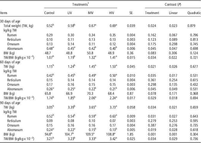
Stomach weight, ratios of different stomach to total stomach weight, BW and ratio of total stomach to BW are shown in Table 2. Total stomach weight increased linearly (P<0.05) in pre- (30 and 60 days of age) and post-weaning (90 days of age) dairy calves with increasing isovalerate supplements, and was greater for MIV and HIV than for LIV and control (P<0.05). The ratio of rumen to total stomach weight increased linearly for calves at 60 and 90 days of age (P<0.05), and was greater for MIV and HIV than for LIV and control (P<0.05), but was unchanged for calves at 30 days of age. Ratios of reticulum to total stomach weight and omasum to total stomach weight were not affected by isovalerate supplements in pre- and post-weaning calves. The ratio of abomasum to total stomach weight decreased linearly (P<0.05) in pre- and post-weaning calves, and was lower for MIV and HIV than for LIV and control (P<0.05). The ratio of total stomach to BW increased linearly in pre- and post-weaning calves (P<0.05) and was greater for MIV and HIV than for LIV and control (P<0.05).
Effects of isovalerate supplementation on total stomach weight and the ratio of different stomach to total stomach weight in pre- and post-weaning dairy calves

a,bMeans with different superscripts in each row differ significantly (P<0.05).
1 Control, low-isovalerate (LIV), moderate-isovalerate (MIV) and high-isovalerate (HIV) with 0, 3, 6 and 9 g isovalerate per calf per day, respectively.
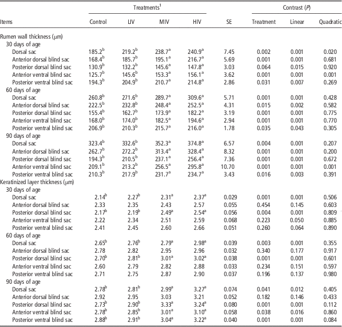
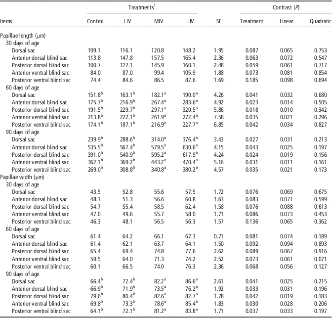
The thickness of rumen wall and keratinized layer is shown in Table 3. The rumen wall thickness increased linearly (P<0.05) in pre- and post-weaning calves with increasing isovalerate supplements, and was greater for MIV and HIV than for LIV and control (P<0.05). The thickness of the rumen keratinized layer in the dorsal sac and posterior dorsal blind sac increased linearly (P<0.05) in pre- and post-weaning calves, and was greater for MIV and HIV than for LIV and control (P<0.05). However, the thickness of the rumen keratinized layer in the anterior dorsal blind sac was not affected by isovalerate supplements in pre- and post-weaning calves. The thickness of the rumen keratinized layer in the anterior and posterior ventral blind was not affected in pre-weaning calves, but increased linearly (P<0.05) in post-weaning calves and was greater for MIV and HIV than for LIV and control (P<0.05). The length and width of rumen papillae are shown in Table 4. Ruminal papillae length was not affected for calves at 30 days of age, whereas increased linearly (P<0.05) for calves at 60 and 90 days of age and were greater for MIV and HIV than for LIV and control (P<0.05). Ruminal papillae width was not affected for pre-weaning calves, whereas increased linearly (P<0.05) in post-weaning calves and were greater for MIV and HIV than for LIV and control (P<0.05).
Effects of isovalerate supplementation on the thickness of rumen wall and keratinized layer thickness in pre- and post-weaning dairy calves

a,bMeans with different superscripts in each row differ significantly (P<0.05).
1 Control, low-isovalerate (LIV), moderate-isovalerate (MIV) and high-isovalerate (HIV) with 0, 3, 6 and 9 g isovalerate per calf per day, respectively.
Effects of isovalerate supplementation on the length and width of rumen papillae in pre- and post-weaning dairy calves

a,bMeans with different superscripts in each row differ significantly (P<0.05).
1 Control, low-isovalerate (LIV), moderate-isovalerate (MIV) and high-isovalerate (HIV) with 0, 3, 6 and 9 g isovalerate per calf per day, respectively.
Functional gene expression of rumen mucosa
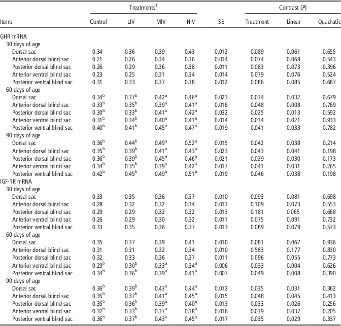
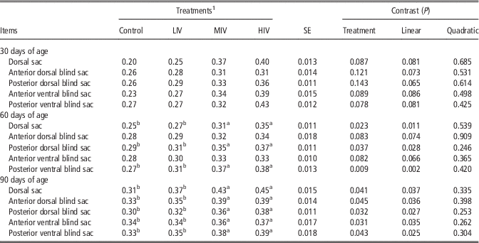
The relative mRNA expression of GHR, IGF-1R and HMGCS1 in rumen mucosa is shown in Tables 5 and 6. The relative GHR mRNA expression in rumen mucosa was not affected for calves at 30 days of age, whereas increased linearly (P<0.05) for calves at 60 and 90 days of age, and was greater for MIV and HIV than for LIV and control (P<0.05). The relative IGF-1R mRNA expression in rumen mucosa for calves at 30 days of age, dorsal sac, anterior dorsal blind sac and posterior dorsal blind sac for calves at 60 days of age was not affected, whereas IGF-1R mRNA expression in anterior ventral blind sac and posterior ventral blind sac for calves at 60 days of age and that of in rumen mucosa for calves at 90 days of age increased linearly (P<0.05), and was greater for MIV and HIV than for LIV and control (P<0.05). The relative HMGCS1 mRNA expression in rumen mucosa for calves at 30 days of age, anterior dorsal blind sac and anterior ventral blind sac for calves at 60 days of age was not affected, but increased linearly (P<0.05) in dorsal sac, posterior dorsal blind sac and posterior ventral blind sac for calves at 60 days of age, and in rumen mucosa for calves at 90 days of age, and was greater for MIV and HIV than for LIV and control (P<0.05).
Effects of isovalerate supplementation on the relative expression of growth hormone receptor (GHR) messenger RNA (mRNA) and IGF-1 receptor (IGF-1R) mRNA of rumen mucosa in pre- and post-weaning dairy calves (proportion of 18S ribosomal RNA)

a,bMeans with different superscripts in each row differ significantly (P<0.05).
1 Control, low-isovalerate (LIV), moderate-isovalerate (MIV) and high-isovalerate (HIV) with 0, 3, 6 and 9 g isovalerate per calf per day, respectively.
Effects of isovalerate supplementation on the relative expression of 3-hydroxy-3-methylglutaryl-CoA synthase 1 (HMGCS1) messenger RNA (mRNA) of rumen mucosa in pre- and post-weaning dairy calves (proportion of 18S ribosomal RNA)

a,bMeans with different superscripts in each row differ significantly (P<0.05).
1 Control, low-isovalerate (LIV), moderate-isovalerate (MIV) and high-isovalerate (HIV) with 0, 3, 6 and 9 g isovalerate per calf per day, respectively.
Blood metabolites
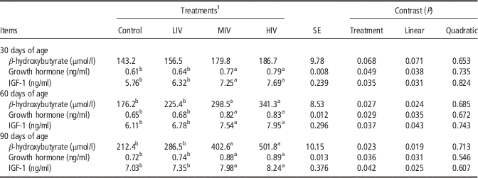
Serum contents of BHBA, GH and IGF-1 are shown in Table 7. Serum BHBA content was not affected for calves at 30 days of age with increasing isovalerate supplements, but increased linearly (P<0.05) for calves at 60 and 90 days of age and was greater for MIV and HIV than for LIV and control (P<0.05). Serum contents of GH and IGF-1 increased linearly (P<0.05) in pre- and post-weaning calves, and were greater for MIV and HIV than for LIV and control (P<0.05).
Effects of isovalerate supplementation on blood parameters in pre- and post-weaning dairy calves

a,bMeans with different superscripts in each row differ significantly (P<0.05).
1 Control, low-isovalerate (LIV), moderate-isovalerate (MIV) and high-isovalerate (HIV) with 0, 3, 6 and 9 g isovalerate per calf per day, respectively.
Discussion
Stomach weight and rumen morphology
The improvements in rumen weight, rumen wall thickness, keratinized layer thickness, papillae length and width with increasing isovalerate supplements were consistent with the increased relative mRNA expression of GHR, IGF-1R and HMGCS1 in rumen mucosa, and indicated that isovalerate supplements stimulated rumen development in dairy calves. The results were consistent with Liu et al. (Reference Liu, Wang, Zhang, Pei, Zhang, Wang, Zhang, Yang, Wang, Guo and Huo2016a) who reported the same results in dairy calves supplemented with 2-methylbutyrate supplements. The proliferation and growth of rumen squamous epithelial cells results in the increase in length and width of papillae, and the thickness of rumen wall and keratinized layer which influence the uptake and transport of nutrients in the rumen epithelium (Baldwin and Jesse, Reference Baldwin and Jesse1992; Lesmeister et al., Reference Lesmeister, Tozer and Heinrichs2004). There is evidence that continuous presence and absorption of VFA could promote cells mitosis, accelerate rumen epithelium cell proliferation, stimulate rumen epithelium development and improve rumen metabolic function (Sakata and Tamate, Reference Sakata and Tamate1978 and Reference Sakata and Tamate1979; Baldwin and McLeod, Reference Baldwin and McLeod2000), and result in the increase in the length and width of papillae (Lane and Jesse, Reference Lane, Baldwin and Jesse1997; Górka et al., Reference Górka, Kowalski, Pietrzak, Kotunia, Jagusiak, Holst, Guilloteau and Zabielski2011). As a special apoptosis inhibitor in rumen epithelium, butyrate is considered as the most important stimulator for papillar growth (Mentschel et al., Reference Mentschel, Leiser, Mülling, Pfarrer and Claus2001). Therefore, the improved rumen development was attributed to increased production of total VFA, butyrate and acetate with increasing isovalerate supplements, and consistent with previous observations in mature steers and dairy calves (Liu et al., Reference Liu, Wang, Huang, Dong, Yang, Zhang and Wang2009 and Reference Liu, Wang, Zhang, Pei, Zhang, Li, Guo, Huo, Yang and Wang2016b). In addition, solid feed intake improved rumen total VFA production, rumen muscularization and rumen volume by stimulating rumen mobility. As a result, rumen weight, rumen wall thickness and keratinized layer thickness increased with increasing DM intake (Zitnan et al., Reference Zitnan, Kuhla, Sanftleben, Bliska, Schneider, Zupcanova and Voigt1998; Baldwin and McLeod, Reference Baldwin and McLeod2000; Baldwin et al., Reference Baldwin, McLeod, Klotz and Heitmann2004). Similarly, the improved rumen development derived from increased solid feed intake in calves has been found in other studies (Shen et al., Reference Smith, Amburgh, Diaz, Lucy and Bauman2004; Zitnan et al., Reference Zitnan, Voigt, Schonhusen, Wegner, Kokardova, Hagemeister, Levkut, Kuhla and Sommer2005; Liu et al., Reference Liu, Wang, Zhang, Pei, Zhang, Wang, Zhang, Yang, Wang, Guo and Huo2016a). Hence, the linear increase in DM intake with increasing isovalerate supplementation observed in the study of Liu et al. (Reference Liu, Wang, Zhang, Pei, Zhang, Li, Guo, Huo, Yang and Wang2016b) may have also contributed to rumen development in dairy calves. It was suggested that the increased rumen development was due to the increased DM intake, which was promoted by isovalerate supplement.
Functional gene expression of rumen mucosa
Growth hormone and IGF-1 could affect the development and function of the gastrointestinal tract after binding to their respective receptors in rumen mucosa, so the development of rumen was influenced by the receptor number (Georgieva et al., Reference Georgiev2003). The increased mRNA expression of GHR and IGF-1R in rumen mucosa was in agreement with increased blood contents of GH and IGF-1, and further confirmed the positive effects of isovalerate supplements on rumen development in calves. Some studies reported the positive relationship between nutrient intake and mRNA expression of GHR and IGF-1 in the liver of bull calves (Smith et al., 2002), IGF-1R mRNA abundance in rumen epithelium of young goats (Shen et al., Reference Smith, Amburgh, Diaz, Lucy and Bauman2004) and IGF-1 mRNA expression in rumen mucosa of newborn calves (Flaga et al., Reference Flaga, Górka, Kowalski, Kaczor, Pietrzak and Zabielski2011). Therefore, the improved nutrient utilization with isovalerate supplements in our previous studies (Liu et al., Reference Liu, Wang, Huang, Dong, Yang, Zhang and Wang2009, Reference Liu, Wang, Pei, Li, Wang, Zhang, Zhang, He, Wang, Yang, Bai, Shi and Liu2014 and Reference Liu, Wang, Zhang, Pei, Zhang, Li, Guo, Huo, Yang and Wang2016b) could support the increased mRNA expression of GHR and IGF-1R in the current study. The result was consistent with Liu et al. (Reference Liu, Wang, Zhang, Pei, Zhang, Wang, Zhang, Yang, Wang, Guo and Huo2016a) who reported the same results in pre- and post-weaning dairy calves supplemented with 2-methylbutyrate supplements.
As the abundance of HMGCS1 mRNA increased in parallel with the ketogenic capacity of rumen epithelium (Lane et al., Reference Lane and Jesse2002), so the increased mRNA expression of HMGCS1 in rumen mucosa suggested that rumen metabolic function was increased by isovalerate supplements. According to Lane and Jesse (Reference Lane, Baldwin and Jesse1997), continuous VFA infusion in physiological concentration increased acetoacetate production and indicated an improvement in the ketogenic capacity of rumen epithelium in lambs. The increased mRNA expression of HMGCS1 in the current study was mainly associated with increased ruminal total VFA concentration with isovalerate supplements, especially butyrate concentration (Liu et al., Reference Liu, Wang, Huang, Dong, Yang, Zhang and Wang2009, Reference Liu, Wang, Pei, Li, Wang, Zhang, Zhang, He, Wang, Yang, Bai, Shi and Liu2014 and Reference Liu, Wang, Zhang, Pei, Zhang, Li, Guo, Huo, Yang and Wang2016b). It has been proposed in another study that increased rumen VFA concentration, earlier exposure or longer period of exposure to VFA might be necessary for improving rumen metabolic function, since delaying the initiation of solid feed consumption results in rumen morphological development rather than rumen metabolic development in sheep (Lane et al., Reference Lane, Baldwin and Jesse2000).
Blood metabolites
The GH regulates IGF-1 production and the growth promoting effects of GH is mediated by IGF-1 production (Brameld et al., Reference Brameld, Atkinson, Saunders, Pell, Buttery and Gilmour1996). The IGF-1 influences morphology and function of rumen mucosa by promoting cell proliferation and differentiation as well as inhibiting cell apoptosis (Roffler et al., Reference Roffler, Fah, Sauter, Hammon, Gallmann, Brem and Blum2003; Baldwin et al., Reference Baldwin, McLeod, Klotz and Heitmann2004; Georgiev, Reference Georgieva, Georgiev, Ontsouka, Hammon, Pfaffl and Blum2008). The positive response of papillae length, papillae width and tunica muscularis thickness of rumen mucosa to plasma IGF-1 concentration was observed in calves (Zitnan et al., Reference Zitnan, Voigt, Schonhusen, Wegner, Kokardova, Hagemeister, Levkut, Kuhla and Sommer2005; Georgiev, Reference Georgieva, Georgiev, Ontsouka, Hammon, Pfaffl and Blum2008; Flaga et al., Reference Flaga, Górka, Kowalski, Kaczor, Pietrzak and Zabielski2011). Therefore, the increased blood GH and IGF-1 content in per- and post-weaning calves was consistent with the increased rumen weight, thickness of rumen wall and keratinized layer as well as the length and width of papillae with isovalerate supplements. The increase in blood BHBA content in post-weaning calves was also observed in the study of Liu et al. (Reference Liu, Wang, Zhang, Pei, Zhang, Wang, Zhang, Yang, Wang, Guo and Huo2016a) with 2-methylbutyrate supplements. The results from in vitro studies have also shown that total ketone bodies production of rumen epithelium tissue increased with the increased total VFA concentration (Bush, Reference Bush1988; Lane and Jesse, Reference Lane, Baldwin and Jesse1997). Similarly, serum BHBA concentration increased with the supplements of coated calcium butyrate in calves (Nazari et al., Reference Nazari, Karkoodi and Alizadeh2012). Therefore, the increase in plasma BHBA concentration in the current study was derived from the improved ketogenic capacity of rumen epithelium, which was stimulated by increased total VFA production, especially butyrate production with isovalerate supplements (Liu et al., Reference Liu, Wang, Huang, Dong, Yang, Zhang and Wang2009 and Reference Liu, Wang, Zhang, Pei, Zhang, Li, Guo, Huo, Yang and Wang2016b). No further change of stomach weight, rumen morphology and functional gene expression of rumen mucosa with isovalerate supplementation from MIV to HIV was consistent with its effects on growth performance and ruminal fermentation (Liu et al., Reference Liu, Wang, Zhang, Pei, Zhang, Li, Guo, Huo, Yang and Wang2016b), and indicated that a high dose of isovalerate (9 g/calf per day) supplementation did not provide any additional benefit for ruminal development.
Conclusion
For pre-weaning calves, total stomach weight, rumen wall thickness, keratinized layer thickness and blood concentration of GH and IGF-1 increased linearly with isovalerate supplements. For post-weaning calves, total stomach weight, ratio of rumen to total stomach weight, rumen wall thickness, keratinized layer thickness, length and width of rumen papillae, relative mRNA expression of GHR, IGF-1R and HMGCS1 in rumen mucosa and blood concentration of BHBA, GH and IGF-1 increased linearly with isovalerate supplements. It is suggested that the development and metabolic function of the rumen in calves is improved by isovalerate supplements in a dose-dependent manner, and the optimum supplemental dose was about 6.0 g of isovalerate per calf per day.
Acknowledgments
This work was supported by a grant from Natural Science Funding projects of National Natural Science Foundation of China (31272471). The authors thank the staff of Shanxi Agriculture University beef unit for care of the animals.