Introduction
The introduction of image-guided radiation therapy (IGRT) has made the delivery of external beam radiotherapy more accurate.Reference Verellen, De Ridder and Storme1 IGRT refines the delivery of therapeutic radiation by applying image-based target re-localisation,Reference Potters, Gasper and Kavanagh2 which involves imaging the patient's anatomy before and even during their treatment and altering the patient's position or treatment plan, accordingly.Reference Gwynne, Webster, Adams, Mukherjee, Coles and Staffurth3 Pre-treatment images can be taken using electronic portal imaging devices (EPIDs), on-board imagers (OBIs) and cone-beam computed tomography (CBCT). EPIDs use megavoltage (MV) radiation, and OBIs use kilovoltage (kV) radiation to capture planar images. These imaging systems can be used to minimise set-up errors by using skeletal anatomy to verify the treatment positionReference Gwynne, Webster, Adams, Mukherjee, Coles and Staffurth3 and assess translational shifts. Some correction of yaw (rotation about the vertical axis) is also possible. However, because of couch-positioning limitations, these systems do not allow for the correction of pitch (rotation about the transverse axis) or roll (rotation about the longitudinal axis; Figure 1). CBCT uses kV radiation to capture a volumetric dataset, which can provide detail of internal anatomy and soft tissue delineation; however, the radiation dose delivered is increased.Reference Gwynne, Webster, Adams, Mukherjee, Coles and Staffurth3 Nijkamp et al.Reference Nijkamp, de Jong, Sonke, van Vliet and Marijnen4 found there was inter-observer variation when matching the mesorectum on CBCT and its use in IGRT for rectal carcinoma remains an area of active research.Reference Gwynne, Webster, Adams, Mukherjee, Coles and Staffurth3
This image demonstrates the direction of pitch, roll and yaw when patient is in the prone position.

Current clinical target volume (CTV) to planning target volume (PTV) margins used in radiotherapy are empirically derived or calculated from an assessment of measured systematic and random variations.Reference Van Herk, Remeijer, Rasch and Lebesque5 Van Herk'sReference Van Herk, Remeijer, Rasch and Lebesque5 margin recipe was calculated for a spherical CTV and does not account for rotational variations. For rectal carcinoma treatment, the CTV is not spherical, with the treatment volume being large and irregular in shape.
The delivery of radiation to the target volumes is limited by the surrounding normal tissue tolerances. The main dose limiting structure for rectal carcinoma treatment is the small bowel. Acute small bowel toxicities are diarrhoea and abdominal pain.Reference Baglan, Frazier and Yan6 The long-term complications include obstruction, perforation and fistula.Reference Emami, Lyman and Brown7 When 50 Gray (Gy) is delivered to one-third of the small bowel volume, there is a 5% chance of a late obstruction occurring in 5 years.Reference Emami, Lyman and Brown7
Despite advancements in treatment delivery, there remains a sparcity in literature on the impact of pelvic rotational errors in radiation therapy. The degree of inter-fraction rotationReference Aubry, Beaulieu and Girouard8 and its effect on dosimetryReference Cranmer-Sargison9 have been investigated for prostate carcinoma. There has also been one study quantifying the degree of pitch for rectal carcinoma.Reference Kasabasic, Faj and Ivkovic10 However, this study was limited by its use of portal films and manual measurements taken from defined points on the sacrum, as portal films have been found to be less accurate for the detection and quantification of treatment errors.Reference Kruse, Herman and Hagness11
Therefore, this study aims to:
• Quantify the degree of inter-fractional pitch demonstrated by the rectal carcinoma patients at the Andrew Love Cancer Centre (ALCC).
• Model inter-fractional pitch on CTV coverage by the prescribed dose.
• Determine a tolerance for which no correction is needed.
Materials and methods
This study consisted of two phases: the first was a retrospective analysis of pre-treatment images to investigate pitch, and the second involved dosimetric modelling to reproduce pitch on three-dimensional (3D) computed tomography (CT) datasets.
Patient cohort
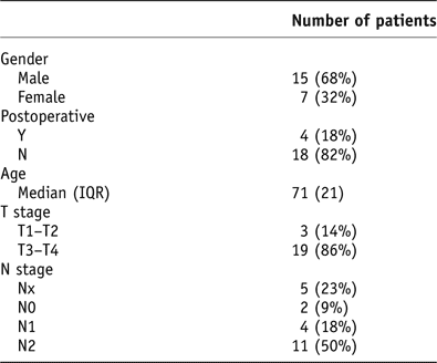
Phase 1 of this study had a convenience sample of 22 rectal carcinoma patients treated at the ALCC in 2010. The pre-treatment images of these cases were analysed for pelvic pitch. Patient characteristics are presented in Table 1.
Patient characteristics
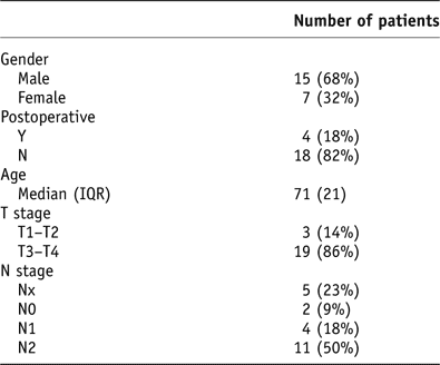
Abbreviation: IQR, interquartile range.
Phase 2 consisted of the patients from phase 1 that had a 3D conformal plan and small bowel contoured by the radiation oncologist. Patients treated with intensity-modulated radiation therapy (IMRT) or those who had bulky diseases were excluded from the 3D simulation. IMRT cases were excluded, as simulation of a complex IMRT plan was not possible with the current software. Cases with bulky diseases were excluded, as they were atypical and had the potential to confound or skew the results.
Planning and treatment
The patients were CT simulated and treatment planned. For the CT, patients were in a prone position, on an institution-produced bellyboard device with a custom Vak-Loc™ Cushion (Civco Medical Solutions, Orange City, IA, USA) under their upper torso and head, to reduce the volume of small bowel irradiated.Reference Koelbl, Richter and Flentje12, Reference Kim, Chie and Kim13 The inferior edge of the bellyboard hole was aligned with the L5–S1 junction, which aligns with the upper level of the treatment field. The patients were simulated and treated with a full bladder to displace small bowel from the treatment fields.Reference Kim, Chie and Kim13, Reference Nuyttens, Robertson, Yan and Martinez14 The patients were instructed to empty their bladder and then drink 400 mL of water 30 minutes before the procedure. CT slices were taken at 2 mm intervals.
The CTV to PTV expansion for these patients was typically 1 cm, on the basis of a complex mix of clinical requirements (with a variation of no greater than 0·3 cm); however, it was beyond the scope of this study to explore this further. The standard CTV typically encompassed the gross disease, mesorectum, internal iliac, pre-sacral and peri-rectal lymphatics.Reference Myerson, Garofalo and Naqa15 The isocentre was located in the centre of the CTV.
Routine treatment typically consisted of a 3D conformal plan, using a three-field technique (Figure 2), using a posterior and two lateral fields. The lateral fields had Enhanced Dynamic Wedges of 45°or 60° (as required) with the ‘hot edge’ anterior. A four-field box plan was used for one patient whose volume extended anteriorly, and a nine-field IMRT plan was used for one patient with an irregularly shaped volume encompassing the small bowel (Figure 2). The use of volumetric-modulated arc therapy (VMAT) in the treatment of rectal carcinoma had not been implemented at the time of this study.
This figure shows the field arrangements for rectal cases at the ALCC where the patients were in the prone position, with three-field arrangement (left) and the nine-field IMRT field arrangement (right). The right image (IMRT planned) demonstrates a larger amount of small bowel around the treatment area.
Abbreviations: ALCC, Andrew Love Cancer Centre; IMRT, intensity-modulated radiation therapy.
From the CT dataset, digitally reconstructed radiographs (DRRs) were created. Daily planar pre-treatment images were taken by a Varian AS1000 (Varian Medical Systems, Palo Alto, CA, USA), using both kV and MV radiation each fraction. The images were assessed using 4D Treatment Console version 8·6 (Varian Medical Systems), and a ‘zero action’ threshold was used when assessing the images. Pre-treatment online translational shifts were made for all daily variations in the left–right, anterior–posterior and superior–inferior directions, using surrogate match anatomy outlining the anterior aspect of the sacrum, L5 and Upper Boarder Pubis (UBP; Figure 3). Yaw could be measured during pre-treatment imaging, and was corrected by rotating the treatment couch when a variation of >3° was found. Roll and pitch could not be measured for online images, and the translational shifts applied were a best fit to assist in correcting for rotation. During images were taken during the treatment when the gantry was at 0° and 270°, if the field length was <19 cm. If the field length was >19 cm; taking during images was not possible as there are sections of the EPID that are radiosensitive making image acquisition not possible.
This image displays a lateral DRR, with the sacrum, L5 and UBP outlined as surrogate match anatomy.
Abbreviations: DRR, digitally reconstructed radiograph; UBP, upper boarder pubis.
Image analysis (phase 1)
A total of 576 pre-treatment images were analysed using Offline Review version 8·6 (Varian Medical Systems). The pre-treatment images were aligned with the planned DRRs. The degree of pitch during treatment was measured using surrogate match anatomy, outlining the anterior aspect of the sacrum, L5 and UBP as demonstrated in Figure 3.
3D simulation (phase 2)
The dosimetric impact of pitch was simulated, using Eclipse treatment planning software version 8·6 (Varian Medical Systems). Pitch was simulated by rotating the collimators for the lateral fields, and rotating the couch and gantry for the posterior field to maintain field geometry, as the gantry cannot be angled alone to match for pitch (Figure 4). Pitch was simulated in 5° increments, ranging from −10° to +10°. Positive degrees of rotation were defined as the superior end of the sacrum moving anteriorly in the prone position. Dose distributions were calculated using an Anisotropic Analytical Algorithm version 8·6 (Varian Medical Systems) on a 0·2 cm grid. The CTV and small bowel dosimetry were assessed using dose–volume histograms (DVH).
This image is a left lateral view of a patient, demonstrating the field geometry when 0° and −10° were simulated.
Results
Image analysis (phase 1)
A total of 576 pre-treatment images were evaluated for the 22 rectal carcinoma patients. The pitch values for the 576 pre-treatment images had a mean (μ) of 0·27°, a standard deviation (SD) of 2·23° and a standard error of 0·09°. The data were also analysed by the patients. The pitch values for each patient are shown in a box and whisker plots in Figure 5. Patients A and B are outliers, as the patients’ mean pitch values during treatment were greater than two SDs (−4·2° to 4·7°) of the sample mean (i.e. the mean of the pitch values of 576 pre-treatment images). Patient A had a mean pitch value of −4·8, and patient B had a mean of 4·9°.
Box plot of pitch split by patient. ±2 SD (+4·7,−4·2) of sample mean is marked. The box contains 50% of all data points, the median is represented by line within the box. The whiskers include all values excluding outliers. Outliers are represented by circles and are beyond 1·5 box lengths form the edge of the box. The extreme outliers are represented by stars, and are beyond 3 box lengths from the edge of the box.
Abbreviation: SD, standard deviation.
3D simulation (phase 2)
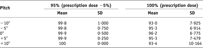
Pitch was simulated on the 16 patients’ CT datasets, in 5° increments, ranging from −10° to +10°. Dosimetric coverage of the CTV was analysed using DVHs. The volume of CTV receiving the prescription dose, as well as 95% of the prescription dose, was analysed, in accordance with ICRU guidelines16 for minimum target volume coverage. Table 2 demonstrates the average volume of CTV receiving dose for each simulated pitch value. The volume of CTV receiving 95% of the prescription dose varied by 0·1% when there was 10° of pitch, in either a positive or negative direction. Fifteen (93·8%) of the 22 patients investigated had 100% of the CTV receive 95% of the prescription dose for all pitch values simulated.
Volume of CTV, in percentage (%), receiving dose relative to the prescription dose
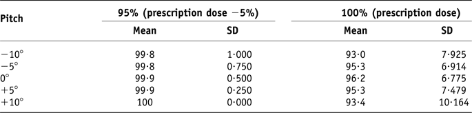
The mean and standard deviation of the sample (n = 16) are reported in this table.
Small bowel dose
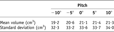
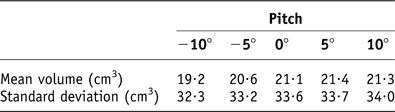
The volume of small bowel receiving 15 Gy was evaluated, as it has a direct relationship with toxicity.Reference Baglan, Frazier and Yan6 Small bowel volumes above 150 cm3 increase the probability of grade two and three toxicities from 10% to 70%.Reference Baglan, Frazier and Yan6 There was minimal increase in small bowel volume irradiated; with the maximum increase 0·3 cm3 when 5° of pitch was simulated (Table 3). Six patients simulated with a volume <1 cm3 receive 15 Gy, and no patient with a volume >150 cm3 receive 15 Gy; the maximum volume recorded was 124 cm3.
Mean volume and standard deviation of the sample (n = 16) receiving 15 Gy to the small bowel
Discussion
Image analysis (phase 1)
The pre-treatment images were matched using the sacrum, L5 and UBP as a surrogate for PTV/CTV. As planar imaging was utilised, it was not possible to see soft tissue, and therefore the bony anatomy of the pelvis was used as a surrogate for the PTV/CTV position. CBCT is not currently used at the ALCC, and Gwynne et al.Reference Gwynne, Webster, Adams, Mukherjee, Coles and Staffurth3 reports its use in rectal carcinoma treatment to be a topic of ongoing investigation.
The unusually large pitch values for patients A and B were investigated to determine whether there were any confounding factors that needed to be considered. The large pitch values for these two cases can be attributed to poor positioning and pain during treatment. Because of a pre-existing injury, patient A had their arms bent underneath their chest during treatment, which differed from our institution's protocol of arms crossed above the head. With their arms in this position, the patient was able to support their weight using their arms to potentially suspend their abdomen above the bellyboard. On reviewing patient B's treatment review notes, the large pitch values correlated with increasing pain. The introduction of pain control medication resulted in a reported improvement in pain and a decrease in pitch values for the final seven radiotherapy treatments.
Before this study, there was a commonly held belief that significant pitch occurred in many instances. This study has found that only two of the 22 patients had a mean pitch value equal to or above 4·8°. From the findings of this study, we can report that the anecdotally held belief was not supported by the evidence and was disproved.
3D Simulation (phase 2)
This phase of the study assessed the dose coverage of the CTV in conjunction with the pitch. The purpose of the PTV is to account for possible internal and geometric variations and inaccuracies to ensure that the prescribed dose is delivered to the CTV.16 The CTV was assessed rather than the PTV, as the CTV contains as the gross palpable or visible tumour and surrounding subclinical microscopic diseases.16
The pitch values recorded from the pre-treatment images did not exceed 10°, and 95% of the values were below 5°. The dosimetric effect of pitch rotations were modelled using 5° increments, ranging from −10° to +10°. The CTV to PTV margin expansions are largely based on a minimum dose coverage of 95% to the CTV for 90% of patients, as referred to by Van Herk et al.Reference Van Herk, Remeijer, Rasch and Lebesque5 In this study, 93·8% of patients received a minimum of 95% dose coverage of the CTV. An average CTV volume of 99·9% received 95% of the dose when there was no pitch; this volume only decreased by 0·1% when −10° of pitch was simulated, and increased by 0·1% when 10° was simulated. This decrease in volume by 0·1% had no clinical significance as more than 90% of patients still received 95% dose coverage of the CTV.
Limitations
The use of rotating the collimators to assess the resultant dosimetry from change in pelvic pitch is an approximation. This technique does not take into account tissue deformation caused by the change in position. Soft tissue deformation cannot be assessed using the current software available. The future development of deformation software would improve the accuracy of the 3D simulation in phase 2. Another limitation of this study was the use of sacral pitch as a surrogate for CTV pitch. This assumes that the position of the sacrum is indicative of the CTV position. This is in line with our current imaging protocol, where the sacrum, L5 and UBP are used to approximate the CTV pitch during treatment.
This study was conducted on patients with 3D conformal plans; IMRT and VMAT are now also used for rectal carcinoma patients in our institution when 3D conformal planning fails to protect the critical structures. IMRT and VMAT techniques frequently use smaller CTV to PTV margins. The effect of margin reductions and steep IMRT and VMAT that dose gradients have on CTV coverage needs further investigation.
Conclusion
This study consisted of two phases: the first involved retrospective pre-treatment image analysis, and the second involved a 3D dosimetric simulation. In phase 1, the pitch values measured had a mean (μ) of 0·27° and the SD was 2·23°. From our 3D simulation study (phase 2), we found that there was no clinically significant change in CTV coverage when up to ±10° was modelled.
As a result of this study, our institution has adopted a ±10° tolerance for pitch, when daily pre-treatment imaging is used, during conformal radiation therapy of the rectum. Further investigations are required to determine the effect of pitch for margin reductions, IMRT and VMAT treatment techniques.
This study assessed the impact of the pitch on CTV dosimetry. The study assumes that there is no anatomical deformation, and daily pre-treatment shifts are made to correct for all translational variations. The dosimetric impact of combined pitch and translational variations would need to be further investigated in institutions where the ‘action threshold’ on pre-treatment imaging is above 0 mm for translational shifts.
It is strongly recommended that each institution reviews their practice protocols and techniques. The tolerance determined by this study is individual to our institution, where for rectal carcinoma patients CTV is <16 cm in length, and standard practice for these patients is to:
• Be treated prone on a custom bellyboard with personalised Vak-Loc™ bag;
• have a full bladder;
• be CT simulated with 2 mm slices;
• have a typical CTV to PTV expansion of 1 cm; and
• have daily pre-treatment imaging with 0 mm action threshold for translational shifts.
Acknowledgements
The authors acknowledge the staff at the Andrew Love Cancer Centre, Geelong, Australia, for their assistance during this study.