A 41-year-old woman with a history of complex cyanotic congenital cardiac disease was referred for cardiac magnetic resonance imaging.Reference Mc Mahon, Taylor and Vick1 She was diagnosed in infancy with tricuspid atresia. Her segemental anatomy is as follows: visceral situs solitus, levocardia, left aortic arch, absent right atrio-ventricular connection, concordant ventriculo-arterial connection with pulmonary atresia, hypoplastic right ventricle, unrestrictive membranous ventricular septal defect, large secundum atrial septal defect, normal systemic venous return, and normal pulmonary venous return.
She was palliated with an aorta to left pulmonary connection (Potts shunt) as a newborn and later had her pulmonary blood flow augmented with a superior caval vein to right pulmonary artery connection (classic Glenn shunt) at the age of 12 years. She refused any additional surgical procedure and was therefore left with chronic cyanosis and a hypertensive left pulmonary artery due to systemic flow through the Potts shunt.
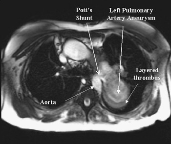
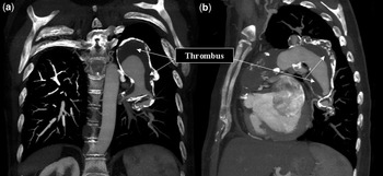
Her cardiac magnetic resonance imaging showed the segmental anatomy, as described above. The Glenn anastomosis was patent and the Potts shunt was unobstructed. There was a markedly aneurysmal left pulmonary artery measuring approximately 6.2 centimetres in the anterior–posterior dimension and 5.3 centimetres in the right–left dimension with layered thrombus posteriorly (Fig 1). Contrast-enhanced cardiac computer tomography was arranged to exclude acute pulmonary emboli. This study showed layered thrombus at the distal portion of the left pulmonary artery with extension to the segmental branches of the left upper, left lower, and lingual lobes, with sparing of the sub-segmental branches. Extensive calcification was noted. (Fig 2a and b).
Axial steady state free precession cine image at the level of the left main pulmonary artery (LPA). The surgical connection between the descending thoracic aorta and LPA is identified. The LPA is aneurismal and contains low signal material consistent with layered thrombus (see text).

Coronal (a) and sagittal (b) maximum intensity projections of a contrast-enhanced computed tomography data set. The left main pulmonary artery (LPA) is again noted to be significantly dilated. Large quantities of intraluminal thrombus are present. Note the areas of high attenuation within the thrombus. These represent calcification and are indicative of a chronic in situ thrombosis rather than acute pulmonary embolism. Such appearances are relatively common in pulmonary hypertensive patient with chronic thrombus formation and relate to alterations in Virchow’s triad (altered vessel, altered flow, and altered coaguability) as in this case.

Acknowledgements
The authors have not received any financial support for this study.