Baby SM (female) was born prematurely at 24 weeks of gestation in a tertiary neonatal intensive care unit, with a birth weight of 640 g. Her mother went into spontaneous labor and developed peripartum sepsis. The baby developed severe hyaline membrane disease and received two doses of endotracheal surfactant, and also experienced a pulmonary hemorrhage. In total, she required mechanical ventilation for 375 h followed by 789 h of continuous positive pressure ventilation. She also needed supplemental oxygen for another 1706 h. In the initial period, she received nutritional support via parenteral nutrition and full enteral feeding was achieved by the 3rd week of life.
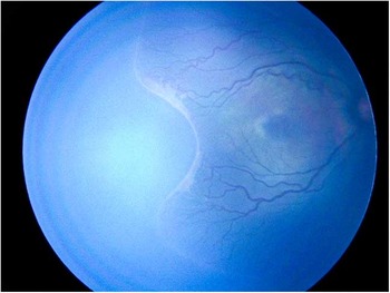
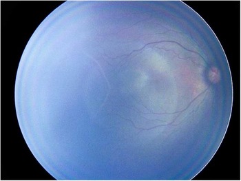
This neonate developed severe retinopathy of prematurity (ROP) (Stage 3 Zone 2, Plus Disease) (Fig. 1) and was treated with 0.012 ml of an intravitreal vascular endothelial growth factor (VEGF) antagonist (Bevacizumab) in both eyes. Posttreatment, the infant underwent weekly retinal assessment for the rest of the hospital stay. Figure 2 is a retinal image taken 2 weeks after this treatment, acquired using a neonatal retinal camera (RetCam III, Clarity Medical System, Pleasanton, CA, USA). The neonate remained in the neonatal unit for 127 days before being discharged home. She was followed up 6 months after discharge from hospital and did not receive a second dose of the VEGF-antagonist. The neonate presented in this case is part of an ongoing observational study, and written parental consent and ethics committee approval were obtained. Treatment of ROP is NOT part of this study. Treatment is based on clinical indication and determined by the ophthalmology team.
Retinal microvascular appearance before treatment with a vascular endothelial growth factor antagonist.

Reduction in retinal microvascular tortuosity 2 weeks after the treatment with the vascular endothelial growth factor antagonist.

Discussion
Although arteries are straight conduits in normal conditions, in certain other disease states they take a tortuous path. In an adult, retinal microvascular tortuosities are linked to ageing, atherosclerosis, hypertension and diabetes mellitusReference Han 1 , although the mechanism that causes a normal vessel to undergo these changes in not well understood. The development of retinal microvascular tortuosities in premature neonates is different to those seen in adults. In premature neonates, the retinal arteries and veins are tortuous from the onset of development, especially in neonates born <32 weeks of gestation. A combination of high blood flow, angiogenesis and blood vessel congestion have been proposed to be the cause of tortuosity.Reference Turior, Onkaew, Uyyanonvara and Chutinantvarodom 2 Animal studies indicate that VEGF is a key molecule in the pathogenesis of retinal microvascular tortuosities in prematurity.Reference Hartnett, Martiniuk and Byfield 3 Dilation and tortuosities of retinal vessels are the hallmarks of severe ROP.Reference Good 4 The stages of ROP are defined by vessel appearance at the interface between the vascular and avascular retinal areas.Reference Mintz-Hittner, Kennedy and Chuang 5 This interface resembles a line for stage 1, a three-dimensional ridge for stage 2, and a ridge with neovascularization extending into the vitreous gel for stage 3 (treatment is indicated). Treatment is also indicated if there is evidence of Plus disease (dilated veins and tortuous arteries near the optic disk in two or more quadrants of the eye).Reference Mintz-Hittner, Kennedy and Chuang 5 These retinal changes are never seen in term neonates.
In recent years, intravitreal VEGF-antagonists are increasingly being used as ‘off-label medication’ to treat this condition, with some success.Reference Sankar, Sankar, Mehta, Bhat and Srinivasan 6 There is mounting evidence that intravitreal anti-VEGFs quickly enter the bloodstream and have a deleterious effect on organogenesis.Reference Morin, Luu and Superstein 7 In a recently published systematic review on the use of VEGF inhibitors compared with conventional treatment, the reviewers concluded that there is insufficient medium- and long-term safety data to recommend routine use of this medication.Reference Pertl, Steinwender and Mayer 8 Another recently published study showed that preterm neonates treated with intravitreal VEGF-antagonists tend to have an increased risk of developing severe neurodevelopmental disabilities at 18 months of age.Reference Morin, Luu and Superstein 7 To date, there are 12 human studies that have investigated the association between systemic VEGF and ROP in premature infants, and the findings are inconsistent.Reference Kandasamy, Hartley, Rudd and Smith 9 As such, more studies are required to determine the mechanistic relationships between systemic VEGF and ROP in premature infants.
In our patient after receiving the intravitreal VEGF antagonist, vessel tortuosity decreased. However, 4 weeks later, at a time when the circulating VEGF-antagonist levels would be expected to decline, the tortuosity re-appeared. We believe the observed changes demonstrate the inherent plastic properties in the retinal microvasculature of a premature neonate. A few different models of microvascular plasticity have been described,Reference Jacobsen, Hornbech and Holstein-Rathlou 10 but this is the first time that it has been subjectively observed in a premature neonate.
Conclusion
With improved survival of extremely premature neonates and the availability of retinal imaging technology, we are now able to demonstrate microvascular plasticity in premature neonates. As this condition is never observed in term neonates, it is likely that the human fetus has this property before delivery, but loses it late in gestation. The long-term effect of retinal microvascular tortuosity on later health is yet to be determined. Our data raise the possibility that administration of VEGF-antagonists to preterm infants with retinopathy may affect other vascular beds that also demonstrate plasticity at this stage of development.
Acknowledgments
The authors are grateful to Prof. Ian MR Wright and the Townsville Hospital’s Ophthalmology team.
Conflicts of Interest
None.
Ethical Standards
The Hospital’s Human Research Ethics Committee permission has been obtained to publish this report.