Scimitar syndrome and VACTERL association are both rare congenital malformations in human infants, with severe consequences for those affected. In this case report, we present a second patient who incorporates both diagnoses,Reference Takawira and Omar 1 and therefore want to raise perinatal awareness for the association of Scimitar syndrome in children with VACTERL association.
Case report
The male newborn was delivered at 33+6 weeks of gestation because of premature rupture of the membranes by caesarean. Prenatal ultrasound showed a single umbilical artery, wedge-shaped lumbal vertebra, suspicion of an imperforate anus based on the dilated intestines, and hyperechogenic kidneys with small cortical cysts, leading to the suspicion of VACTERL association. Furthermore, mesocardia and a small left ventricle with a small aortic arch with suspicion of foetal coarctation were seen. The mother had a history of Hashimoto thyroiditis and a non-insulin-dependent gestational diabetes. Prenatal foetal karyotyping and fluorescence in vitro hybridisation analysis for 22q11.2 deletion syndrome revealed normal results.
Postnatally, the diagnosis of VACTERL association was confirmed, but the cardiac anatomy remained puzzling.
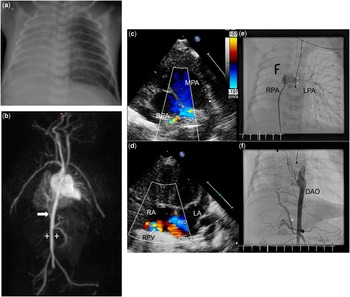
Echocardiography depicted cardiac dextroposition with right ventricular hypertrophy and a small apex forming the left ventricle. The aortic valve and aorta were small without coarctation (z-scores: −1.4 for aortic valve, −2.8 for transverse arch, −1.6 for aortic istmus). The normal-sized main pulmonary artery split up into a wide left pulmonary artery and a very small right pulmonary artery of coronary artery size (Fig 1c). The right lung seemed to be supplied by several major aortopulmonary collateral arteries and a large artery from the subdiaphragmatic abdominal aorta. The left pulmonary veins showed a pronounced perfusion. At least one right pulmonary vein seemed to empty directly into the right atrium (Fig 1d). Furthermore, an atrial septum defect was seen.
Chest x-ray, magnetic resonance imaging angiographic, echocardiographic, and angiocardiographic findings: ( a ) Pronounced opacification of the complete right lung with no discernible sign of a scimitar-shaped vein. ( b ) Clearly visible artery (white arrow) originating from the descending aorta (white crosses), supplying the right lower lung. ( c ) Hypoplastic right pulmonary artery (1.2 mm, z-score −7) and enlarged left pulmonary artery (6 mm, z-score +2.5) in a high parasternal short axis. ( d ) Right pulmonary vein (RPV, arrow) draining in the right atrium (RA). ( e ) Hypoplastic right pulmonary artery (RPA, small white crosses), supplying the upper part of the right lung, normal-sized left pulmonary artery (LPA, large white crosses), and dextrocardia on angiography. ( f ) Supply of the pulmonary vascular bed by a large collateral (asterix) from the descending aorta. ASD=atrial septal defect; LA=left atrium; MPA=main pulmonary artery; RPA=right pulmonary artery; SVC=superior vena cava.

Chest x-rays showed a completely shadowed right lung with no improvement at ventilation. A scimitar vein was not detectable (Fig 1a). Magnetic resonance imaging of the thorax was suggestive of pulmonary sequestration of the right lower lobes with a systemic arterial blood supply from the distal abdominal aorta and a venous drainage into the inferior vena cava. Neither a right pulmonary artery nor a right pulmonary vein could be reliably detected (Fig 1b).
Because of the typical radiologic findings of a pulmonary sequestration, thoracotomy was performed. During the procedure, the supposed pulmonary sequestration transpired to be atypical and immature lung tissue, which could be inflated and fully ventilated. Therefore, the tissue was left in situ.
To further elucidate the underlying anatomy of the right pulmonary blood supply, a cardiac catheterisation was performed. On angiography, the right pulmonary artery and its vascular bed was very hypoplastic (Fig 1e). In addition, several major aortopulmonary collateral arteries supplied the right upper lung, whereas the lower part of the lung was supplied by the large subdiaphragmatic artery previously found on echocardiography (Fig 1f). Venous drainage of the lower part of the right lung happened directly into the right atrium.
These findings were consistent with the diagnosis of Scimitar syndrome.
As no sign of congestive heart failure or pulmonary hypertension was detected, no interventional treatment was necessary.
On follow-up examination 2 months after discharge, the child showed minimal tachypnoea with no echocardiographic signs of relevant pulmonary hypertension.
When reviewed, the prenatal ultrasound video clips showed, that it had not been possible to depict the drainage of the right pulmonary veins into the left atrium, whereas two left pulmonary veins were connected to the left atrium.
Discussion
To the best of our knowledge, this is the only second reported case of a patient that incorporates the two rare diagnoses of VACTERL association and Scimitar syndrome.Reference Takawira and Omar 1
The VACTERL association is a rare diagnosis with an estimated frequency of 1:10.000 to 1:40.000 in live-born infants, describing vertebral defects, anal atresia, cardiac malformations, tracheo-oesophageal fistula with or without oesophageal atresia, and radial, renal, and limb anomalies.Reference Quan and Smith 2 , Reference Temtamy and Miller 3 , Reference Solomon 4 Clinical diagnosis requires three of six possible component features. No single cause for this group of congenital malformations has been found yet,Reference Solomon 4 but the popular theory of the “developmental field defect” explains the multiple birth defects of various organ systems by malfunctions or malformations occurring during blastogenesis and resulting in polytopic anomalies.Reference Opitz 5 , Reference Martínez-Frías, Frías and Opitz 6 While cardiac malformations are part of the VACTERL association, the combination of the above described cardiac malformation qualifies for a separate syndrome on its own, the Scimitar syndrome.
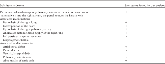
Scimitar syndrome is a very rare congenital anomaly of the heart with an incidence of 2:100.000 in live-born infants.Reference Gudjonsson and Brown 7 Familial clustering has been rarely reported.Reference Gudjonsson and Brown 7 , Reference Ruggieri, Abbate and Parano 8 The pathogenesis is unclear, but maldevelopment of the lung bud or the pulmonary vascularisation during early embryogenesis has been assumed.Reference Gudjonsson and Brown 7 Associated with the partial anomalous pulmonary venous return are other malformations as given in Table 1.Reference Gudjonsson and Brown 7 , Reference Levent, Esen and Memnune 9 These accompanying malformations tend to be far more severe and occur far more often (75%) in the infantile form, leading to the development of pulmonary hypertension and heart failure, which are the main reasons for the high mortality of up to 45% in this group, compared with the child/adult form (36%),Reference Levent, Esen and Memnune 9 which is often asymptomatic or associated with a history of recurrent pulmonary infections.Reference Gudjonsson and Brown 7 , Reference Levent, Esen and Memnune 9
Scimitar syndrome: defining finding and associated malformations and cardiac anomalies.

(+): Symptom seen in our patient; (−): symptom not seen in our patient
As cardiac malformations represent one of six component features of the VACTERL association Scimitar syndrome fits well into the phenotypic spectrum of its associated cardiovascular malformations. Therefore the existence of a rare but nevertheless closer association between the VACTERL association and Scimitar syndrome seems possible. This notion is further reinforced by the report of another case with Scimitar syndrome and VACTERL association.Reference Takawira and Omar 1 If taken into account that Scimitar syndrome is a rather rare cardiovascular anomaly and that there are also mild forms of Scimitar syndrome being diagnosed only by chance, it seems possible that the association might have been previously overlooked in cases of VACTERL association. We therefore propose that the combination of Scimitar syndrome and VACTERL association is not a random occurrence of two rare diagnoses in one patient but a further expansion of VACTERL-associated symptoms.
Hence, we hope to raise the perinatal awareness for the possible occurrence of Scimitar syndrome in children with VACTERL association, leading to an earlier prenatal or postnatal diagnosis and therapy with consecutive improvement of the clinical outcome of those afflicted.
Especially as prenatal diagnosis of Scimitar syndrome is possible but by far not routinely feasible,Reference Seale, Carvalho and Gardiner 10 awareness of this possible combination should be raised. Cardiac anomalies such as mesocardia, a small left ventricle, or a small aortic arch in prenatal diagnostics of children with suspicion of VACTERL association should lead to a thorough search for possible partial anomalous pulmonary venous return into the right atrium/vena cava.
Similarly, Scimitar syndrome should be taken into account in neonates with suspicion of VACTERL association and additional signs of atelectasis or lung sequestration, a hypoplastic pulmonary artery, or signs of partial anomalous pulmonary venous return from the right lung. Furthermore, the right pulmonary veins might be very hypoplastic, making a diagnosis by means of echocardiography, chest x-ray, or even magnetic resonance imaging difficult. We therefore conclude that Scimitar syndrome might be part of the cardiac malformations of the VACTERL association. Hence, its presence should be considered in a patient with suspicion of VACTERL association and cardiac anomalies.
Acknowledgements
The authors would like to thank Professor U. Gembruch, MD, head of the Department of Obstetrics and Prenatal Medicine at the University Hospital Bonn for his help and advice with the prenatal Diagnostics, Haitham Bachour, MD, head of the department of pediatric surgery at the University Hospital of Bonn, for his help and advice in treating the presented patient, Professor J. Breuer, MD and Professor P. Bartmann, MD, PhD, for their help treating the patient and their assistance in the preparation of the case report.
Financial Support
This research received no specific grant from any funding agency, commercial, or not-for-profit sectors.
Conflicts of Interest
None.