Implications
Growth, carcass characteristics and meat quality are important components of an efficient lamb production system. Vitamin E plays an important role in enhancing meat stability and improving meat quality. This study shows that intramuscular injections of vitamin E, given to growing lambs, reduced collagen maturity, which could increase meat tenderness, but decreased the percentage of pelvic limb.
Introduction
Vitamin E is the generic term used for all tocol and tocotrienol derivatives that exhibit the activity of α-tocopherol. Vitamin E has been known for its roles as the principal lipid-soluble chain-breaking antioxidant in biological membranes (Chan, Reference Chan1993) and in improving the quality characteristics of meat such as color, flavor, texture and nutritional value, and also extending its shelf-life (Morrisey et al., Reference Morrisey, Buckley, Sheehy and Monahan1994; Kasapidou et al., Reference Kasapidou, Wood, Richardson, Sinclair, Wilkinson and Enser2012). Contradictory data regarding the effect of vitamin E on growth traits and collagen synthesis in lambs (reviewed in the study by Maiorano et al., Reference Maiorano, Cavone, McCormick, Ciarlariello, Gambacorta and Manchisi2007) and pigs (reviewed in the study by Maiorano et al., Reference Maiorano, Manchisi, Salvatori, Filetti and Oriani1999) exist. Some works have demonstrated a beneficial effect of vitamin E treatment on growth traits in lambs (900 IU DL-α-tocopheryl acetate, Gentry et al., Reference Gentry, Ross, Oetting and Birch1992; 15 mg vitamin E/lamb per day, Macit et al., Reference Macit, Aksakal, Amsen and Aksu2003a). Conversely, other authors (1500 IU α-tocopherol, Birch et al., Reference Birch, Thomas and Ross1994; 1200 IU DL-α-tocopheryl Maiorano et al., Reference Maiorano, Cavone, McCormick, Ciarlariello, Gambacorta and Manchisi2007) observed a lower carcass weight and a negative effect on carcass wholesale cut weights in lambs injected with vitamin E. Several other studies provide evidence that vitamin E influences intramuscular collagen (IMC) characteristics, improving collagen solubility (50 or 100 μM α-tocopherol in vitro, Archile-Contreras et al., Reference Archile-Contreras, Cha, Mandell, Miller and Purslow2011) or reducing IMC maturity (Maiorano et al., Reference Maiorano, Cavone, McCormick, Ciarlariello, Gambacorta and Manchisi2007), which could affect meat tenderness. On the other hand, it has been reported that the use of 1000 IU of vitamin E, administered daily for 104 days before slaughter, effectively decreased toughness of longissimus steaks from heifers (Carnagey et al., Reference Carnagey, Huff-Lonergan, Trenkle, Wertz-Lutz, Horst and Beitz2008). However, according to the available information, no research has yet been conducted to study the effect of vitamin E on proteoglycan decorin. The extracellular matrix of muscle is composed mostly of the protein collagen with lesser quantities of other constituents such as proteoglycans also present (McCormick, Reference McCormick1999). It has been suggested a potential role for the proteoglycan decorin (a small dermatan sulfate) in regulating collagen fibrillogenesis, ordering the spatial arrangement of collagen molecules and, thus, influencing crosslinking patterns (McCormick, Reference McCormick1999).
The objective of the present study was to determine the effect of DL-α-tocopheryl acetate injections on growth, carcass traits and extracellular matrix in growing lambs.
Material and methods
Animals
Animal handling followed the recommendations of European Union directive 86/609/EEC and Italian law 116/92 concerning animal care. Lambs were reared in Molise, on a farm situated near the Abruzzi, Lazio and Molise National Park (Italy) at 560 m above sea level (latitude: 41°29'12"12 N, longitude: 14°28'25"68 E).
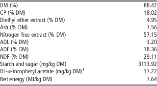
The trial was carried out on 24 15-day-old Ile de France suckling male lambs (average BW of 7.20 kg), born as singles in late February from 3-year-old dams of the same weight (58±1.8 kg). The ewes selected for the experiment were homogeneous in terms of parity and of milk yield and milk protein and fat contents of previous lactations. During the experiment, all dams, reared indoors, were fed with 1.4 to 1.7 kg vetch/oat alfalfa and polyphitic hay and 0.5 to 0.7 kg of concentrate and had free access to water. The lambs were randomly allotted to two groups: control (C, n =12) or vitamin E-treated (V, n=12). From the beginning of the study (15 days of age) until day 64 of age, each lamb of the V group received intramuscular injections of DL-α-tocopheryl acetate (left gluteus, 150 IU/week) in aqueous solution (Vitalene E; Fatro, Bologna, Italy) for 8 weeks for a total dose of 1200 IU. The C group lambs received injections of physiological saline. All animals received only maternal milk until the 21th day of age and, until weaned at 28th day of age, they had free access to a starter feed (18% CP and 6.89 MJ/kg dry matter) for an adaptation period of 7 days. The lambs stayed with their respective mothers from 1800 to 0700 h. From weaning, lambs were housed into six pens of 4 m2 with four animals per pen (three pens per treatment) and concentrate (Table 1) was offered ad libitum (in two daily meals at 0800 and 1600 h). The pens were cleaned weekly. Lambs had free access to water during the experiment. To calculate average daily weight gain (ADG), lambs were individually weighed at 15, 29, 43 and 57 days of age (in the morning after an overnight fast), and at slaughter. Moreover, average feed consumption for each experimental group was calculated; amounts of feed offered were recorded and refused feed was weighed daily.
Chemical composition of concentrate fed to lambs

DM=dry matter.
1 17.22 IU DL-α-tocopheryl acetate/kg DM.
Slaughter surveys
At 71 days of age, after 12 h fasting, lambs were electrically stunned, exsanguinated and processed at a local slaughterhouse. Hot and cold carcass weights were recorded and dressing percentages were calculated after dressing and chilling at 2°C to 4°C for 24 h. Carcass shrink losses, calculated as the difference between hot and cold carcass weights, were expressed as a percentage of hot carcass weight. Longissimus muscle (LM) pH was measured between the 12th and 13th ribs at 45 min (pH1) and 24 h (pHu) postmortem using a portable HI 9625 pH meter (Hanna Instruments, Padova, Italy) equipped with a penetrating glass electrode. In addition, LM area was measured, between the 12th and 13th ribs, by manually tracing muscles outlines onto acetate sheets and measuring areas by planimeter (Haff-Planimeter no. 317E). After the refrigeration period (24 h at 2°C to 4°C), from the right side of the carcass the pelvic limb was removed and its percentage was calculated based on cold carcass weight.
Collagen analysis
The LM, between the 11th to 12th thoracic vertebrae and the 4th to 5th lumbar vertebrae, and semimembranosus muscle (SM) were removed from the carcass (after 24 h at 2°C to 4°C); ~150 g of each muscle (wet weight) was collected, vacuum packaged and stored frozen (−40°C) until IMC and decorin analyses.
For IMC analyses, muscle samples were thawed, trimmed of fat and epimysium, lyophilized for 48 h, weighed and hydrolyzed in Duran tubes in 6 N HCl at 110°C for 18 to 20 h for determination of hydroxyproline (Woessner, Reference Woessner1961) and crosslinking. IMC concentration was expressed as μg hydroxyproline/mg of lyophilized sample. Hydroxylysylpyridinoline (HLP) concentration, the principal non-reducible crosslink of muscle collagen (McCormick, Reference McCormick1999), was determined using a modified HPLC procedure developed by Eyre et al. (Reference Eyre, Koob and van Ness1984). A Kontron HPLC (Kontron Instruments, Milan, Italy) model 535, equipped with a Luna C18 column (250×4.6 mm×5 µm; Phenomenex, Torrance, CA, USA) was used. The concentration of HLP residues in the samples was calculated based on the concentration of collagen in each hydrolyzate, assuming that the molecular weight of collagen was 300 000 Da and the molar fluorescence yield of pyridoxamine (internal standard) was 3.1 times that of HLP (Eyre et al., Reference Eyre, Koob and van Ness1984). The HLP was expressed as moles of HLP per mole of collagen.
Decorin analysis
Decorin blots were determined by the method of Velleman (Reference Velleman1995). Muscle portions were homogenized in Tris-EDTA buffer (10 mM Tris and 1 mM EDTA; pH 8.0; Sigma-Aldrich, St. Louis, MO, USA). Total protein concentrations were assessed using the Bio-Rad protein assay kit II (Bio-Rad Laboratories, Hercules, CA, USA). Protein (10 mg) from each sample was spotted onto nitrocellulose (Trans-blot transfer medium; Bio-Rad Laboratories) using the mini-Protean system (Bio-Rad, Hercules, CA, USA). Blots were exposed to the rabbit antidecorin polyclonal antibody and to the goat anti-rabbit IgG with an alkaline phosphatase conjugate. Blots were then exposed to the 5-bromo-4-chloro-3-indolyl phosphate/nitro blue tetrazolium color system, and decorin concentrations within each blot were quantified to a relative scale by color intensity after scanning with a digital color scanner (GS-800 Calibrated Densitometer; Bio-Rad) (Velleman, Reference Velleman1995).
Statistics analyses
Growth, slaughter performance and pH data were analyzed by one-way ANOVA using the SPSS package (SPSS/PC1Statistics 18.0; SPSS Inc., Chicago, IL, USA, 2010). IMC and decorin data were evaluated by ANOVA, in a 2×2 factorial design. The model included DL-α-tocopheryl acetate treatment (C v. V) and muscle (LM v. SM) and their interaction as fixed effects and individual animal as a random effect. Scheffé’s test was used for comparing mean values. BW at slaughter was included as a covariant. Pen was the experimental unit for the analysis of dry matter intake data and each individual lamb was considered as the experimental unit for growth, slaughter performance and pH.
Results and discussion
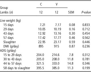
In the present study, α-tocopherol content was not measured in the muscle. However, it has been reported that there is a higher α-tocopherol accumulation in skeletal muscle of lambs intramuscular injected (Salvatori et al., Reference Salvatori, Pantaleo, Di Cesare, Maiorano, Filetti and Oriani2004) or supplemented (Ripoll et al., Reference Ripoll, González-Calvo, Molino, Calvo and Joy2013) with vitamin E when compared with control lambs. Lamb growth performance are presented in Table 2. Neither lamb growth nor amount of DMI was affected by DL-α-tocopheryl acetate administration, a finding in accordance with the results of Maiorano et al. (Reference Maiorano, Manchisi, Salvatori, Filetti and Oriani1999) who reported no effect of vitamin E administration on weight of lambs slaughtered at 40 days. Conversely, Maiorano et al. (Reference Maiorano, Cavone, McCormick, Ciarlariello, Gambacorta and Manchisi2007) observed that 1200 IU of DL-α-tocopheryl acetate intramuscular injected in Ile de France lambs, slaughtered at 71 days, did not influence final live weight, but increased ADG significantly during suckling (day 15 to 22), whereas it did not affect the DMI. Similarly, Gentry et al. (Reference Gentry, Ross, Oetting and Birch1992) observed increased gains from birth to 30 days of age in Suffolk lambs injected with 900 IU DL-α-tocopheryl acetate. Our results are also in accordance with other studies conducted in different breeds, with vitamin E supplemented in the concentrate (Álvarez et al., Reference Álvarez, De la Fuente, Díaz, Lauzerica, Pérez and Cañeque2008; Ripoll et al., Reference Ripoll, González-Calvo, Molino, Calvo and Joy2013) or in grass silage-based feed (Kasapidou et al., Reference Kasapidou, Wood, Richardson, Sinclair, Wilkinson and Enser2012). However, several other studies have demonstrated a beneficial effect of vitamin E treatment on growth traits in lambs (Macit et al., Reference Macit, Aksakal, Amsen and Aksu2003a).
Mean values for growth performance of Ile de France lambs

C=intramuscular injection of physiological saline; V=intramuscular injection of 1200 IU DL-α-tocopheryl acetate; DMI=dry matter intake; ADG=average daily weight gain.
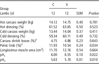
The treatment with vitamin E did not affect (P>0.05) the hot and cold carcass weight, the dressing percentage or LM area (Table 3). A similar result was obtained in other studies involving lambs of the same breed (Maiorano et al., Reference Maiorano, Manchisi, Salvatori, Filetti and Oriani1999, Reference Maiorano, Cavone, McCormick, Ciarlariello, Gambacorta and Manchisi2007) and on Awassi breed (Macit et al., Reference Macit, Aksakal, Amsen, Esenbuga and Aksu2003b). On the contrary, Birch et al. (Reference Birch, Thomas and Ross1994) observed a lower carcass weight and a negative effect on carcass wholesale cut weights in fine-wool wether lambs injected with a single-dose of vitamin E (1500 IU); while Njeru et al. (Reference Njeru, McDowell, Wilkinson, Linda, Williams and Lentz1992) did not find any effect of different doses (ranging from 125 to 1000 IU) of DL-α-tocopherol injection.
Mean values for slaughter performance and pH of Ile de France lambs

C=intramuscular injection of physiological saline; V=intramuscular injection of 1200 IU DL-α-tocopheryl acetate.
1 Calculated on hot carcass weight.
2 Calculated on cold carcass weight.
Carcass shrink losses were not influenced (P>0.05) by the treatment (Table 3). However, contrasting findings are reported in literature. Maiorano et al. (Reference Maiorano, Cavone, McCormick, Ciarlariello, Gambacorta and Manchisi2007), noted a higher value in the control group than in DL-α-tocopheryl acetate-treated lambs; they attributed this results to the treatment with vitamin E and its antioxidant effect on muscle cell membranes. Differently, Macit et al. (Reference Macit, Aksakal, Amsen, Esenbuga and Aksu2003b) found a higher value (+1.9%) of carcass shrink losses of lamb receiving a supplement of 45 mg vitamin E/lamb per day for a 75 days fattening period when compared with control animals.
The treatment with vitamin E reduced (−1.39%; P<0.05) the pelvic limb percentage (Table 3), with a potential negative impact on lamb market weight. This finding is in agreement with an earlier study on the same breed (Maiorano et al., Reference Maiorano, Cavone, McCormick, Ciarlariello, Gambacorta and Manchisi2007), but in contrast with the result of Macit et al. (Reference Macit, Aksakal, Amsen, Esenbuga and Aksu2003b). However, it has also been reported that vitamin E treated lambs had lower leg and shoulder weights (Birch et al., Reference Birch, Thomas and Ross1994) compared with control lambs. Hatfield et al. (2000) suggested that this might be due to vitamin E stimulation of the immune system that, in turn, caused a partitioning of energy away from growth and promoted muscle catabolism.
Vitamin E treatment clearly influenced both pH1 and pHu in LM, which were higher (P<0.05) in treated animals compared with control ones (Table 3). These findings are consistent with study involving injected lambs (Maiorano et al., Reference Maiorano, Cavone, McCormick, Ciarlariello, Gambacorta and Manchisi2007). As reported by Cheah et al. (Reference Cheah, Cheah and Krausgrill1995) and Castellini et al. (Reference Castellini, Dal Bosco and Bernardini1999), the capability of vitamin E to stabilize membranes is presumably achieved by decreasing Ca++ level release (either mitochondria or the sarcoplasmic reticulum) that reduces phospholipase A2 activity and, hence, phospholipid hydrolysis. Decreased levels of cytosolic Ca++ reduce the rate of postmortem glycolysis resulting in a higher postmortem pH. However, the pHu values observed in this study (mean 5.66; SEM 0.01) varied within the pH range accepted for commercial meats, with no evidence of pre-slaughter stress and meat defect.
IMC and decorin properties of LM and SM muscles are shown in Table 4. Administration of vitamin E did not affect (P>0.05) IMC amount but slowed (P<0.05) collagen maturation (HLP/collagen). These results may have implications on meat tenderness since it is reported that an increase in collagen maturation leads to an increased IMC thermal stability, which has been related to shear force (Monin and Ouali, Reference Monin and Ouali1991; Hopkins et al., Reference Hopkins, Allingham, Colgrave and van de Ven2013) and with an undesirable changes in eating quality of meat (McCormick, Reference McCormick1999; Purslow, Reference Purslow2005; Lepetit, Reference Lepetit2008).
Mean values for collagen and decorin of LM and SM muscles of Ile de France lambs

LM=longissimus muscle; SM=semimembranosus muscle; C=intramuscular injection of physiological saline; V=intramuscular injection of 1200 IU DL-α-tocopheryl acetate; T=treatment; M=muscle; IMC=intramuscular collagen; HLP=hydroxylysylpyridinoline.
1 Of lyophilized tissue.
2 Of fresh muscle tissue.
A marked muscle effect on IMC properties was apparent (Table 4). The SM muscle had higher (P<0.01) values of IMC amount as well as collagen maturation (HLP/collagen). Differences in IMC properties between muscles could be due to functional and structural differences between locomotor and postural muscles (Maiorano et al., Reference Maiorano, Ciarlariello, Cianciullo, Roychoudhury and Manchisi2009). In fact, it has been documented that locomotor muscles possess more crosslinking than postural muscles (McCormick, Reference McCormick1999). The difference between the two studied muscles evidenced, in agreement with the literature (McCormick, Reference McCormick1999), that variation in IMC properties with muscle type and function leads to the well-known differences in background toughness among meat cuts.
Compared with lambs of the C group, those of the V group had higher decorin concentration (+28.8%; P<0.05). Furthermore, it was recorded a marked difference in decorin concentration between muscles: LM had higher (+45.2%) values than that of SM (P<0.01). To our knowledge, no studies are available on the effect of vitamin E on decorin concentration in lamb muscles. Fewer studies have focused on how α-tocopherol might regulate the extracellular matrix. Villacorta et al. (Reference Villacorta, Azzi and Zingg2007) recently proposed that vitamin E is able to protect the damaged vascular wall, not only by limiting cell proliferation, but also by mediating the process that leads to the stabilization of a fibrous cap by influencing components of the extracellular matrix. In previous study, Schwartz (Reference Schwartz1979) reported that a supplementation of α-tocopherol, in human articular cartilage, stimulated sulfate proteoglycan biosynthesis and, in addition, it inhibited the degradation activity of lysosomal enzymes (arylsulfatase A and acid phosphatase) on proteoglycans. However, as already observed, the proteoglycan decorin plays a key role in regulating cell proliferation, growth factor activity and collagen organization (Velleman et al., Reference Velleman, Liu, Eggen and Nestor1999). Albrecht et al. (Reference Albrecht, Liu, Yang, Zhao, Jones and Maak2011) suggested that an increase in decorin level could affect the formation of collagen fibers and therefore negatively influence meat quality. However, McCormick (Reference McCormick1999) reported that the temporal gap between increased decorin expression and the measured levels of elevated crosslinking is consistent with the concept that development of crosslinking is a time-dependent process. As fibrillogenesis proceeds, crosslinks form as reactive residues align; initial reducible crosslinks condense, ultimately forming the mature trivalent crosslinks (McCormick, Reference McCormick1999).
Conclusion
The results of the present study indicate that the DL-α-tocopheryl acetate treatment did not affect growth, hot and cold carcass weights, carcass shrink losses and area of LM, but increased pH values. However, it should be noted that intramuscular vitamin E administration has the potential of impacting negatively on pelvic limb mass, in agreement with some previous findings. Vitamin E treatment reduced collagen maturity and increased decorin in the LM and SM muscles of growing lambs. Although the treatment effects noted in this study were subtle, further research is warranted to elucidate, in particular, the effect of vitamin E on extracellular matrix.
Acknowledgment
This study was supported by a Grant from University of Molise, Italy.