Introduction
Vertebrates, including mammals, are considered to have evolved by whole genome duplications (Ohno, Reference Ohno1970), which is called the 2R hypothesis. This hypothesis is supported by analyses of the genomic sequences of various animals (Brodsky and Uryvaeva, Reference Brodsky and Uryvaeva1985; Holland et al., Reference Holland, Garcia-Fernàndez, Williams and Sidow1994; Panopoulou and Poustka, Reference Panopoulou and Poustka2005). Polyploid fish and amphibians are present and some polyploid fish have already been applied to industrial aquaculture (Piferrer et al., Reference Piferrer, Beaumont, Falguière, Flajšhans, Haffray and Colombo2009). In mammals, polyploids do not naturally exist at the individual level, although somatic polyploid cells physiologically exist at a cellular level in some organs.
Mouse polyploidy cannot survive due to embryonic lethality (Kaufman and Webb, Reference Kaufman and Webb1990; Eakin et al., Reference Eakin, Hadjantonakis, Papaioannou and Behringer2005). Polyploid mammalian embryos might exhibit lethality for the following reasons: (1) developmental disorder due to abnormal gene expressions; and (2) extended cell cycles and reduced number of cells constructing the embryos. In mouse tetraploid embryos, abnormal expression of Wee1 is associated with cell division and increased apoptosis via p53 were observed (Horii et al., Reference Horii, Yamamoto, Morita, Kimura, Nagao and Hatada2015; Wen et al., Reference Wen, Li, Cheng, Li, Zhang, Xiang, Wang and Han2017). Abnormalities in epigenomic modifications including genomic imprinting and histone modification also occur (Yamazaki et al., Reference Yamazaki, Amano, Bai, Takahashi and Kawahara2016; Wu et al., Reference Wu, Zhao, Zhu, Chen, Wei, Bao, Sun and Li2017). In addition, preimplantation polyploid embryos show a decrease in the number of cells constructing the embryos due to extended cell cycles (Kawaguchi et al., Reference Kawaguchi, Kano and Naito2009; Imai et al., Reference Imai, Fujii, Kusakabe, Kiso and Kano2019), and postimplantation polyploid embryos degenerated, leading to a reduction in pregnancy rate (Gazzo et al., Reference Gazzo, Peña, Valdéz, Chung, Velit, Ascenzo and Escudero2020).
Polyploid cells physiologically are present in some organs of mammals. In the liver, polyploid hepatocytes and hepatocytes fused with bone marrow cells play an important role in liver regeneration (Duncan et al., Reference Duncan, Taylor, Hickey, Hanlon Newell, Lenzi, Olson, Finegold and Grompe2010; Wang et al., Reference Wang, Chen, Li, Liu, Zhang, Xia, Yu, You, Xiang, Lu, Yao, Borjigin, Yang, Wangensteen, He, Wang and Hu2014). In the placenta, syncytial cells are involved in the maintenance of pregnancy via endocrine and paracrine systems (Hay, Reference Hay1988; Hu and Cross, Reference Hu and Cross2010). Furthermore, the metabolism and function of polyploid cells are different from those of diploid cells in the heart and liver (Anatskaya and Vinogradov, Reference Anatskaya and Vinogradov2010). However, the elucidation of cellular behaviour and functions in mammalian polyploid cells in vivo is inhibited by the lethality of polyploid embryos after implantation and the small cell number of polyploid somatic cells.
The purpose of this present study was to investigate whether the cellular function is maintained after polyploidization in mammals. To this end, we analyzed the differentiation potency and behaviour of tetraploid embryonic stem cells (ESCs) in vivo. Our present study will generate important reference data for in vitro experimental systems for producing polyploid mammals in the future.
Materials and methods
Cell culture and differentiation induction
The diploid and tetraploid ESCs used in this study were established from embryos recovered from BDF1 (Japan SLC) mice and maintained in ESGRO medium (Merck) supplemented with KSR. The establishment method details and its efficiency have been described previously (Imai et al., Reference Imai, Kano, Fujii, Takasawa, Wakitani, Hiyama, Nishino, Kusakabe and Kiso2015). Embryoid body (EB) formation was performed by culturing for 5 days in DMEM supplemented KSR (10% final concentration), MEM non-essential amino acids solution 100× (1× final concentration) and 2-mercaptoethanol (1× final concentration) using low adherence plates (MS-9096U, Sumitomo Bakelite). The medium was replaced every 2 days. The diploid and tetraploid EB-derived cells were obtained by culturing in DMEM supplemented with 10% FBS and 10% KSR on gelatin-coated dishes. FACS analysis, chromosomal spreads and growth rate followed our previous report (Imai et al., Reference Imai, Kano, Fujii, Takasawa, Wakitani, Hiyama, Nishino, Kusakabe and Kiso2015). In brief, chromosomal spreads were prepared using Giemsa staining of colchicine-treated cells. The number of chromosomes was counted from more than 10 field images. Flow cytometric analysis was performed using BD Accuri C6 (BD) on propidium iodide (PI)-stained cells. TC20 cell counter (Bio-Rad) was used for the calculation of the proliferation rate.
Differentiation into neural lineage was performed by culturing diploid and tetraploid ESCs using NDiff227 (TaKaRa Bio). The medium was replaced every 2 days and analyzed 14 days later. Differentiation into osteocyte lineage was performed using αMEM + 10% FBS supplemented with dexamethasone (Sigma, 0.1 μM final concentration), β-glycerophosphate (Nacalai Tesque, 10 mM final concentration) and ascorbic acid (Nacalai Tesque, 50 µg/ml final concentration). The medium was replaced every 2 days and analyzed 14 days later. Mouse embryonic fibroblasts (MEF) were used as a negative control.
Chimera formation and analysis
Two-cell stage embryos were collected from BDF1 mice and cultured in M16 (Sigma) until the morula stage. After removing the zona pellucida with acidic Tyrode’s solution, morulae adhered to EGFP-expressing tetraploid ESCs in aggregation holes prepared on a dish with an aggregation needle (DN-9, BLS) and cultured until the blastocyst stage. Thereafter, chimeric embryos were transplanted into the uterus of pseudopregnant mice (eight embryos per uterine horn), and analysis of chimeric embryos was performed at E12.5. Chimeric embryos were observed and dissected under a fluorescence stereomicroscope, and embedded in paraffin according to a standard method after fixation. Chimeric embryonic limb buds were cultured in DMEM + 10% FBS.
All procedures were approved by the Experimental Animal Care and Use Committee as protocol number: 268 at Yamaguchi University.
Staining
For prepare sections, tissues fixed in 4% PFA were embedded in paraffin (Histosec, Merck) according to the standard method, and 4-µm thick sections were prepared using a microtome (Leica Biosystems RM2245, Leica).
For immunofluorescence, cells were fixed in 4% PFA at 4°C for 30 min, permeabilized with 0.2% Triton-X100–PBS at room temperature for 30 min. After blocking with 2% FBS–PBS for 15 min, samples were incubated with primary antibodies at 4°C overnight, and then incubated with secondary antibodies for 1 h under light protection. The antibodies used in this study were as follow: NANOG (ab80892; abcam, 1:100) and Alexa Fluor 488 conjugated goat anti-rabbit IgG (ab150077; abcam, 1:300), NESTIN (012-26843; Wako, 1:200) and Alexa Fluor 594 conjugated goat anti-mouse IgG (ab150116; abcam, 1:400), TUJ1 (GTX631836; Gene Tex, 1:200) and Alexa Fluor 488 conjugated goat anti-rat IgG (ab150157; abcam, 1:400), EGFP (#598; MBL, 1:500) and FITC conjugated goat anti-rabbit IgG (#234; MBL, 1:160). After washing with PBS, samples were stained with 5.0 µg/ml DAPI–PBS and observed using an ECLIPSE Ti2 (Nikon), BZ9000 (Keyence) or LSM710 (Zeiss) microscope.
For alizarin red staining, cells were fixed with 100% ethanol for 10 min and stained by adding 0.1% alizarin red S solution.
For von Kossa staining, cells were fixed in 4% PFA at 4°C for 30 min, and calcium substitution was performed with 0.5% silver nitrate for 2 h under light protection. Thereafter, cells were incubated in 3% sodium carbonate formalin for 2 min and observed after washing with PBS.
Cytospin preparations were performed according to the manufacturer’s protocol (Cytospin4; Thermo Fisher Scientific) and stained with 5.0 µg/ml Hoechst 33342–PBS.
Nucleic acid extraction and analysis
Total RNA was purified using ReliaPrep RNA Cell MiniPrep System (Promega) according to the manufacture’s protocol. The purity and concentration of the extracted RNA were measured using a NanoDrop 2000 spectrophotometer (Thermo Fisher Scientific). The cDNA was synthesized from 1 µg of RNA using a QuantiTect Reverse Transcription Kit (Qiagen). Genomic DNA was isolated from a part of the embryos and paraffin-embedded embryonic sections (4-µm thick × ∼20 sheets) using Nucleospin TissueXS (TaKaRa Bio) according to the manufacture’s protocol. The PCR was performed using PrimeSTAR HS (TaKaRa Bio) or BIOTAQ (Bioline). The number of cycles for RT-PCR was 30 and for PCR was 35. Primer sequences are shown in Table S1.
Statistical analysis
Mann–Whitney test was used for statistical analysis.
Results
Characteristics of tetraploid ESCs and differentiated cells
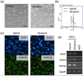
The mouse diploid and tetraploid ESCs used in this study were established previously (Imai et al., Reference Imai, Kano, Fujii, Takasawa, Wakitani, Hiyama, Nishino, Kusakabe and Kiso2015). The tetraploid ESC lines showed the same naive type colony as diploids, despite altering the ploidy (Fig. 1a, b), and also detected the expression of pluripotency marker genes (Fig. 1c, d).
Characteristics of mouse diploid and tetraploid embryonic stem cells (ESCs). (a) Morphology of mouse diploid and tetraploid ESCs is shown. Bars, 50 µm. (b) Ploidy of diploid tetraploid ESCs was compared using flow cytometric analysis with PI staining. (c) Immunofluorescence images of NANOG. Bars, 200 µm. (d) Expression of pluripotent marker genes was analyzed using RT-PCR.

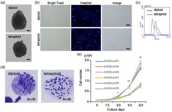
Differentiation induction was performed on these diploid and tetraploid ESC lines, and EB formation by suspension culture was observed (Fig. 2a). For handling differentiated cells easily, diploid and tetraploid EB-derived cells were induced by the adherent culture of the embryoid bodies (Fig. 2b). Subsequently, the ploidy of diploid and tetraploid EB-derived cells was examined. As a result, tetraploid EB-derived cells had twice as much DNA volume as diploid EB-derived cells using flow cytometric analysis with PI staining (Fig. 2c) and maintained 80 chromosome sets using chromosomal spreads with Giemsa staining (Figs 2d, S1). Furthermore, tetraploid EB-derived cells had a significantly reduced cell growth rate compared with diploid EB-derived cells (Fig. 2e).
Formation of embryoid bodies (EB) and EB-derived cells. (a) Morphology of diploid and tetraploid EB induced by suspension culture is shown. Bars, 50 µm. (b) Morphology of diploid and tetraploid EB-derived cells induced by adherent culture of embryoid bodies is shown. Bars, 50 µm. (c) Ploidy of diploid and tetraploid EB-derived cells was compared using flow cytometric analysis with PI staining. (d) Number of chromosomes of diploid and tetraploid EB-derived cells was compared using chromosome spreads and Giemsa staining. (e) Growth rates of EB-derived cell lines were compared. The data represented the mean and standard error of the mean (SEM). *, P < 0.05; **, P < 0.01.

Induction of differentiation to specific cell lineage
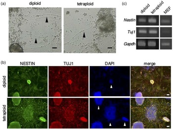
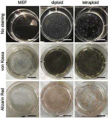
Next, differentiation into lineages of three germ layers was performed using these ESCs and EBs. First, we analyzed the expression of endodermal marker genes in EBs. RT-PCR showed the expression of the endodermal marker genes Eomes, Gata4, Sox7 and Sox17 in these EBs (Fig. S2). Next, differentiation into neural cell lineage was performed. After 14 days of culture in the differentiation medium, axon-like structures that extended from colonies of cells were observed (Fig. 3a). Immunostaining revealed that cells and axon-like structures were positive for neural lineage markers NESTIN and TUJ1 (Fig. 3b). RT-PCR also revealed the expression of these neural marker genes both in diploid and tetraploid cells (Fig. 3c). These results indicated that tetraploid ESCs could be induced to a neural cell lineage the same as diploid ESCs in vitro. Furthermore, differentiation into osteocyte lineage was performed. After differentiation culture of diploid and tetraploid EB-derived cells for 14 days, low transparency cells appeared (Fig. 4 upper). These cells were evaluated for osteocyte differentiation as an index of calcification by von Kossa and alizarin red staining to detect calcium phosphate and Ca2+, which are the major components of the bone matrix. These staining patterns showed that calcifications were detected both in diploid and tetraploid EB-derived cells (Fig. 4, middle and lower). These results indicated that tetraploid cells could be induced into an osteocyte lineage containing calcium salts.
Differentiation into neural lineage. (a) Cell morphology was shown 14 days after differentiation of ESCs into neural lineage. Arrowheads: Axon-like structures extending from the colony. Bars, 50 µm. (b) Immunostaining of neural markers NESTIN and TUJ1. DAPI was used to stain DNA. White arrowheads indicate colonies of cells. Bars, 50 µm. (c) Expression of neural marker genes was analyzed using RT-PCR.

Differentiation into the osteocyte lineage. The unstained (upper row), von Kossa-stained (middle row) and alizarin red-stained images (lower row) after differentiation into the osteocyte lineage using diploid and tetraploid EB-derived cells are shown. Mouse embryonic fibroblasts (MEF) were used as a negative control. Bars, 1.0 cm.

Analysis of tissue contribution in chimeric embryos
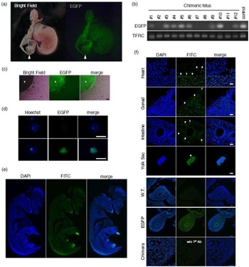
Tetraploid ESCs maintained pluripotent state and differentiation potency into specific cell lineages despite the alternation of ploidy. Subsequently, the chimeric embryo formation was performed to investigate whether tetraploid cells can contribute to embryonic tissue formation. Our previous report showed that tetraploid ESCs contribute to the inner cell mass of the host embryos in the blastocyst stage (Imai et al., Reference Imai, Kano, Fujii, Takasawa, Wakitani, Hiyama, Nishino, Kusakabe and Kiso2015). In this present study, we analyzed E12.5 chimeric embryos. In total, 240 embryos were used for the embryo transferred experiment, the fluorescence of EGFP derived from tetraploid ESCs was localized in the embryo and yolk sac (Figs 5a, S3A). In addition, EGFP was observed in each organ of the dissected chimeric embryo (Fig. S3B). Moreover, PCR targeted EGFP using genomic DNA revealed that chimerism was detected in eight of the 12 obtained embryos (Fig. 5b). EGFP was also observed in the tissue piece and its migrating cells after culturing a part of the chimeric embryonic limb buds (Fig. 5c). Cytospin preparation of migrating cells from chimeric embryonic tissue showed cells expressing EGFP (Fig. 5d). Immunofluorescence staining was performed to analyze tetraploid ESC-derived cells for more detailed tissue localization with control groups. EGFP-positive cells were uniformly distributed in embryonic tissues (Fig. 5e, f). These results indicated that tetraploid ESCs could contribute to embryonic tissue development in vivo.
Analysis of chimeric embryos using tetraploid ESCs. (a) Fluorescence observation images of chimeric embryos using EGFP-expressing tetraploid ESCs. White arrowheads indicated the yolk sac. (b) Genotyping using PCR targeting EGFP for obtained embryos. (c) Primary culture cells migrated from chimeric embryonic limb buds are shown. The upper left * indicates a tissue mass from a chimeric embryo. Bar, 50 µm. (d) Fluorescence images of the cytospin preparation of cultured cell from the chimeric embryonic limb buds. Both EGFP-negative (upper) and EGFP-positive cells (lower) were observed in migrating cells. Bars, 10 µm. (e) Immunofluorescence of sagittal section of whole chimeric embryo. (f) Immunofluorescence of different tissues from the chimeric embryo. Heart, gonads, intestine, yolk sac are shown in the upper to lower rows. The wild-type (WT) embryo was used as a negative control, and the EGFP-expressing embryo was used as a positive control. The chimeric embryo without the primary antibody was used as a secondary antibody control. White arrows indicate EGFP-positive cells. Bars, 50 µm.

Discussion
In this present study, tetraploid ESCs can give rise to differentiated cell types that are derived from germ layers in vitro. We also found that tetraploid ESCs could differentiate into various cell lineages in vivo overcoming the embryonic lethality. Our results suggested that mammalian stem cells maintained the plasticity for differentiation after polyploidization.
Tetraploid embryoid bodies and embryoid body-derived cells showed the same morphology as diploids. In addition, its ploidy was maintained after differentiation induction. Although haploid ESCs were established, many haploid cells would be diploidized after differentiation (Elling et al., Reference Elling, Taubenschmid, Wirnsberger, O’Malley, Demers, Vanhaelen, Shukalyuk, Schmauss, Schramek, Schnuetgen, von Melchner, Ecker, Stanford, Zuber, Stark and Penninger2011; Leeb and Wutz, Reference Leeb and Wutz2011). In addition, the pluripotency of various polyploid ESC lines established via cytostatics treatment maintained LIF independently, but the ploidy collapsed in these cell lines (Fujikawa-Yamamoto et al., Reference Fujikawa-Yamamoto, Miyagoshi, Luo and Yamagishi2011, Reference Fujikawa-Yamamoto, Ota, Miyagoshi and Yamagishi2012, Reference Fujikawa-Yamamoto, Miyagoshi and Yamagishi2013). From these reports, mouse ESCs are considered to be stable in diploid; however, ploidy of our established tetraploid ESCs was stable as tetraploid cell lines after differentiation (Figs 1c, d and S1). The electrofusion method might provide stable maintenance of tetraploid ESCs through chromosomal remodelling in the early tetraploid embryo (Park et al., Reference Park, Lee, Bui, Park, Park, Cho, Song, Kim, Nguyen and Kim2011).
The proliferation rate of tetraploid EB-derived cells was significantly lower compared with diploid EB-derived cells, which is similar to our previously reported growth rates in the undifferentiated state (Imai et al., Reference Imai, Kano, Fujii, Takasawa, Wakitani, Hiyama, Nishino, Kusakabe and Kiso2015). In the undifferentiated state, the cell cycle is prolonged uniformly (Imai et al., Reference Imai, Fujii, Kusakabe, Kiso and Kano2016) and, even after induction of differentiation, the time required for one cycle of the cell cycle might be prolonged by altering the ploidy. The proliferation rate of tetraploid cells produced by electric cell fusion is reduced compared with that before fusion (McGill et al., Reference McGill, Lalley, Leach, Johnson and Von Hoff1992), and the proliferation rate is also reduced due to the introduction of foreign DNA into cells (Von Hoff et al., Reference Von Hoff, Forseth, Clare, Hansen and VanDevanter1990), suggesting that the ploidy in the nuclei is related to the cell growth rate. A comprehensive analysis of cell cycle-related genes or ultrastructural analysis of nuclei might reveal the relationship between nuclear DNA content and cell growth.
To identify the ability to differentiate to specific cell lineage in tetraploid cells, we performed induction to ectoderm-derived neural cell lineage and mesendoderm-derived osteocyte lineage. As some neurons have been known to be physiologically tetraploidization in association with p75 (López-Sánchez and Frade, Reference López-Sánchez and Frade2013), we considered that neurons could accept ploidy alternation and differentiation according to previous reports (Okada et al., Reference Okada, Matsumoto, Shimazaki, Enoki, Koizumi, Ishii, Itoyama, Sobue and Okano2008). Axon-like structures that extended from the tetraploid cell colonies emerged and expressed neural cell lineage specific markers, indicating that differentiation potency into neural lineage was maintained after tetraploidization. Recent studies showed that osteocyte was generated from axial stem cells or mesoderm (Takemoto et al., Reference Takemoto, Uchikawa, Yoshida, Bell, Lovell-Badge, Papaioannou and Kondoh2011; Kondoh and Takemoto, Reference Kondoh and Takemoto2012). In this study, we confirmed that embryoid body-derived cells differentiated into osteocytes containing calcium salts. Although our previous study detected mRNA for marker genes (Imai et al., Reference Imai, Kano, Fujii, Takasawa, Wakitani, Hiyama, Nishino, Kusakabe and Kiso2015), we confirmed the characteristics of differentiated cells including proteins, cell structure and calcium salts. These results suggested that the ability to differentiate into both mesodermal and ectoderm-derived cell lineages was maintained despite altering ploidy.
Furthermore, this present study clarified that tetraploid ESCs contributed to the fetal tissue. In chimeric mice produced by combination with diploid embryo and tetraploid embryos, tetraploid cells contribute only to placental tissue (Nagy et al., Reference Nagy, Rossant, Nagy, Abramow-Newerly and Roder1993). This has been universally applied to a tetraploid complementation method to produce efficient knockout mice (Wang et al., Reference Wang, Kiefer, Urbánek and Wagner1997). The positioning of tetraploid embryonic cells during the formation of chimeric blastocysts was different from that of diploid cells (MacKay and West, Reference MacKay and West2005), which might be related to the establishment of the tetraploid complementation method. Tetraploid embryos had fewer cells in their inner cell masses (ICM) than diploid embryos (Liu et al., Reference Liu, Czerwiec and Keefe2004; Park et al., Reference Park, Lee, Bui, Park, Park, Cho, Song, Kim, Nguyen and Kim2011), diploid cells may predominate over tetraploid cells in chimeric embryos. The mechanism of the tetraploid complementation method may become clearer by elucidation of the change in molecular characteristics of the cell constructing the embryos after polyploidization, through comprehensive analysis or time-lapse observation using multi-colour fluorescence. Furthermore, it is unclear how the cells themselves are affected by the alternation in ploidy before the tetraploid complementation method is elucidated.
In this present study, as tetraploid ESCs were derived from tetraploid ICM, tetraploid ESCs might be fated into ICM lineage and maintain differentiation potency into ICM lineage not into placental tissues and contribute ICM lineage in chimeric embryos. Hybrid tetraploid cells between mouse ESCs and somatic cells showed pluripotency to the fetal lineage (Tada et al., Reference Tada, Takahama, Abe, Nakatsuji and Tada2001) suggesting that pluripotency in the contribution to chimeric tissues might be determined by cellular lineage rather than by ploidy.
In summary, we first showed that mouse tetraploid ESCs had high differentiation potential similar to diploid ESCs despite increment in the ploidy, suggesting that mammalian embryos have differentiation potency into somatic cells even after polyploidization. Furthermore, tetraploid ESCs could be a useful experimental model in vitro for clarifying mammalian polyploid embryogenesis.
Supplementary material
To view supplementary material for this article, please visit https://doi.org/10.1017/S0967199421000800
Acknowledgements
This work was supported by JSPS KAKENHI (Grant-in-Aid for Challenging Exploratory Research; JP25660254 and JP15K14880 to KK, Grant-in-Aid for JSPS Research Fellow; JP17J07902 and Young Scientists; 20K15664 to HI) and Kaibara Morikazu Medical Science Promotion Foundation (to HI). This research was also supported by the research programme on development of innovative technology grants from the Project of the Bio-oriented Technology Research Advancement Institution (BRAIN) (to KK, HI, and WF).