INTRODUCTION
The main objective of radiation therapy is to deliver the maximum possible dose to the target tumor with minimum dose to the normal surrounding tissues.Reference Wieslander and Knöös 1 To achieve this aim, a good understanding is needed from the dose distribution in irradiated tissue and most importantly, experimental verification of dose distribution. During the actual radiation treatment planning in clinics, dose distribution is calculated by treatment planning systems (TPS). Any deviation between calculated dose distribution and true value would lead to change in patient's dose that would have an important effect on the quality and effectiveness of the radiotherapy treatment.
Some methods of dose calculation algorithms are not enough accurate in term of dose calculation due to electron disequilibrium at radiation small field boundaries or at the inhomogeneities interfaces.Reference Arnfield, Siantar, Siebers, Garmon, Cox and Mohan 2 – Reference Jones, Aird and Colyer 5 It is known that the Monte Carlo (MC) is the most accurate method for dose calculationReference Sharpe and Battista 6 – Reference Li, Pawlicki, Deng, Jiang, Mok and Ma 10 and with the advancement of computer technology, dose calculation algorithms based on the MC method has found the necessary potential for dose calculation. But, due to the time consuming process of the full MC calculations and some of the limitations in speed of computers,Reference Bielajew and Rogers 11 it is still impossible to use full MC in routine clinical calculations for heavy traffic radiotherapy departments. However, it is still the most powerful technique for verification of other calculation algorithms in TPS's engines.Reference Verhaegen and Seuntjens 12 – Reference Rogers 14
In the present work, we compared two dose calculation algorithms employed in CorePLAN TPS for the special clinical case of pituitary adenoma. The algorithms are equivalent tissue-air ratio (ETAR) and collapsed cone convolution (CCC) which are routinely used in TPS of our radiotherapy departments. The project divided in to two parts: (1) Validation of MC model in homogeneous and heterogeneous volumes and, (2) Comparison of ETAR and CCC algorithms with MC as a gold standard. After validation of simulated head of the medical linear accelerator (Linac) in water phantom, it was used for pituitary gland irradiation in Rando (human-like) phantom.
MATERIALS AND METHODS
MC simulations
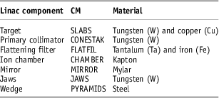
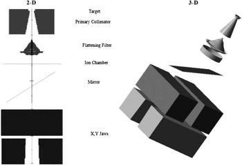
The EGSnrcReference Kawrakow 15 user code BEAMnrcReference Rogers, Walters and Kawrakow 16 was used to model an 18 MV beam from a Varian 2100C/D (Varian Medical Systems, Palo Alto, CA, USA). The Linac was modelled with different component modules (CM). Table 1 shows components and their materials. The schematic geometries of the CMs are shown in Figure 1. The 3D image was created by EGS_WINDOWS 17 program.
Used CMs for Linac modelling in BEAMnrc code

Abbreviation: CM, component modules.
Schematic geometry of modelled Linac. 2D view created by BEAMnrc and 3D view created by EGS_WINDOWS.

The incident electron beam was modelled by ISOURC = 19 module. This source is a circular beam with 2D Gaussian distribution of particles. ECUT and PCUT parameters which are used to define the global electron and photon cutoff energies were set to 0.7 and 0.01 MeV. Also, Electron Range Rejection with ESAVE value of 0.7 MeV in the target and ESAVE-GLOBAL =2 MeV and Directional Bremsstrahlung Splitting with NBRSL = 750 were used to minimise the simulation time.
Phase Space data were created for open 10 × 10 cm2 photon beam and the percentage depth dose (PDD) and profiles of measured and simulated data were used for verification of the beam energy and full width at half maximum (FWHM) of the incident electron beam in 30 × 30 × 30 cm3 water phantom using DOSXYZnrc code.Reference Ma, Reckwerdt, Holmes, Rogers and Geiser 18
The method introduced by Sheikh-Bagheri and RogersReference Sheikh-Bagheri and Rogers 19 were used to drive the best estimates for the energy and FWHM of the incident electron beam. For comparison between calculations and measurements, all curves were normalised to the center of each dose profile and for the PDD curve to the depth of maximum dose. This procedure is suggested by Pemler et al.Reference Pemler, Besserer, Schneider and Neuenschwander 20 for MC calculated dose distributions of single electron fields. Differences between the calculated and measured curves of dose profiles were explained in terms of dose difference and distance to agreement (DTA) in millimetre (mm) for the low and high dose gradient areas, respectively.
For both of the Rando phantom and patient studies, CT images were used by CTcreate program to make *.egsphant file to be irradiated by ISOURC = 8 in DOSXYZnrc code.
Radiation treatment planning
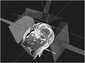
The study was done on the Rando phantom as well as on a real clinical case CT images. Initially, the dose distribution in pituitary gland was calculated by means of ETAR implemented in CorePLAN (Seoul C&J Inc., www.coreplan.com) treatment planning software for radiotherapy. The two other dose algorithms were CCC and MC programs (BEAMnrc and DOSXYZnrc codes). The dose calculation algorithms were compared for two lateral parallel opposed and one fronto-occipital 45° oblique 3 × 3 cm2 18 MV photon fields employing 30° physical wedges. Figure 2 shows the plan for Rando phantom and the patient.
The axonometric image of treatment plan for Rando phantom and patient shows three non-coplanar 18 MV photon beams (two lateral parallel opposed and one fronto-occipital fields).

Dose distributions
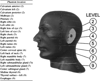
In MC simulation, dose distributions were calculated with DOSXYZnrc and the results of MC were cross validated by film dosimetry. For this purpose, both two radiographic (Kodak EDR2) and radiochromic (Gafchromic EBT2) films were used to obtain planar dose distributions in Rando phantom study. All films scanned with Microtek 9800XL scanner. Gafchromic EBT2 films scanned 24 hours after irradiation.Reference Cheung, Butson and Peter 21 As the pituitary gland is placed in third level of Rando phantom (see Figure 3),Reference Kihlen and Rudén 22 , Reference Ludlow and Ivanovic 23 dose distributions were obtained for the surfaces between levels 2 and 3, 3 and 4, and finally 4 and 5. All measurements were repeated three times.
Rando phantom. The pituitary gland is in third level.Reference Kihlen and Rudén 22 , Reference Ludlow and Ivanovic 23

RESULTS
Validation of MC
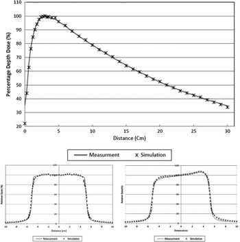
For validation of MC simulation, results were tuned against measurements in water phantom and finally, the incident electron beam with energy of 18.2 MeV and 1.5 mm FWHM showed the best match with measurements. Figure 4 shows PDD and dose profile for above mentioned energy and FWHM. For PDD curve; dose difference was below 1%, and for dose profile; dose difference and DTA were 0.97 ± 0.65% and 1.71 ± 1.08 mm for open field and 1.23 ± 1.09% and 1.79 ± 0.96 mm for wedged field, respectively.
Percentage depth dose (PDD) and dose profiles for open field and 30° wedged field for E = 18.2 MeV and full width at half maximum (FWHM) = 1.5 mm in water phantom.

Evaluation of MC simulation in Rando phantom
Simulated Linac head was evaluated against EBT2 and EDR2 film dosimetry in heterogeneous Rando phantom for the pituitary critical region where it is impossible to do in vivo dosimetry. This part of the study was performed to estimate the accuracy and compatibility of modelling within a heterogeneous mimicking human head phantom.
Overall, the mean difference between MC and film measurements were 4.93 ± 0.87% for all of the levels. The differences were 4.62 ± 1.37% for EBT2 and 5.03 ± 0.49% for EDR2 film dosimeters. Also, we found 1.2% difference between EBT2 and EDR2 results.
Results of the patient study
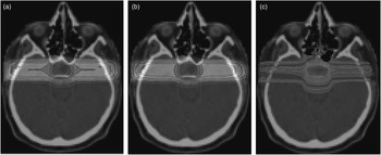
The final purpose of this study was evaluation of ETAR and CCC dose calculation algorithms in small size complex radiation wedged fields. The MC simulation was used as the gold standard to be compared with the various algorithms. Figure 5 shows the results of the radiation treatment planning using MC method, ETAR and CCC dose calculation algorithms.
The plans of three diffferent methods of dose calculation are compared: (a) collapsed cone convolution (CCC), (b) equivalent tissue-air ratio (ETAR) and (c) Monte Carlo simulation. The planning was performed on the patient CT data after voxelisation.

The average dose calculation differences between MC and proposed algorithms (CCC and ETAR) were 6.40 ± 3.44% (3.8–10.3%) and 10.36 ± 4.37% (5.5–13.9%), respectively.
DISCUSSION
Dose calculation algorithms in TPS have an important role in radiation treatment outcome. Any inaccuracy in predicting dose distribution in target volume can also change the patient quality of life. So, it is important to find the suitable algorithm for dose calculation customised to the departmental demands and irradiation fields. New algorithms which commonly used in TPSs, beside of their improvements in calculation, may have appreciable inaccuracies in some clinical situations such as small fields, electron disequilibrium and interfaces between different densities.Reference Vanderstraeten, Reynaert and Paelinck 7
In this study, the differences between CCC and ETAR algorithms were compared with MC simulation and experimental film dosimetry results. The study was done on the Rando phantom and a real clinically diagnosed case of pituitary adenoma.
Results of simulated head in water phantom showed that the best match between simulated results and measurement data will appear when The optimum energy and FWHM of incident electron beam is obtained to be 18.2 MeV and 1.5 mm, respectively, those are well in the range of other researchers in previous MC studies.Reference Sheikh-Bagheri and Rogers 19 , Reference Ding 24 – Reference Jaffray, Battista, Fenster and Munro 26
There are several recommendations for evaluate the accuracy of dose calculations in various areas with high or low dose gradients.Reference Venselaar, Welleweerd and Mijnheer 27 – Reference Mohan, Brewster and Barest 32 Our results for open and wedged fields in water phantom were in agreement with the values recommended by Venselaar et al.Reference Venselaar, Welleweerd and Mijnheer 27 for dose profiles. The differences between measured and simulated results were also <2% that is situated well within the recommended level in related studies for PDD curves.Reference Verhaegen and Seuntjens 12 , Reference Sheikh-Bagheri and Rogers 19 , Reference Venselaar, Welleweerd and Mijnheer 27 , Reference Andreo 33
Evaluations in Rando phantom study showed 4.93 ± 0.87% for all of the EBT2 and EDR2 levels in comparison to the MC simulation. This difference was <7% of the discrepancy reported by Brualla et al.Reference Brualla, Palanco-Zamora, Steuhl, Bornfeld and Sauerwein 34 and was more than Dobler et al.Reference Dobler, Walter and Knopf 35 results which reported 3% difference between MC and film dosimetry in heterogeneous medium. There would be two reasons for this difference; the first one can be the gaps between the different levels of Rando phantom which is created after placing the film between them. The gap is larger for EDR2 film because of its cover thickness. The gap affects the dose distribution on the film, while its effect is not existed during MC calculation.
The second reason may arise from the different electron densities on the various tissues interface region. In high density tissues such as bone the number of the secondary electrons increases per photon interaction that is well considered by MC simulation power. When a film is placed between the levels of the phantom, because the film density is equal to the soft tissue density, this may lead to a lower dose impartation to the cavity that finally causes disagreement between MC and film dosimetry.
Film dosimeters have uncertainties pertinent to several factors such as non-uniform thickness of the sensitive layer, temperature effects, scanner uncertainty and its warm up effect.Reference Todorovic, Fischer, Cremers, Thom and Schmidt 36 – Reference Devic, Seuntjens and Hegyi 38 There was 1.2% difference between EBT2 and EDR2 films. EDR2 films are light sensitive and are cut in a dark room and under safe light, however, it may be better to consider the effect of low level light. Also, EDR2 film processing may have remarkable effects on the readout while there is no need for processing with EBT2 films.
Present findings showed about 6.4% difference for CCC algorithm and about 10.3% for ETAR algorithm in comparison to MC simulation. Chow et al.Reference Chow, Jiang and Leung 39 evaluated the anisotropic analytical algorithm and CCC in heterogeneous phantom for tangential photon beam. They showed that the mean dose differences between MC and CCC was about 4.6% for 15 MV photon beam with 7 × 7 cm2 field size. Polednik et al.Reference Polednik, Abo Madyan and Schneider 40 in a comparison between pencil beam and collapsed cone algorithms in an anthropomorphic phantom, reported that there is about 6% difference between collapsed cone algorithm calculations and measurements. Our results are close to their findings and also Calvo et al.Reference Calvo, Gutiérrez, Stathakis, Esquivel and Papanikolaou 41 results which reported about 5.6% differences for CCC in comparison with MC.
Figure 5 shows that MC is superior concerning dose absorption within a non-homogenous volume, especially, in the bone–fat (brain tissue) interface. There is pronounced change in dose distribution after skull that can effectively modify the dose distribution in the pituitary adenoma and the difference is larger for ETAR method. In fact, the ETAR algorithm uses the ratio of two tissue-air ratio (TAR) for inhomogeneity correction and in definition, TAR is ratio of absorbed dose in a given depth in absorbent material to the same depth in a small air region in electron equilibrium situation. Therefore, this algorithm assumes that there is electron equilibrium in all points. So, in bone-air and/or tissue interfaces which there is an imbalance in electron equilibrium, ETAR will have fault in dose calculations.Reference Podgorsak 42 Also, ETAR only considers primary and scattered photons and does not consider the secondary electrons. Hence, it cannot evaluate the electron disequilibrium.Reference Verhaegen and Seuntjens 12 , Reference da Rosa, Cardoso, Campos, Alves, Batista and Facure 43 – Reference Ahmad, Hussain, Muhammad, Rizvi and Matiullah 47 While, CCC models electron transport and will predict the effects of electron disequilibrium in heterogeneous interfaces.Reference Ahnesjö 48 Our results showed that CCC algorithm as a model based dose calculation algorithm, have a better agreement with MC simulation in pituitary radiation treatment and the results of this study confirms the previous studies.Reference Wieslander and Knöös 49 – Reference Roberts 54
In conclusion, as differences between algorithms may have effects on quality of treatment, it is important to evaluate algorithms to choose the best one for use in clinical situations. MC method is a great evaluation tool for comparison of clinical dose calculation algorithms.
Acknowledgements
The data presented here are provided from the Kaveh Tanha's MSc thesis. The authors would like to thank Dr M. Sohrabpour from Sharif University of Technology for his support on this study and physics staff of radiotherapy department of Pars Hospital at Tehran, Iran for their assistance.
Financial Support
This work was financially supported by grant no. 90-04-30-15948 to S. R. Mahdavi from the deputy of research of Tehran University of Medical Sciences.
Conflicts of Interest
None.