Introduction
The genus Troglotrema Odhner, 1914 consists of three species: T. acutum (Leuckart, 1842), T. mustelae Wallace, 1932, found in the American mink Neovison vison (Schreber, 1777) in North America, and T. srebarni Genov, 1964, found in the muskrat Ondatra zibethicus (L., 1766) in Bulgaria (Skrjabin, Reference Skrjabin1949). Troglotrema acutum is widespread in Europe (Skrjabin, Reference Skrjabin1949; Yamaguti, Reference Yamaguti1971) and its main host is the western polecat Mustela putorius (L., 1758) (Koubeck et al., 2004). According to Koubek et al. (Reference Koubek, Baruŝ and Koubkovā2004), the Eurasian badger Meles meles L. 1758 is an accidental host. This hypothesis is supported by the fact that, despite its broad Palaearctic distribution (Revilla et al., Reference Revilla, Casanovas, Virgos, Palomo and Gisbert2002), there are few reports of T. acutum parasitizing this host. Hanák & Beneš (Reference Hanák and Beneš2001) did not detect this trematode in 16 badger skulls from the Czech Republic.
In the Iberian Peninsula T. acutum was recorded in the western polecat, from an unknown locality and with an undetermined prevalence (Torres et al., Reference Torres, Feliu, Miquel, Casanova, García-Perea and Gisbert1996), and was reported in an American mink from northern Spain (Torres et al., 2006). This trematode has also been reported in south-western France in European mink Mustela lutreola (L., 1761) (2.27%), western polecat (3.57%) and American mink (33.33%) (Torres et al., Reference Torres, Miquel, Fournier, Fournier-Chambrillon, Liberge, Fons and Feliu2008). It has not been detected in southern Europe in the Eurasian otter Lutra lutra (L., 1758) (Torres et al., Reference Torres, Feliu, Fernández-Morán, Ruíz-Olmo, Rosoux, Santos-Reis, Miquel and Fons2004), and studies of badger helminths have likewise not detected this species in the Iberian Peninsula (Torres et al., Reference Torres, Miquel and Motje2001; Rosalino et al., Reference Rosalino, Torres and Santos-Reis2006). Nevertheless, none of the above-mentioned studies included an examination of skulls, where the worm is found.
The life cycle of Troglotrema involves a prosobranch snail (genus Bythinella) as the first intermediate host and an amphibian as the second, which is where the metacercaria is found and eaten by the definitive host, that is, the carnivore in which the adult stage of Troglotrema develops in the nasal sinuses (Koubek et al., Reference Koubek, Baruŝ and Koubkovā2004). Once in the nasal sinuses, the destruction of bone tissue has been reported (Koubek et al., Reference Koubek, Baruŝ and Koubkovā2004). Studies of the damage to mammalian skulls caused by helminthiasis are rare. The nematode Crassicauda sp. (Crassicaudidae) had been shown to parasitize Odontocetes (Montes et al., Reference Montes, Chavera, Van Bresem, Perales, Falcón and Van Waerebeek2004), producing lesions, while in terrestrial mammals the nematode Skrjabingylus nasicola (Leuckart, 1842) (Metastrongylidae) causes visible damage to mustelid skulls, as reported by Prigioni & Boria (Reference Prigioni and Boria1995) in the weasel Mustela nivalis (L., 1766). The only study to date of damage by T. acutum was performed in the Czech Republic by Koubek et al. (Reference Koubek, Baruŝ and Koubkovā2004) and was based on the examination of skulls, but did not employ axial computed tomography (CT) or examine fresh heads as a means of isolating the adult stage of the trematode. Moreover, this latter study was mainly focused on lesions in the western polecat and provided no details of lesions in badgers. To our knowledge, the application of CT in cranial helminth detection in wild mammals has so far only been used to detect the nematode Crassicauda grampicola (Johnston & Mawson, 1941) in Risso's dolphin Grampus griseus (Cuvier, 1812) (Zucca et al., Reference Zucca, Di Guardo, Pozzi-Mucelli, Scaravelli and Francese2004).
The aim of this study was thus: (1) to try and detect for the first time the presence of T. acutum in badgers in Catalonia, Spain; (2) to identify the geographical distribution of this trematode in the badgers sampled in the study area; (3) to describe the damage caused in affected skulls; and (4) for first time, to study with CT the damage caused by a trematode in the skull of a mammal.
Materials and methods
Collection and examination of skulls
The analysed hosts (badger carcasses) consisted of badgers killed on roads that were collected by a number of different institutions and the authors and then kept frozen until dissection. Animals were collected between 1982 and 2010 and skulls were catalogued and stored in the collection of the Granollers Museum of Natural Sciences.
A total of 109 skulls of M. meles from Catalonia (north-eastern Iberian Peninsula) were studied. Of the studied skulls with presumptive lesions, six originated from the Montseny Natural Park (Barcelona province): two from El Brull (both 31TDG4528); three from Sant Celoni (31TDG6015, 31TDG6313 and 31TDG6413); and one from Tagamanent (31TDG4325). In addition, we studied individual skulls from L'Espunyola (31TCG9756) (Barcelona province) and from Sant Llorenç de la Muga (31TDG8285), Celrà (31TDG9152) and Olot (31TDG6070) (all Girona province).
Three methodologies were used in the study: (1) an examination for surface lesions; (2) CT and (3) fresh skull dissection.
Surface lesions
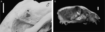
A total of 109 skulls were studied, including the above-mentioned dissected specimens. After manually removing most of the soft tissue, the skulls were left in the open air in a bucket of water until the inner soft parts had rotted away (a minimum of 3 months between early spring and autumn in a Mediterranean climate). The buckets were refilled regularly so that the skulls were always covered by water. Subsequently, the skulls were whitened with hydrogen peroxide and screened for surface lesions (see fig. 1).
(A) Detail of the orbital zone, showing the ‘net-shaped’ pattern, including two large holes where all bone has been lost (scale bar = 1 cm). (B) Composition of images from two photographs of the same skull, illuminated first overhead and then from behind, to improve transparency and the visualization of lesions (scale bar = 1 cm). Photo: Siqui Sanchez; photos combined with Photoshop.

Axial computed tomography (CT)
Nine skulls with surface lesions were analysed using CT. Five-millimetre-thick slices of the sagittal, dorsal and transverse planes were taken (Toshiba Aquilion TSX-101A: kVP, 120; mA, 150; ms, 500; mAs, 75; Tkh, 0.5 mm). To compare anatomic and imaging findings, a negative control was performed on one skull.
Fresh skull dissection
Forty-four complete heads from animals frozen immediately after death were examined for parasites. Skulls were cut open via the nasal and frontal bone with a radial saw (Dremel©). Once dissected, the nasal sinuses were extracted using a forceps and placed in a Petri dish until examination for helminths with a binocular stereo-microscope. Skulls were then repeatedly cleaned with a saline solution and their content was also examined under a binocular microscope. The only worms detected were trematodes and all were isolated and preserved in alcohol before being refixed in Bouin solution, stained in Semichon acetocarmine and then mounted in Canada balsam. Finally, worms were identified following Skrjabin (Reference Skrjabin1949) and Yamaguti (Reference Yamaguti1971).
Results
Surface lesions
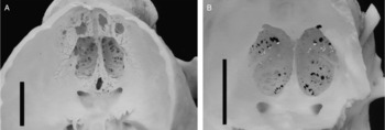
Nine skulls (8.25%) had surface damage, although the one skull in which a trematode was isolated in fact showed no surface bone lesions. Lesions were mainly characterized by mild-to-severe bilateral hyperostosis, along with osteolysis of the frontal bone and multiple small bone perforations measuring less than 1 mm deep (see fig. 1A). Two skulls also had single bigger holes with maximum widths of 3.09 and 3.45 mm. Osteolysis and defects in the bone tissue of the frontal bone led to the formation of a ‘net-shape’ structure. All the affected animals had lesions on the frontal bone, although three individuals also exhibited rarefaction hyperostosis of the palate, with perforations of the nasal sinus (fig. 1B) reaching maximum widths of between 3.55 and 13.18 mm. In four individuals the cribiform plate was completely destroyed (fig. 2) and, of these, in three there was serious damage and, in two, small lesions (fig. 3).
Internal view of cribiform plate (after removing parietal bones and partially the occipital bone): (A) parasitized, loss of bone is observed, including in the area surrounding the nasal sinus; (B) non-parasitized cribiform plate with natural symmetrical perforations and surrounding area of the nasal sinus (scale bar = 1 cm).

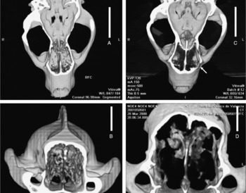
Tomography images of healthy (A, B) and parasitized (C, D) individuals. The arrow indicates the perforation of the frontal bone. In the parasitized animals, the black areas in the nasal sinus show the leakage of the bone tissue (osteolysis) (scale bar = 3 cm).

Externally, the rough bone surface showed evidence of hyperostosis of the maxillary and nasal bones in most damaged animals, indicating that bone apposition was concurrent with osteolysis. Maxillary and frontal surfaces were also enlarged and there was leakage in the surface structure (but not in the negative individual).
Fresh skull dissection
Of the fresh examined skulls (n = 44), one individual contained the digenean trematode T. acutum, (prevalence of 2.27% and intensity 8). All further analyses of surface lesions were negative for these skulls.
Axial computed tomography (CT)
All planes of the CT revealed internal lesions in the damaged skulls, specifically in the sinus and ethmoid regions (fig. 3). Osteolysis was revealed by the presence of black regions in the CT image (fig. 3). Bone rarefaction and the formation of cavitary tracts, along with the dissolution of the bone tissue causing leakage in the structure of the cribiform plate of the ethmoid, the ethmoid labyrinth and the nasal sinus lamella, were all characteristic lesions observed in the CT images.
In some parasitized individuals, osteolysis had caused perforations of the outer table of the frontal, nasal and maxillary bones. In one individual, there was also evidence of perforating osteolysis in the inner table of the maxillary bone. All the parasitized individuals also had hyperostosis, bone surface erosion and thickness irregularities in the affected bones (frontal, nasal and maxilla). As well, in some individuals the external surface of the frontal bone had increased in volume.
Discussion
Despite a considerable number of previous studies of badger helminths (Torres et al., Reference Torres, Miquel and Motje2001; Millán et al., Reference Millán, Sevilla, Gerrikagoitia, García-perez and Barral2004; Rosalino et al., Reference Rosalino, Torres and Santos-Reis2006), this is the first time that this trematode species has been reported in the badger in the Iberian Peninsula. The above-cited authors did not check skulls for helminths (the reason why this parasite was not detected) and so we believe that it is worth stressing the need to include carnivore skulls in future helminthological studies.
In Europe, Koubek et al. (Reference Koubek, Baruŝ and Koubkovā2004) analysed 5282 skulls belonging to 14 carnivore species from the Czech Republic and, of the 184 badger skulls analysed, two had lesions (1.1% prevalence). Fresh material consisted of 225 individuals of seven species, the commonest of which was the western polecat (7.1% prevalence); the badger was considered only as an accidental host. In the study by Torres et al. (Reference Torres, Feliu, Miquel, Casanova, García-Perea and Gisbert1996) of 99 western polecats in Spain, skull analysis was performed irregularly and no data are provided on how many skulls were analysed. These authors provide no details on prevalence or mean intensity and so, on the basis of their results, it is impossible to evaluate the importance of this trematode in this host. Thus, from a purely faunistic angle, the present study is useful as a means of rectifying the lack of previous information from the Iberian Peninsula.
Despite performing cranial dissections that would have revealed the presence of this parasite, Torres et al. (2006) did not detect cranial lesions in the 12 N. vison parasitized by T. acutum, probably because the existence of lesions is only related to an advanced stage of trematodiasis (as in our positive case from the fresh dissection methodology). In the study by Torres et al. (2006), 58 American mink from Catalonia (our study area) were shown to be negative for T. acutum; previously the presence of this trematode had only been confirmed in the Iberian Peninsula for Álava province (NW Spain).
The American mink could play a role in the maintenance of this parasite in the native mink species, since high prevalences (33.33%) have been detected in southern France (Torres et al., Reference Torres, Miquel, Fournier, Fournier-Chambrillon, Liberge, Fons and Feliu2008) in this invasive carnivore that has colonized many aquatic habitats (Bravo, Reference Bravo, Palomo and Gisbert2002). The western polecat has not been reported from the Montseny Natural Park (Palazón et al., Reference Palazón, Pérez, Batet, Arjona, Rafart, Malo and Ruiz-Olmo2010), where we found the majority of the parasitized badgers, and so we suspect that it may be the American mink (common in our study area) that is ensuring the maintenance of this parasite. Vogel & Voelker (1978) showed that at sea level Bythinella snails (first intermediate host) are only present in cold freshwater. Since the American mink occupies a wide variety of habitats, including areas where Bythinella snails are not found, the American mink will only help maintain T. acutum in environments where snails are present.
In previous work a focal distribution of this parasite was observed (Koubek et al., Reference Koubek, Baruŝ and Koubkovā2004; Torres et al., 2006, Reference Torres, Miquel, Fournier, Fournier-Chambrillon, Liberge, Fons and Feliu2008), which is confirmed by our results: six of the nine damaged skulls were from the same area, El Montseny Natural Park, with, moreover, two from the same locality. This could be explained by the fact that the Montseny Natural Park lies in an area of Eurosiberian climate, with an abundance of cold water, which is a good habitat for Bythinella spp. (Vilella-Tejedo, Reference Vilella-Tejedo2002). The diet of the badger is generalist in the Mediterranean area: they mainly feed on fruit, but also on a variety of other items such as invertebrates (insects, snails, earthworms, etc.) and occasionally rodents and amphibians (Virgós et al., Reference Virgós, Mangas, Blanco-Aguilar, Garrote, Almagro and Viso2004; Rosalino et al., Reference Rosalino, Loureiro, Macdonald and Santos-Reis2005), the latter being the second intermediate host of this parasite.
The use of CT in the present study is a novelty in mammalian studies and previously has only ever been used by Zucca et al. (Reference Zucca, Di Guardo, Pozzi-Mucelli, Scaravelli and Francese2004) in a study of a nematode in cetaceans. Thus, CT is used here for the first time to detect cranial trematodiasis in a terrestrial wild mammal.
Surface cranial lesions differed: in the western polecat marked perforations were found (between 1 and 13 mm in the study by Koubek et al., Reference Koubek, Baruŝ and Koubkovā2004) and some lesions were over 1 cm wide (measurements taken from photographic interpolation). In the badger, these perforations were smaller, but present in greater numbers (fig. 1A) in the thickest parts of the bone (upper part of nasal and frontal bones). In addition, large perforations were found in the thickest bones (lateral part of frontal, ethmoid and cribiform plate). Koubek et al. (Reference Koubek, Baruŝ and Koubkovā2004) noted that the Eurasian otter and badgers they examined had a ‘net-shaped deformation’ in their frontal and parietal bones, although they only examined two skulls with this type of pathology and no photos are provided. Here we provide photographic evidence of nine skulls with these ‘net-shaped’ lesions, thereby confirming a typical pattern caused by T. acutum in the badger. We believe that this type of pattern could derive from certain characteristics of the badger's skull, which is more robust than those of other mustelids (e.g. genus Mustela and Martes) and more difficult to perforate.
In the image of the nasal sinuses taken by CT, the dark colour corresponds to the area of bone tissue destruction, where asymmetrical and irregular damage is obvious. In the front of the skull, large perforations are visible and are similar to the external lesions previously described in the western polecat by Koubek et al. (Reference Koubek, Baruŝ and Koubkovā2004); however, since badgers' skulls are thicker, the externalization of lesions did not occur. Trematode prevalence could be underestimated during the first stages of helminthiasis if studies are limited to external lesions. CT techniques, however, enabled us to detect infection even in its first stages.
Transverse sections of the skulls allow us to build up a complementary vision of lesions, and bone tissue destruction in the inner structure of the sinuses is shown by a darker colour (fig. 3). CT provides a more accurate picture of the extent of lesions than conventional external inspections, especially when lesions affect the inner parts of the skull, such as the ethmoid.
Since most lesions are located in the sinuses, nasal endoscopy could facilitate the early detection of lesions caused by Troglotrema in living animals. This diagnostic tool could establish parasitization prevalences and permit the study of host reactions to parasites in free-living or captive animals, or in animals brought to recovery centres. Even so, to strengthen the diagnosis of parasitization by T. acutum, we still recommend the examination of faeces, a method that Torres et al. (2006) have shown to be effective.
Lesions observed in parasitized animals seem to occur partly as a reaction in the host to the damage caused by the parasite or to a secondary bone infection (osteomyelitis). Further research regarding host histological and physiological responses to T. acutum is still needed in order to evaluate the effects of this parasite on the nervous system and on behaviour in badgers.
Acknowledgements
The collection, dissection and cleaning of skulls was partially supported by a grant from project 2006 ACOM 00057. A.R. was supported by grant 2008BP A 00045 (Generalitat de Catalunya). We are grateful to the network of natural parks of the Barcelona Provincial Council (Diputació de Barcelona) and of the Catalan government (Generalitat de Catalunya) for providing badger carcasses. We are also extremely grateful to the rangers of El Montseny, Montnegre, La Garrotxa Volcanic Zone and Collserola Natural Parks, and the Wild Fauna Centres of the Generalitat de Catalunya, the police of La Vall del Tenes and Galanthus Associació. Many thanks also to the following people who provided us with material: Misael Arrizabalaga, Marc Bosch, Jordi Brau, Pere Casals, Jaume Casas, Enric Fàbregas, Belen Fernández-Villacañas, Lídia Freixas, Marta Pladevall, Xavier Junqueras, José Martí, Marta Miralles, Èlia Montagud, Francesc Pou, Xevi Puig, Joan Manuel Riera, Jaume Rodri, Octavi Serra and Elies Valls.