Introduction
Cardiovascular diseases, including severe cardiac valve disorders, are the leading global cause of disability and death, causing more than 17.5 million deaths a year (Ref. 1); the prevalence of valvular heart disease in the USA is estimated at more than 5 million people. Moreover, aortic valve disease, which affects 2.5% of the USA adult population to some extent, is responsible for more than 25 000 deaths each year (Ref. Reference Go2). Also, 3% of sudden deaths in the European Union (EU) are caused by cardiac valve defects. Over the preceding decades valvular heart diseases were not considered a major public health problem. However, Nkomo et al. (Ref. Reference Nkomo3) demonstrated in 2006 that the high prevalence of these disorders, together with their boost with ageing, indicated the burden of these conditions would only likely increase in the future.
The advent of organ transplantation has brought about the greatest progress in cardiac valve disease treatment so far however, the number of patients who need heart valve (HV) replacements is rising by an average of 5% per year (Ref. Reference Hoerstrup4). This number is likely to triple over the next five decades due to the continuously increasing aging of the population in developed countries (Ref. Reference Sewell-Loftin5). According to the Global Alliance in Transplantation, the USA is currently the world's leader in HV replacements, reaching 95 000 cases per year (Ref. Reference Groth and Chapman6) but this is not an exception. By the year 2020, 20.2% of EU population will be older than 65. The growing pool of older people living in the area with age related HV disorders will continue to boost demand for HV repair and replacement procedures. These medical procedures, only in EU countries, generated $827.5 million expenses in 2010, and the estimate for 2015 is to reach $1375.3 million (Ref. Reference Julie and Greenfield7).
Even more dramatic is the case of children and young adults where cardiac valve defects represent approximately 30% of all congenital cardiovascular malformations. Only in the USA, the incidence is estimated at 1–2/1000 births, and globally is around 8/1000 births (Refs Reference Loffredo8, Reference Kalter and Kalter9). Congenital heart defects are relatively common and serious malformations have significant impact on morbidity, mortality and healthcare costs. Homografts are used for their long durability in these patients but they show a higher immunological response compared with adults (Ref. Reference Rajani, Mee and Ratliff10). Moreover post-surgical calcification and the ultimate destruction of the valve replacement always occur more frequently in children and young adults than in old patients. In addition, the durability of the homografts in this case is also limited because of their inability to grow with concomitant growth of child organism. Consequently, the need for reoperation is higher in younger patients (Refs Reference Rajani, Mee and Ratliff10, Reference O'Brien11). Further efforts focused on paediatric recipients of cardiac valves are required.
Although organ replacement saves many lives the need for more donors, together with the collateral problems caused after transplantation, encourage the search for new alternatives (Refs Reference Vahanian and Alfieri12, Reference Hasan13). Cardiac valve replacement is not an exception to the rule and several problems have to be solved: the administration of lifelong immunosuppressive drugs, poor durability and resulting short lifespan of the mechanical prostheses, structural degradation of the biological valve replacement, risk of rejection and the risk of zoonosis from xenotransplants. Even with the significant innovations currently reached, a perfect valve substitute has still not yet been developed (Ref. Reference Vahanian and Alfieri12), and approaches based on tissue engineering (TE) are now being considered for developing artificial HVs (Refs Reference Murphy and Atala14, Reference Mayer15).
Tissue-engineered heart valves (TEHVs) have the potential to become the best solution for treating valvular heart diseases because of their non-thrombogenicity, decreased risk of infections and low structural deterioration over time (Ref. Reference Murphy and Atala14). There are two crucial challenges derived from three major components (cells, scaffolds and signalling molecules) in TE field; the former is the selection of potential candidate cell types and their related issues concerning ethics, safety and effectiveness. The latter is the design of appropriate scaffolds that let for cell growth and differentiation as well as host tissue integration.
In this review we evaluate the current treatment strategies for valvular heart disease and their limitations with a view to provide valuable information in selecting the best approach for heart valve tissue engineering (HVTE), the challenges that HVTE faces today and the current status of the research in the field. This review focuses on the aortic valve because of the fact that aortic diseases have the poorest prognosis, even the higher incidence of mitral valve diseases in the population (Ref. Reference Bonow16). However, research on pulmonary valve is also taken into account since pulmonary autograft represents a proof of principle for clinical effectiveness of TEHVs (Refs Reference Ross17, Reference El-Hamamsy18).
Understanding of HV biology
Substructural elements
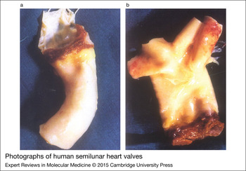
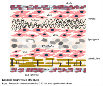
Mammalian hearts comprise four HVs: pulmonary, aortic, mitral and tricuspid, whose primary function is to maintain a unidirectional blood flow during the cardiac cycle. We focus on the first two types, which are shown in Figure 1a and b, but especially our main interest is the aortic valve. Pulmonary and aortic valves, also called semilunar valves, are commonly affected by pathological processes, but with distinct mechanisms of failure (Ref. Reference Jana19) that we will treat later. Both share similar structure containing three semicircular leaflets, or cusps, joined to a fibrous annulus called the root. According to Stradins et al. (Ref. Reference Stradins20), aortic and pulmonary valve tissues have similar mechanical characteristics and composition, but exhibit slight differences in their layer density and thickness. The cross-sectional structure of the leaflets is divided into three layers, namely the ventricularis, spongiosa and fibrosa (Ref. Reference Hasan13) whose structure and composition are detailed in the cross-sectional diagram in Figure 2. These layers are dynamic tissues with complex structure mainly composed of valvular- interstitial cells (VICs) and-endothelial cells (VECs) combined with extracellular matrix (ECM). This makes them capable of remodelling themselves in response to the changes generated by local forces (Ref. Reference Rabkin-Aikawa22). The semilunar valves also possess blood vessels. The majority of them are present in the proximal portion near the myocardium, with few found in the cusps, which are nourished by diffusion from the blood in the heart because of their thinner structure.
Photographs of human semilunar heart valves: aortic (a) and pulmonary (b).

Detailed heart valve structure. It is composed of three inner layers (ventricularis, spongiosa and fibrosa) and an outer layer formed by valvular endothelial cells (VECs). The three inner layers contain mainly proteoglycans (PGs), glycosaminoglycans (GAGs), collagen type I and type III, elastin and valvular interstitial cells (VICs). Adapted from (Ref. Reference Rajamannan21).

The creation of a functional TEHV requires profound knowledge of the healthy valve structure, the biological processes associated with their development and also their functional features and requirements for their formation. The main components that comprised the aortic valve are described in the following subsections.
VICs
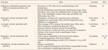
VICs, the most abundant cell type in HVs, are a heterogeneous population embedded in their own ECM. Liu et al. (Ref. Reference Liu, Joag and Gotlieb23) classified these cells in five different types: embryonic endothelial progenitor cells (EPCs) (Ref. Reference Markwald, Fitzharris and Manasek24), quiescent (Ref. Reference Yoshida25), activated (Refs Reference Desmouliere and Gabbiani26, Reference Tamura27), progenitor (Refs Reference Paruchuri28, Reference Sata29, Reference Visconti30) and osteoblastic VICs (Ref. Reference Rajamannan31). They have particular characteristics and functions regulated by environmental conditions that are summarised in Table 1. VICs properties are also different in health and disease. For example VICs from aortic and mitral valve are stiffer than those from pulmonary and tricuspid, suggesting that VICs respond to local tissue stress by altering their stiffness (Ref. Reference Sacks and Yoganathan33).
The different phenotypes and roles of the valvular interstitial cells (VICs)

From the morphological point of view VICs have a fusiform and fibroblastic shape with prominent cytoplasmic extensions and an incomplete basal lamina (Refs Reference Johnson, Hanson and Helgeson34, Reference Filip, Radu and Simionescu35). They are mainly characterised by their cytoplasm, which is rich in mitochondria, rough endoplasmic reticulum and frequently exocytic vesicles (Ref. Reference Hinton36). VICs mediate matrix remodelling and also maintain valve homeostasis. They are crucial for supporting normal valve physiology, playing an important role in some pathological valve processes (Ref. Reference Taylor37).
VECs
VECs are in charge of maintaining a non-thrombogenic surface, nutrient transport, and transduction of biochemical and mechanical signals in the HV. They are located in parallel to the ECM collagen fibres of valves and perpendicular to the blood flow direction (Ref. Reference Butcher38). VECs release factors that act on the underlying VICs, inhibiting their proliferation and increasing ECM synthesis (Ref. Reference Butcher and Nerem39). They share some similarities with vascular endothelial cells (ECs). They present cobblestone-like morphology when cultured in vitro and also exhibit contact inhibition. Preliminary studies indicated that subpopulations of these cells were also able to transdifferentiate into smooth muscle–like phenotype (Ref. Reference Butcher38). Moreover, VECs dysfunction seems to be involved in certain cardiovascular diseases (Ref. Reference Poggianti40).
ECM
The ECM is considered to be the main responsible for the durability of HVs. As shown in Figure 2, its principal components are collagens type I and type III, elastin, glycosaminoglycans (GAGs), and proteoglycans (PGs). Collagens are fundamental for valve's biomechanical functions, providing stiffness and elasticity; moreover, they have a strong influence on cell adhesion and migration (Ref. Reference Baaijens, Bouten and Driessen41). Microscopically, the three layers that make up the valve have different ECM composition. For instance the thinnest layer, the ventricularis, is mainly composed of elastin which provides elasticity (Ref. Reference Vesely and Noseworthy42), whereas the thickest layer, the fibrosa that is in charge of providing mechanical strength and load resistance to the valve, mainly contains collagen I and III (3:1) and 2% of collagen type IV (Refs Reference Hinton36, Reference Schoen43).
The spongiosa layer, responsible for the compressible stiffness required for proper HV function, contains mainly GAGs and PGs. GAGs are largely responsible for the huge water-absorbing capacity of this layer thanks to the high concentration of negative charges that they carry. Furthermore, GAGs are also required for hydration of the spongiosa layer and absorb compressive forces, reducing flexural deformation (Ref. Reference Lovekamp44). Given their nature, GAGs can also reduce the calcification process by chelating calcium ions. All of these features are important factors which go towards explaining the mechanical behaviour of HV tissues. In particular GAGs are crucial components that must be considered when creating TEHVs. For instance, after the process of decellularisation with detergents, GAG loss was observed in valve tissues, compromising their structure and functions (Ref. Reference Cigliano45).
HV ECM is subjected to dynamic remodelling, which is induced by VICs. The quantity and quality of ECM essentially depends on the nature of the VICs (Refs Reference Hinton36, Reference Schoen43). The relationship between ECM organisation and VIC distribution is also crucial during development and in pathological states (Ref. Reference Hinton36). Walker et al. (Ref. Reference Walker46) observed that various genes associated with ECM are mutated in congenital valve diseases, highlighting its importance in the morphology and functionality of HVs.
HV biomechanics and related illnesses
Mitral and aortic valves are in charge of pumping the blood, experiencing intense pressure and supporting higher biomechanical stress than pulmonary and tricuspid. Therefore, they are more affected by pathologies and degenerative processes while alterations in pulmonary or tricuspid valves are less common and mainly related with genetic or foetal defects (Ref. Reference Hasan13). Valve pathologies are characterised within two functional groups: stenosis and regurgitation. Haemodinamically, the pulmonary valve flow profile is similar to that of the aortic, but the velocity magnitude is lower (Ref. Reference Sacks and Yoganathan33). The slightly difference in the biomechanical and ultrastructural properties aforementioned in the subsection ‘Substructural elements’ strongly constraints the type of disease developed. Pathologies of the aortic valve can be triggered by inflammation or by degenerative valve disease, caused by aging coupled with rheumatic and infective endocarditis (Ref. Reference Yacoub47). On the contrary, congenital stenosis is the most common pulmonary valve pathology, mainly caused by a defect during foetal development; this alteration is normally solved by surgery during infancy. Finally, mitral regurgitation, which can be caused by rheumatic fever, congenital abnormalities, ischemic heart disease or cardiomyopathies, is the most common functional abnormality of this valve.
Cardiac valve problems and clinical strategies for their treatment: the importance of valve tissue banks
As we mentioned above, different illnesses can compromise and damage the delicate structure of HVs, triggering their alteration and causing the three main valve problems: regurgitation, stenosis and calcification. In the past the inflammation caused by rheumatic heart disease and endocarditis induced by bacterial infections were the root causes of valve alterations. Although these problems remain a major health roadblock in developing countries (Ref. Reference Nkomo3), valve dysfunctions in the industrialised world are almost always caused by degenerative diseases, mostly as a result of aging and congenital defects (Ref. Reference Vahanian and Alfieri12). Several drugs can be used as short-term therapies to improve the health and condition of patients with severe valvular pathologies, however in most cases the best option for correcting the problem is to proceed with valve repair surgery and/or replacement. The choice of surgical procedure is adapted to the team's experience, the presence of an aneurysm, the characteristics of the leaflets, and also to the patient's condition and life expectancy (Ref. Reference Vahanian and Alfieri12).
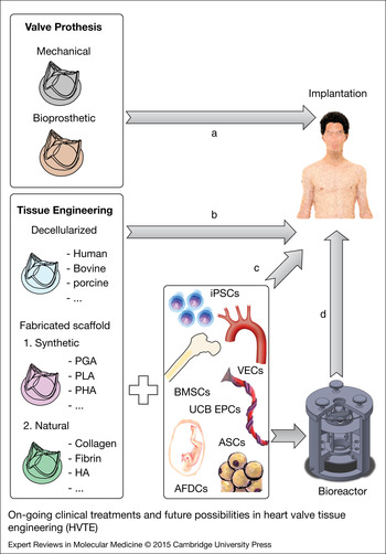
Current alternatives for HV replacements are mechanical and biological (xenogeneic and allogeneic) implants (strategy A in Fig. 3).
On-going clinical treatments and future possibilities in heart valve tissue engineering (HVTE). The methods to create autologous constructs are a potential alternative to overcome the main drawbacks of traditional bioprosthetic and mechanical valve prostheses (Strategy A). The creation of tissue-engineered heart valves starts from the decellularised or synthetic scaffold, which represents the basic platform where cells proliferate. This can be directly implanted without (Strategy B) or with reseeding cells (Strategies C and D). Different cell types could be candidates for this purpose: vascular endothelial cells (VECs), bone marrow mesenchymal stem cells (BMSCs), induced pluripotent stem cells (iPSCs), chorionic villi-derived mesenchymal progenitor cells, umbilical cord blood (UCB) derived endothelial progenitor cells (EPCs), and adipose-derived stem cells (ASCs), among others.

Mechanical implants
Since 1960, when Harken et al. (Ref. Reference Harken48) and Starr and Edwards (Ref. Reference Starr and Edwards49) reported the first successful surgical implantations of a mechanical HV, their use has widespread and they are now the option selected in more than 50% of replacement surgeries. The main advantage is their high mechanical strength; but although mechanical implants are very resistant and work satisfactorily for years, they carry with them the risk of thrombosis generated by high shear stress, blood alterations or non-physiological flows (Ref. Reference Mol50). Consequently, they generally imply the use of anticoagulant medication for the rest of the patient's life (Ref. Reference Hammermeister51).
Bioprosthetic valves
The bioprosthetic valves can be obtained from animal (xenografts) or human origin (autografts and allografts/homografts). Compared with their mechanical counterparts, bioprostheses are affected by structural dysfunction due to tissue degeneration, so they need to be replaced within 10–15 years. For this reason current clinical guidelines recommend the use of bioprosthetic replacements in patients over 65 years (Refs Reference Bonow16, Reference Mol50).
Xenografts have usually bovine or porcine origin. Once extracted, they are fixed with glutaraldehyde (GA) to prevent rejection (by masking immunogenic epitopes) and zoonosis (Ref. Reference Klepetko52). Animal valves do not induce clot formation but they entail a risk of xenoinfection and degrade faster (Refs Reference Sacks and Schoen53, Reference Jamieson54). Their dysfunction is motivated due to tissue damage, mainly by calcification and collagen degradation.
Those detrimental processes seem to be triggered by the use of GA during fixation. Fixation with GA preserves the integrity of membrane phospholipids, which remain in the valve after the treatment. These lipids react with the surrounding extracellular calcium, creating aggregates and promoting valve calcification. Because calcification evolves faster in young patients, xenogeneic replacements are mainly prescribed in patients with high death risk or advanced age (Ref. Reference Jamieson54). Moreover, GA degrades animal's VICs and VECs of valves, affecting also to the collagen matrix and destroying the functional structure. Once implanted, the receptor's cells are not able to restore the functionality, and they start to secrete altered ECM proteins profile, increasing the damage (Ref. Reference Sacks and Schoen53). Several alternatives to solve this problem have been tried however, without great success (Refs Reference van den Heever55, Reference Mathapati56).
Given their better hemodynamic profile, human allografts, or autografts in the case of Ross procedure, are currently the optimal solution, but because of their importance and peculiarities, they deserve a detailed study (Ref. Reference Dohmen57).
Human homografts and the importance of HV tissue banking
Since the last century, thousands of HV homografts (especially aortic and pulmonary) have been used in reconstructive cardiac surgery and tissue replacement (Refs Reference Ross17, Reference Yacoub47, Reference Barratt-Boyes58, Reference O'Brien11). One of the main differences between cardiac valve homografts and other tissues for transplantation (e.g. bone) is that the valve conduits need to perform at its best immediately after surgery. Additionally, the clinical outcome of a homograft can be conditioned by the physiologic process of inflammation, the biomechanical stress, the immune response and other molecular events.
Once the tissue has been transplanted, the analysis of factors which could play a significant role in its deterioration also remains as controversial issue. Cell viability is one of the most controversial topics at this point. Several authors have reported that maintaining the viability of cells residing within the graft have beneficial effects (Ref. Reference O'Brien59); but donor cell viability is drastically few months after transplantation. As Hilbert and Koolbergen documented, instead of the donor cells, the valvular surface is covered by a fibrous sheath (Refs Reference Hilbert60, Reference Koolbergen61).
It is known that homograft immunogenicity is directly related to cell viability (Ref. Reference Hawkins62). Cryopreserved homografts have been shown to elicit a donor-specific immune response at humoral and cellular levels (Refs Reference Hogan63, Reference den Hamer64). The necessity to address or not cross-match for blood group is a clear example of contradictory opinions on this issue (Refs Reference Christenson65, Reference Jashari66, Reference Shaddy and Hawkins67). In any case, since endothelium is especially pointed for valve immunogenicity (Ref. Reference Johnson68), a strategy based on the elimination of this layer during tissue processing could be used to reduce this risk. Nevertheless, although host cells can colonise the matrix, endothelium reconstitution seems to be difficult (Refs Reference Hawkins62, Reference Koolbergen69) and the loss of this layer may lead to detrimental effects on the ECM (Ref. Reference Koolbergen61). Additionally, to what extent the immunological response affects homograft durability is still unclear (Refs Reference Johnson68, Reference Mitchell, Jonas and Schoen70, Reference Bechtel71, Reference Troost72).
Other factors noted above can lead to apoptosis of donor cells in the transplanted homograft: prolonged ischemia, addition and removal of cryoprotectant solution, cooling and warming rates, ischemia-reperfusion injury (Refs Reference Hilbert60, Reference Lu73). Homograft degeneration probably reflects the cumulative damage of several factors.
The use of proper manufacturing and storage practices to preserve the homografts until the moment of their transplant is also a key issue in the success of the implant. For this reason, cell and tissue banks and transplant organisations are the key guardians for preserving safety. The critical issues that may affect the safety and efficacy of the homografts are:
-
• Disease transmission: Rigorous donor screening is actually the best available method to improve allograft safety (Refs Reference Jashari74, Reference Herberts75).
-
• Tissue assessment: Quality codes are assigned to homografts, depending on the presence of calcification, atheromas or fenestrations a morphological status is determined (Refs Reference Jashari74, Reference van Wijk, van den Bogaerdt and Bokhorst76). These classifications are crucial during the decision-making process for tissue allocation.
-
• Contamination: Tissue microbial contamination may arise from several sources such as the operation team (Ref. Reference Segur77), the own donor, especially if death is caused by trauma or the heart has been recovered late after death. In order to prevent contamination, the control of processing area is also crucial. The homograft extraction needs to be performed in an aseptic clean room with grade B background and under sterile conditions (air quality class A) (Ref. Reference Caselli-Fernández and Terkola78). If the replacement needs to be frozen, storage in liquid nitrogen needs to be strictly controlled. Liquid nitrogen is not sterile and microorganisms survive at this ultra-low temperature (Ref. Reference Mirabet79). A liquid environment acts as an efficient vehicle for microbial diffusion. For this reason, the bag containing the tissue is doubly wrapped using another bag (so called double-bagging), and sealed. It is common practice in cardiac valve banks, to ensure the immersion of tissues in an antibiotic cocktail immediately following collection. The length and temperature of incubation, as well as the concentration and type of antibiotics used, vary among banks (Ref. Reference Heng80).
-
• Cell and tissue damage: HV processing is not harmless and some important factors must be controlled to enhance cell viability (Ref. Reference Lu73). Those factors are ischemia (Refs Reference Fischlein81, Reference Gall82), freezing and thawing conditions (Ref. Reference Mirabet83), or disinfection and storage of the samples before their use.
Native HVs are dynamic structures that are in continuous remodelling to adapt the hemodynamic necessities. As we already mentioned, interstitial cell viability may enable continuous remodelling (Refs Reference Johnson68, Reference Messier84) and fibroblastic matrix cells are immunologically well-tolerated. Koolbergen et al. found fibroblastic donor cells in vessel walls several years post-transplantation (Ref. Reference Koolbergen69).
None of the current replacements is able to restore completely the capacities and functions of native valves, so in the end, after several years cardiac function may be compromised. As we already mentioned, this is especially crucial for children and young adults suffering cardiac valve problems. In those cases, as well as in particularly active older people, the most successful valve replacement approach is the Ross procedure (Ref. Reference Ross17). This procedure is a specialised valve surgery where the diseased aortic valve is replaced by patient's own pulmonary valve, and then this is substituted with a cadaveric pulmonary homograft. Ross procedure offers several advantages over replacements with biological or mechanical prosthesis.
First, in the case of paediatric patients, the pulmonary autograft can continue growing in the aortic position according with the recipient with higher longevity than xenografts. Anticoagulation treatment, which is required for mechanical replacements, is not necessary after Ross procedure. This is especially beneficial for women who want to have children because anticoagulation drugs are contraindicated during pregnancy. However, especially in the case of children, this replacement procedure may fail. After the relocation in aortic position, pulmonary valve composition can be altered due to the higher biomechanical demand that aortic valve supports. Additionally, the pulmonary autograft transplanted during Ross procedure, needs to be replaced at some point (Refs Reference Shaddy and Hawkins67, Reference Clarke and Bishop85). Despite the fact that human homografts seem to be the most physiologically suitable substitutes (Ref. Reference Vahanian and Alfieri12), the number of cadaveric donor valves cannot meet current demands. For this reason finding a suitable TE solution is especially challenging in paediatric or young patients.
TEHVs
HVTE represent a promising strategy to create prostheses with similar functional and biomechanical properties to native HVs (Ref. Reference Harken86). They should be able to withstand the repetitive changes in shape, dimensions and stress generated by the opening and closing cycles they are subjected to during cardiac beating (Ref. Reference Chang87). Similarly, the TEHVs should solve some of the common limitations that still remain in commercially available prostheses and have a direct impact on the clinical outcome (Ref. Reference Sutherland88): thrombogenicity, calcification and high immunogenicity, among others. Poor biomechanical properties or inadequate ECM composition of TEHVs could induce stenosis, valve regurgitation, or low transvalvular pressure gradient of the incumbent valve.
Regarding paediatric patients, their implanted TEHVs should be capable of growing and remodelling according to the stage of patient's life, responding to biological signals and initiating self-repair operations after small damaging events in the same way that native valves respond. Eventually, the TEHVs should be able to integrate themselves into the patient's tissue. Taken together, these requirements should be accomplished while integrating all individual components of the TEHV into a synergistic design.
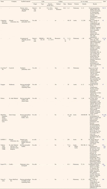
The fabrication of functional, viable and human implantable prostheses depends on three substantial components widely known as TE triad: cells, scaffolds and signalling molecules (Ref. Reference Deme89). The following sections focus on two of these building blocks and the decisions affecting them during the creation of TEHVs: the selection of the cell type and the fabrication of the scaffold. Clinical on-going solutions and future possibilities based on TE are summarised in Figure 3 (options A, B and C). Additionally, the progresses reached in TE through in vitro studies, preclinical studies and clinical trials are summarised in Tables 2–4, respectively.
Summary of reported or currently conducting clinical trials using tissue-engineered heart valves (TEHVs)

LV, left ventricle; PBMSCs, peripheral blood-derived mesenchymal stem cells; PET, polyethylene terephthalate.
All the trials followed a tissue engineering (TE) strategy of decellularisation.
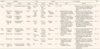
Summary of representative in vitro studies in different heart valve tissue engineering (HVTE) approaches

BMSC, bone marrow mesenchymal stem cells; EC, endothelial cells; ECM, extracellular matrix; FB, fibroblasts; HA, hyaluronic acid; HUVEC, human umbilical vein endothelial cell; Me-HA/Me-Gel methacrylated hyaluronic acid (Me-HA) and methacrylated gelatin (Me-Gel); MF, myofibroblasts; NR, non-reported; PGA, polyglycolic acid; PHA, polyhydroxyalkanoate; P4HB, poly-4-hydroxybutyrate; PLA, polylactic acid; P(L,DL)LA, Poly(L-lactide-co-D,L-lactide); PU, polyurethane; SMC, smooth muscle cells; UCC, umbilical cord blood-derived progenitor cells; VEC, valvular endothelial cells; VIC, valvular interstitial cells.
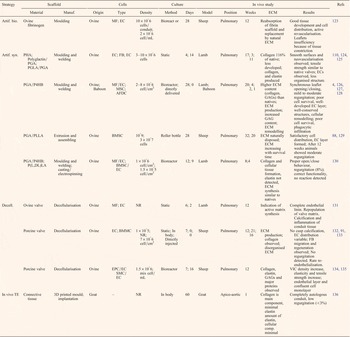
Summary of pre-clinical investigations with different heart valve tissue engineering (HVTE) strategies

AFDC, amniotic fluid-derived cells; BMSC, bone marrow mesenchymal stem cells; EC, endothelial cells; ECM, extracellular matrix; EPC, endothelial progenitor cells; FB, fibroblasts; GAG, glycosaminoglycans; HA, Hyaluronic acid; HUVEC, human umbilical vein endothelial cell; Me-HA/Me-Gel methacrylated hyaluronic acid (Me-HA) and methacrylated gelatin (Me-Gel); MF, myofibroblasts; MSC, mesenchymal stem cell; NR, non-reported PGA, polyglycolic acid; PHA, polyhydroxyalkanoate; P4HB, poly-4-hydroxybutyrate; PLA, polylactic acid; P(L,DL)LA, Poly(L-lactide-co-D,L-lactide); PU, polyurethane; SMC, smooth muscle cells; UCC, umbilical cord blood-derived progenitor cells; VIC, valvular interstitial cells.
Challenge 1: selecting biomaterials for scaffolds
The success of the TEHV strongly depends on the biocompatible materials selected to construct the scaffold and the resulting physicochemical properties (Ref. Reference Khademhosseini137). The scaffold is the three-dimensional (3D) structure that allows cells to adhere, generate ECM, grow, migrate and also differentiate; crucial processes that guarantee the full integration of the implant into the heart.
The scaffolds of TEHVs face important challenges in terms of integrity, mechanical strength, calcification resistance and thrombosis risk; but even more important, they must be prepared to deal with the cardiac environment conditions immediately upon implantation (Ref. Reference Driessen138). As shown in Figure 3, there are mainly two types of scaffold for HVTE: acellular native valves and artificially engineered scaffolds; in case of the latter, its constituents need to be carefully considered, since the aim is to mimic the native valve anatomy and functionality.
Finally, the materials selected must be suitable for different manufacturing processes and reasonably accessible, enabling mass production that caters to the important demand in a shorter time than the currently available (Ref. Reference Legg, Mathews and Pelzer139).
Decellularised cardiac valve scaffolds from allogeneic/xenogeneic sources
Traditionally, decellularised HVs and pericardium tissues from sheep, cows and pigs have been selected as scaffolds because of the shortage of human valve allografts (Ref. Reference Cebotari120). The decellularisation methods use detergents, chelating agents, proteolytic enzymes, or combinations thereof (Ref. Reference Gilbert, Sellaro and Badylak140). Other alternatives applied include the use of electrical pulses, hydrostatic pressure, or freeze-thaw cycles (Ref. Reference Jana19).
The main advantages of decellularisation are the retention of the original structure as well as many ECM proteins, preservation of the mechanical properties and a relatively low cost. However, there are serious limitations such as the possibility of disease transmission, or excessively high density and stiffness. Moreover, the porosity is especially low on valves decellularised and treated with cross-linking agents. A failure in the decellularisation process has detrimental consequences for patients as shown in the SynerGRaft trial (Ref. Reference Simon141). However, the necessity of re-seeding or not a valve after decellularisation is still the subject of an intense debate. Some authors argue that in vitro recellularisation is not a realistic strategy that can be translated into routine clinical practices because of its high financial and time costs (Ref. Reference Emmert126). Indeed, some studies demonstrate that the seeded cells do not survive when the valve is introduced into the patient and that the host's cells may be the origin of the observed repopulation (Ref. Reference Cebotari120). A summary of reported clinical experiences is given in Table 2. It should be noted that this strategy, which is labelled as B in Figure 3, is the one that has gone further of all TEHV approaches concerning clinical trials (Refs Reference Cheung103, Reference Sadowski104, Reference Bobylev106).
Important differences in cell growth when compared with human-source matrices were demonstrated after seeding human cells over them (Ref. Reference Iop142). The use of multilayer vascular endothelial growth factor (VEGF)/heparin or chitosan/heparin films to cover the decellularised valve has demonstrated to increase in vitro adhesion and proliferation of EPCs. This has resulted in an improved decellularised valve haemocompatibility and a reduced risk of thrombosis (Refs Reference Ye143, Reference Ye144).
Although it has been proved that host cells can recolonise the scaffold, a study comparing native HVs with decellularised and recolonised ones, concluded that the cell types present in the manipulated structures are different from the native ones. This may interfere in the normal function and/or stability of the valve. Additionally, the limited re-endothelisation that can be achieved in decellularised matrices can compromise their integration and long-term viability. In vivo recellularisation studies have stated that pre-treatment with host cells can improve scaffold's biocompatibility reducing calcinosis (Ref. Reference Francoise145). Therefore, a short-term incubation of the scaffold with cells and/or unprocessed cell preparations from the patient seems to help in both the recellularisation and cell survival outcomes (Refs Reference Emmert126, Reference Weber, Zeisberger and Hoerstrup146). Another alternative is to enhance autologous recellularisation after implantation coating the acellular scaffolds with biological components (Ref. Reference Ye144), or with CD133 antibodies (Ref. Reference Jordan134).
Artificially engineered scaffolds
The alternative to decellularisation seems to be de novo construction of natural or synthetic scaffolds using biocompatible and biodegradable materials. The ideal artificial scaffold should take into account the anisotropy and heterogeneity of native valves. Two types of scaffold for TEHVs are usually created: fibrous and porous. The classical method for manufacture porous scaffolds has been to mould thermoplastic biomaterials into 3D structures (Refs Reference Zund109, Reference Sodian110, Reference Hoerstrup112). In the case of fibrous scaffolds, electrospinning, because of its applicability and simplicity, is by far the most commonly used technique to prepare the nanofibres for the scaffold. This technology is based on the continuous elongation of a viscoelastic flow derived from a polymer solution (Ref. Reference Dan and Younan147). Drops of polymer, created with a syringe, are electrified and ejected by electrostatic repulsions, producing long and thin fibres. They are finally collected with a mould that can have easily the desired shape of the HV (Refs Reference Amoroso148, Reference Kalfa149). Other technique is the knitting, in which 2D flat sheets with well-interconnected pores are created using drawn filaments of polymer. However, current electrospinning moulding techniques and knitting systems may soon be replaced by more flexible additive manufacturing (AM) processes (Ref. Reference Duan115).
Most of the conventional manufacturing procedures generate components with different degrees of stiffness that need to be subsequently joined together by biocompatible suture or glues such as bovine serum albumin- GA (Ref. Reference Tedder116). AM can overcome these limitations. For instance, stereolithography is a technology more suitable to create complex and heterogeneous scaffolds. This technique first can create a scaffold of hydrogel, which is subsequently cross-linked with ultraviolet light (Ref. Reference Hockaday150). Nevertheless, the use of cytotoxic wavelengths and the necessity of additional coating to improve cell adhesion mean that further research in this field is imperative in order to find more realistic alternatives.
Fabricated scaffolds from synthetic materials
The production of synthetic scaffolds can be easily controlled allowing reproducibility, standardisation and stable properties. Theoretically, they are designed to create bioabsorbable matrices that act as transitional platforms until the seeded cells replace the synthetic polymers by natural ECM proteins. The synthetic scaffolds can be produced cost-effectively with enough strength and stiffness, and showing important advantages such as low thrombogenicity, and immunogenicity. However, their structural complexity, shape and elastomeric properties are still far from decellularised valves or native tissue.
Numerous synthetic materials have been tested including, polyglactin (Ref. Reference Shinoka124), polyglycolic acid (PGA) (Refs Reference Shinoka125, Reference Dvorin151), polylactic acid (PLA), copolymer of PGA and PLA, (PLGA) (Ref. Reference Zund109), polyhydroxyalkanoate (PHA) (Ref. Reference Sodian111), polyether urethane (PEU) (Ref. Reference Bernacca152), polyester urethane urea (PEUU) (Ref. Reference Amoroso148), polycarbonate urethane (Ref. Reference Daebritz153), poly-ε-caprolactone (PCL) (Ref. Reference Brugmans154), poly(glycerol-sebacate) (PGS) (Ref. Reference Sant and Khademhosseini155), polydioxanone (PDO) (Ref. Reference Kalfa149), among others. While several combinations of these polymers can be found in the literature (Refs Reference Sodian111, Reference Brugmans154), few studies have systematically compared them with single ones; therefore, it is difficult to establish the prevalence of anyone of them over the rest.
PHA seems to be the biomaterial family most often used for creating highly porous solid scaffolds when preparing scaffolds for 3-leaflet HVs. On the contrary, most of fibrous scaffolds seem to be produced from PGA using mechanical drawing or electrospinning. PHA shows a wide range of mechanical and thermal properties, but, above all, it stands out because it may be manipulated using a wide range of processing techniques (Ref. Reference Sodian110). Mitchell et al. demonstrated that the vascular scaffolds made of polyglactin/PGA copolymer were unstable, inducing aneurysm formation (Ref. Reference Mitchell and Niklason156). PGA combined with PLA improves scaffold properties such as less water absorption and reduced degradation rate (Refs Reference Sutherland88, Reference Emani, Mayer and Emani157, Reference Engelmayr158, Reference Gottlieb129). Stock et al. proved the superior properties of using hydroxyoctanoate (PHO) instead of PLA in terms of cell proliferation and scaffold stiffness (Ref. Reference Stock159). PGA/PHO scaffolds coated with laminin and seeded with ECs and vascular medial cells produced functional TEHVs after implanted in animals for up to 6 months. Also it has been described the thermo-plasticity showed by 3-leaflet valve scaffolds of non-woven PGA mesh coated with poly-4-hydroxybutyrate (P4HB) (Refs Reference Hoerstrup4, Reference Hoerstrup112).
The synthetic scaffolds can be coated with ECM proteins such as those present in plasma or platelet enriched plasma (Ref. Reference Wu160) or other proteins such as growth factors, which enable targeted promotion of particular tissue growth (Ref. Reference Paruchuri28). ECM proteins provide not only additional structural support but also cell receptors, and binding sites (Refs Reference Badylak161, Reference Grant, Twigg and Tobin162). Their use improves the biocompatibility of the scaffold, and provides better cell adhesion, growth and differentiation. Therefore, as a natural evolution to the use of synthetic valves, the scaffolds can also be constructed with biological ECM proteins.
Fabricated scaffolds from natural materials
Various studies have reported the use of collagen, hyaluronic acid, fibrin, chitosan, or combinations thereof, to create biological protein based scaffolds (Refs Reference Tedder116, Reference Sommer163, Reference Anderson and Hinds164). Moulded TEHVs composed of fibrin gel and fibroblasts (FBs) have already been produced with the capacity to withstand backpressure commensurate with aortic valves in regurgitation tests (Ref. Reference Robinson117). In a sheep model, a pilot study demonstrated the potential of fibrinogen to create TEHVs with excellent tissue remodelling and structural durability after 3 months in vivo (Ref. Reference Flanagan123).
A summary of representative in vitro studies using different biomaterials and manufacturing methods is given in Table 3. Note that many of the in vitro studies that highlight the resulting constructions are characterised by weak matrices and poor mechanical properties. This and the fact that it is hard to obtain sufficient amount of the proteins required, indicate that alternative sources, such as cultured human cells, should be found to avoid the risk of zoonosis from animal components (Ref. Reference Wang165). It is certainly clear that human collagen or fibrin-based valves are far from being feasible.
Other approaches for HVTE
Among other AM approaches, 3D printing is one of the most promising technologies that would allow the construction of fully customised TEHVs with accurate geometry, and controlled properties designed to suit patients' specific needs. Enhanced levels of complexity might be achieved by the inclusion of various materials and cell types simultaneously at each step of the manufacturing process (Ref. Reference Hong166). A proof of concept has already been performed by Butcher's group using the open-source 3D printer Fab@home (Refs Reference Hockaday150, Reference Hockaday167). In general, this approach uses cell encapsulated in synthetic and/or natural hydrogels to generate heterogeneous and multi-scaled biological constructions. For instance, inkjet printing, which can fuse scaffold construction and recellularisation into one step, allows the addition of different cell types during valve generation providing a better integration of the components (Ref. Reference Xu168). All of these are still early stage technologies that promise less labour-intensive fabrication of patient-specific TEHVs.
Challenge 2: cell candidates for TEHVs
In all TE systems, the cells are the commanders of regeneration, while the rest of the components act as a biological support (Ref. Reference Vunjak-Novakovic169). For this reason, a proper selection of the cell types is the key part to succeed. An ideal candidate must satisfy the following criteria. First, cells should be in the appropriate differentiation status for re-establishing the desired functions, supporting mechanical loads and synthesising ECM proteins (Ref. Reference Hasan13). Second, they should be immune-compatible. So, the best scenario would be the use of autologous cells because native HV cells have the most suitable phenotype and the potential to synthesise ECM and remodel the bioscaffold (Ref. Reference Dreger170); however, their low availability, and the isolation and maintenance difficulties associated with them are serious impediments.
In this section, we present recent progress in the search for cell candidates as well as some conclusions derived from the studies performed. The cell types selected should be able to produce all cell populations in the HV. To cover these issues we include in this paper the advances made in HVTE using different cell types such embryonic stem cells (ESCs), induced pluripotent stem cells (iPSCs) and even foetal cells, which represent the main candidate to generate prenatal constructs (Fig. 3).
Adult stem cells
Vascular ECs
They play an important role in valvular function, reducing the risk of coagulation and preventing inflammatory complications. If they are maintained in static culture, the vascular ECs share some similarities with VECs but under steady flow, as we mentioned above, the vascular ECs align perpendicularly to flow direction as vascular smooth muscle cells (SMCs) do (Butcher et al., #390) (Ref. Reference Butcher38). For this reason, different vascular ECs have been tried as a source for VECs.
Several groups have used the vascular tissue as a source for extract VECs and recellularise different scaffolds. For example, vascular myofibroblasts (MFs) from human aortic tissue can survive and generate mature collagen matrix in ECM-coated PGA scaffolds (Ref. Reference Ye171), fibrin gel structures (Ref. Reference Ye172) and scaffold-free tissues (Refs Reference Hoerstrup173, Reference Ye174). More accessible venous tissue, such as the saphenous vein, can also be useful for isolating MFs. These produce harder and more mechanically resistant ECM than their arterial counterparts (Refs Reference Mol175, Reference Schnell176).
In some cases, those works have also got ahead towards in vivo studies using animal models. Pulmonary valve leaflets were exchanged by PGA scaffolds and then seeded with arterial FBs and vascular ECs in lambs (Ref. Reference Shinoka124). Additional studies were able to prove that the implanted cells were still alive 8 weeks after transplantation (Ref. Reference Shinoka125). This approach was improved by the addition of a preconditioning step and coating the scaffold with P4HB (Ref. Reference Sodian177).
The same seeding strategy, but with autologous MFs and vascular ECs, was attempted in decellularised sheep HVs. Although a complete restitution was observed, histological signs of inflammatory reaction were observed in the subvalvar muscle (Ref. Reference Steinhoff131). Autologous arterial vascular ECs seeded on fibrin scaffolds were also capable of integrate in adult sheep pulmonary valves; but some degree of leaflet insufficiency after 3 months follow-up was revealed because of tissue contraction (Ref. Reference Flanagan123). This issue may be resolved by the addition of other proteins in the scaffold to increase its resistance and stability. The feasibility of vascular tissue-derived cells has also been evaluated in several clinical trials. A decellularised cryopreserved pulmonary allograft was seeded with autologous vascular ECs isolated from patient's forearm vein to create a viable TEHV; the right ventricular outflow tract was successfully reconstructed after Ross procedure (Ref. Reference Dohmen90). Similar results were observed in 11 patients at 10 years follow-up (Ref. Reference Dohmen93). Same procedure, but with porcine decellularised valves, did not show any statistically significant difference with the allografts, opening the possibility to solve the limitations of allografts (Ref. Reference Dohmen94).
The use of non-valvular ECs in an aortic valve substitute, may sacrifice important functional behaviour. It is currently unknown if the dissimilar responses of vascular ECs and VECs against fluid flow has any effect on overall leaflet function, but different responses may imply altered paracrine signalling to surrounding cells. The main shortcomings of the vascular ECs are their limited availability and low proliferation rate. Moreover, the potential benefits are scarce because isolation of autologous cells requires the destruction of healthy structures and harmful surgical procedures (Ref. Reference Weber, Emmert and Hoerstrup178).
EPCs
Circulating EPCs (Ref. Reference Asahara179) are one of the most promising cell types to solve the problem of the amount of ECs needed for engineering HVs. These cells, characterised by the expression of CD34 and VEGF2, are able to proliferate and generate endothelial structures both in vitro and in vivo (Ref. Reference Yoder180). EPCs are supposed to be recruited towards cardiac tissue in patients with coronal problems such as aortic valve regurgitation, dysfunctional congenitally-bicuspid aortic valves, aortic valve stenosis, or after coronary artery bypass grafting (Refs Reference Matsumoto181, Reference Redondo182, Reference Shimoni183, Reference Smadja184, Reference Vaturi185). EPCs express endothelial markers such as CD31, VE-Cadherin and endothelial nitric oxide synthase (eNOS); and after seeding they also express α smooth muscle (αSM) actin, matrix metalloproteinase 2 (MMP2)/9 and metalloproteinase inhibitors (Refs Reference Sales186, Reference Sales187). Treatments with VEGF and transforming growth factor-β 1 (TGF-β1); can induce EPC endothelial to mesenchymal transition. In fact, some authors suggest that they correspond to the hypothetical group of valvular progenitors that controls normal HV repair (Ref. Reference Bischoff and Aikawa188).
The potential of EPCs in TEHV has been tested in few in vitro (Table 2) and in vivo studies with encouraging results. A summary of some of the most representative pre-clinical investigations involving different HVTE strategies is given in Table 3. Coating a PGA scaffold with P4HB was found to improve EPC adhesion (Refs Reference Dvorin151, Reference Sales186), similarly, the generation of a heparin/VEGF or chitosan/VEGF multilayer film increased EPC proliferation, adhesion and migration in decellularised porcine aortic valves (Refs Reference Ye143, Reference Ye144).
Their feasibility has also been tested in vivo through a clinical study performed by Cebotari et al. (Ref. Reference Cebotari95). A three and a half year follow-up on two paediatric patients who underwent surgical implantation of human pulmonary valves incubated with peripheral mononuclear cells containing EPCs showed some remarkable findings: Both patients’ valves had significantly increased in size as the patients matured without any sign of valvular degradation, and only mild regurgitation was observed. Nevertheless, the exact role of the EPCs in this improvement remained unclear.
Mesenchymal stem cells (MSCs) from bone marrow
Because VICs have an intermediate endothelial-mesenchymal behaviour and phenotype, MSCs are also being widely studied. These cells can be expanded easily in vitro, differentiate into several cell lineages, produce and organise ECM in a very similar way to native HV cells (Ref. Reference Colazzo189), and can also respond to mechanical stimuli, adapting their morphology, marker expression, protein production and migration (Ref. Reference Delaine-Smith and Reilly190). Moreover, they also have important immunomodulatory properties (Ref. Reference O'Neill191). In the two following sections we detail the most important sources of MSCs and their properties.
Probably the most characterised source of adult MSCs is the stromal fraction of bone marrow mesenchymal stem cells (BMSCs) which have non-hematopoietic mesenchymal features and share more than 80% of their markers with VICs (Ref. Reference Latif192). Experiments with chimeric mice transplanted with green fluorescent protein-expressing BMSCs demonstrated that they are recruited to the HV as part of normal homeostasis (Refs Reference Visconti30, Reference Hajdu193), and that their circulation and recruitment is also enhanced in patients who underwent coronary surgery. Similarly granulocyte-colony-stimulating factor treatment in a clinical trial was also shown to promote BMSC mobilisation towards HVs, although no significant functional improvement was observed compared with untreated patients.
BMSCs have a good capacity to adhere, colonise, proliferate and differentiate when seeded into many types of synthetic materials such as PGA/PLA (Refs Reference Sutherland88, Reference Emani, Mayer and Emani157, Reference Engelmayr158, Reference Gottlieb129), PGA/PH4B (Refs Reference Perry194, Reference Perry and Roth195), or poly-propylene carbonate (Ref. Reference Zhang196); but also into biological collagen scaffolds (Ref. Reference Tedder116), fibrin complexes (Refs Reference Kaminski197, Reference Bin198, Reference Vincentelli132, Reference Xin199) and decellularised tissues (Refs Reference Bin198, Reference Vincentelli132, Reference Xin199).
In general, these in vitro studies concluded that BMSCs produced stable structures with an ECM and cellular composition very similar to the native valve. BMSCs have also been used to perform in vivo studies. For example, autologous BMSCs seeded in a biodegradable PGA/PLLA scaffold were implanted in sheep allowing long-term functional recuperation (longer than 4 months) (Ref. Reference Sutherland88). Similar experiments using neonatal BMSCs also concluded that the construct diameter remained unaltered after 20 weeks although pulmonary regurgitation was detected after 6 weeks because of the decrease in the cusp dimension (Ref. Reference Gottlieb129).
The process of BMSCs differentiation towards a VIC-like phenotype has been addressed from various perspectives. The classical epigenetic stimulation has been conducted with VEGF and transforming growth factor β1 and β2 (TGF-β1, TGF-β2) (Ref. Reference Bin198). Apart from these two, it has also been tested the vector-induced expression of the fibroblast growth factor-inducible 14 (Fn14) differentiation factor (Ref. Reference Huang200) and other functional proteins such as eNOS (Ref. Reference Zhang196). Nevertheless, exposure to mechanical stress is essential for their full differentiation toward ECs: after flow-rate stimulation BMSCs showed better integration into the scaffold, differentiation, migration and ECM production (Ref. Reference Lin and Tsai201). This phenomenon is not exclusive to BMSCs and it has also been reported in EPCs and adipose-derived stem cells (ASCs), among others. Moreover, this effect was even higher with combined flex-stretch-flow treatment in special bioreactors (Refs Reference Tedder116, Reference Engelmayr158, Reference Ramaswamy202).
BMSCs can generate cells from many cell lineages. Nevertheless, the complete differentiation of the total population must be addressed carefully, not only to obtain fully functional cells, but also to reduce the possibility of transdifferentiation. For example, BMSC exposure to mechanical tension seems to induce the expression of chondrogenic and osteogenic proteins, promoting valve calcification and compromising graft viability (Ref. Reference Leskela203). Comparative studies with ECs and SMCs revealed that constructions seeded with CD133+ BMSCs showed lower calcification and inflammation accompanied by a higher transvalvular gradient (Ref. Reference Boldt135).
Another important issue is that proliferation and differentiation of BMSCs may be altered in older patients, thus, bone marrow might not be a viable source of autologous cells in many of these patients (Ref. Reference Xin199).
Some authors have worked directly with the bone marrow mononuclear cell (BMMC) fraction. This cell type shows a remarkable potential to adhere, grow and differentiate in GA-fixed valves detoxicated with citric acid (Ref. Reference Kim204), or coated with plasma or platelet-enriched-plasma (Ref. Reference Wu160), but this was not the case of fibrin-coated synthetic scaffolds. BMMCs seeded and incubated with them for short amounts of time did not increase cell integration after implantation neither in sheep (Ref. Reference Emmert126) nor in non-human primates (Ref. Reference Weber127). Compared with BMSCs, the recellularisation of porcine scaffolds with BMMCs produced a higher inflammatory cell infiltration and valve thickness (Ref. Reference Vincentelli132). This reaction is maybe because of the fact that BMMC fraction attracts endogenous cells such as monocytes, promoting regeneration via adjacent blood vessels (Ref. Reference Hibino205).
MSCs from adipose tissue
Among the MSCs obtained from other sources, human ASCs have emerged as strong candidates to play a crucial role in TE and regenerative medicine for several reasons. They can be easily harvested from an abundant source, the fat tissue. The cell yield per gram of fat tissue is about 500-fold that obtained for BMSCs (Refs Reference Fraser, Prockop, Bunnell and Phinney206, Reference Kuhbier, Kasper, van Griensven and Pörtner207). They can be isolated with greater success than in other foetal or adult sources because of their higher colony-forming unit capacity (Refs Reference Kern208, Reference Hannachi, Yamato and Okano209). ASCs show high rate of proliferation in vitro and have multipotent ability to differentiate towards different types of MSC. Therefore, ASCs can be a virtually infinite supply of stem cells (SCs), making them not only a good candidate for HV construction but also for generating other cardiac structures such as vascular bypass grafts (Ref. Reference DiMuzio and Tulenko210).
The ASCs, like their bone marrow counterparts, do not trigger the response of allogeneic lymphocytes in vitro (Ref. Reference Escobedo-Lucea211). They have an immunophenotype which is very similar to other MSCs, however, no studies have been performed to test their potential in vivo. One of the reasons could be that although they are capable of undergoing mesenchymal to endothelial transformation in response to mechanical changes or to the addition of factors like VEGF, current differentiation methods do not produce cells capable of expressing important markers such as eNOS (Refs Reference Colazzo212, Reference Fischer213).
The ASCs have shown a great potential in the preliminary studies in vitro. When their behaviour in PGA/PH4B scaffolds is compared with human BMSCs or VICs, they showed higher elastin, collagen and GAG production; they also have a higher migration rate and are more uniformly distributed within the scaffold, which is thought to be because of their higher expression of α-SM actin (Ref. Reference Colazzo189). Further experiments in order to optimise their final differentiation to adapt them to the cellular requirements must be addressed to test the feasibility of this promising source of cells.
Prenatal extraembryonic cell sources
In addition to the abovementioned adult SCs, foetal and extra-foetal tissues represent additional sources of SCs. As we mentioned above the paediatric problem is one of the challenges in cardiovascular treatments. The finding of autologous, viable and growing replacements such as TEHVs would reduce the insurmountable therapeutic limitations in the treatment of children carrying congenital heart diseases. The majority of these cardiac defects can be detected around week 20 of pregnancy. Autologous stem/progenitor cells can be isolated from several of the foetal annex structures, without damage or compromise the foetus itself. The relevant cells prenatally harvested are amniotic fluid-derived cells (AFDCs), chorionic villi-derived mesenchymal progenitor cells, and umbilical cord blood (UCB) derived EPCs (Ref. Reference Weber, Zeisberger and Hoerstrup146). The cells obtained show a higher expansion rate and plasticity to produce a wider variety of cell lineages that can be used for autologous TEHV implants at birth or prenatally.
AFDCs
Amniotic fluid represents an attractive foetal cell source for the concept of paediatric HVTE since it renders possible prenatal access to foetal cells from all three germ layers via a low-risk procedure (Ref. Reference Weber, Emmert and Hoerstrup178). The amniotic fluid, generally obtained from amniocentesis or amnioreduction, contains a heterogeneous cell population with a small percentage of cells showing stem cell features (Ref. Reference Prusa214). The AFDCs present not only an intermediate growth rate between embryonic and MSCs, but also a phenotype which includes early stemness markers and MSC-like signatures (Refs Reference Benavides215, Reference Moschidou216). In fact, AFDCs can be reverted into a pluripotent state or generating ECs after adequate stimulation (Refs Reference Moschidou217, Reference Moschidou218). The feasibility of generating living autologous HV leaflets in vitro using human amniotic fluid as a single cell source and also by combination of mesenchymal ASCs with amniotic fluid endothelial precursors has been already attempted (Ref. Reference Schmidt219). This research group also demonstrated the in vivo functionality of autologous AFDCs seeded over biodegradable PGA/P4HB scaffolds and transplanted into sheep fetuses before birth (Refs Reference Schmidt220, Reference Weber128).
Chorionic villi-derived MSCs
The chorion is one of the membranes that conforms the human placenta. This membrane is composed for maternal mesoderm tissue embedded between mesenchymal and trophoblastic cells with foetal origin, called chorionic villi. Biopsy of chorionic villi cells is routinely performed for prenatal genetic diagnostics because it provides foetal mesenchymal cells, including foetal progenitor cells, located out of the foetus.
When isolated from an early developmental stage, chorionic villi cells exhibit profound stem cell-like properties, for this reason they are a promising cell source for paediatric TE applications (Ref. Reference Weber, Zeisberger and Hoerstrup146). Schmidt et al. (Ref. Reference Schmidt220) have demonstrated the feasibility of this approach for HVTE by using chorionic villi-derived mesenchymal cells for the fabrication of viable HV leaflets in vitro, with promising results. The greatest roadblock in these initial studies is the overgrowth of maternal cells that sometimes are also present in the samples a fact that needs to be avoided in the future. Chorionic villi samples also contain foetal capillary ECs, which might also be isolated using cell sorting techniques as part of future therapeutic attempts (Ref. Reference Schmidt and Hoerstrup221).
UCB derived-EPCs
The umbilical cord is a common source of hematopoietic cells, but it may also become an invaluable source of non-hematopoietic SCs, the UCB-derived EPCs. They are attractive candidates for enhancing the biocompatibility of mechanical valves. UCB-derived EPCs have already been successfully used to seed decellularised valves (Ref. Reference Weymann121), and mechanical valves made of hydroxyapatite (Ref. Reference Sha222), or titanium (Ref. Reference Tu223). The idea of reseeding a graft using this cell type would be especially useful in cases of foetal cardiopathies that require valve replacement. Furthermore, human UCB-derived FBs and EPCs have been successfully used to generate living patches on PGA/P4HB scaffolds (Ref. Reference Schmidt224) and tissue-engineered blood vessels (Ref. Reference Schmidt225). In order to assure their feasibility, it must be tested whether these cells can acquire the dual mesenchymal-endothelial phenotype, which is required for their proper function in the tissue.
ESCs and iPSCs
ESCs are theoretically capable of recapitulating the whole valvulogenesis process. Indeed, their high expansion rate and pluripotency make them stand out from other cell types. Ethical issues could be overcome using non-destructive embryonic manipulation methods; however, and despite their demonstrated potential, two important shortcomings discourage their use in TE applications: high risk of teratoma formation and xenorejection. The latter could be circumvented by the use of iPSCs. Despite of their adult tissue origin, iPSCs are grouped together with ESCs because of their similarities and pluripotent capacities.
iPSCs are generated by inducing the expression of certain stemness genes in adult differentiated cells extracted from patient's own tissues (Ref. Reference Takahashi226). Recently, they have been proven to be capable of repopulating decellularised mouse HVs (Ref. Reference Lu227), but the generation of functional TEHVs requires further research, especially in controlling cell differentiation. Although the promise of revolutionary improvements in TE, patient-derived iPSCs are still far from clinical practice; they still represent valuable models to study the origin and development of HVs.
Conclusion and future outlook
The ideal replacement for HVs would be an alive and immune-compatible implant containing both endothelial and interstitial cells. These cells must produce and remodel the appropriate ECM to address the growth of the graft in accordance with patient's development. The structure of the TEHVs also needs to be fully functional in terms of hemodynamics and biomechanics immediately upon implantation. For now, none of the available solutions in the market accomplish all these requirements.
Mechanical and biological HV prosthesis, as mentioned before, continue as the gold standard in surgical replacement procedures, saving millions of lives. However, both lack the properties of living replacements, with the subsequent inability to regenerate and growth. Additionally, mechanical substitutes also carry with them the side effects of anticoagulation drugs. For the moment, homografts and autografts appear to be the best clinical solution. But considering the evolution of modern society, the age of potential homograft donors is constantly increasing because of the aging of the population in general. This suggests that the availability and quality of the donated organs is decreasing. In addition, the heterogeneous cellular composition of native valves indicates that different cell types should be seeded sequentially or simultaneously to reach the most realistic approach to create TEHVs. Innovative advances such as non-invasive imaging systems to monitor the tissue regeneration or new flexible manufacturing techniques might be introduced in the equation. For all these reasons we firmly believe that the best solution to this important social-sanitary problem is yet to come and probably remains hidden in the laboratory.
We have come a long way since Vacanti attempted the creation of a TEHV almost 20 years ago. The progress is remarkable in many senses, especially due to recent advances on SC and biodegradable scaffolds, but few advances in HVTE have reached clinical stage. This can be because of several reasons, often interrelated; in our opinion, and considering the heterogeneous cellular composition of the native valves, the complexity of constructions needs to be drastically increased. Current scaffolds also fail to provide the proper properties in terms of morphology and biomechanics that promise artificial implants required to be capable of addressing current limitations of donor supply shortage. They also need to be manufactured with a composition and internal structure that makes them able to convey and release growth factors required in the cell niche of TEHVs.
Additional effort must be given to assess the safety and efficacy of TEHVs before they will be ready to clinical routine. First, based on accumulated experience, it is mandatory to standardise and optimise the combinations of different candidate cells, biomaterials and culture conditions, establishing the criteria to correlate in vitro outcomes with in vivo experiments. Deeper in vitro characterisation of TEHVs at structural and functional levels should be carried out, and additional strategies and criteria are required to this end. Current in vivo models must be replaced by others that better reflects the properties of human cardiac valves. Until now, the ovine model is routinely employed in preclinical studies, but the hemodynamic characteristics of sheep valve significantly differ from human ones. Therefore this might not be a good model to perform preclinical studies.
Finally, it would be very beneficial for the field to join efforts and obtain global conclusions about the on-going research on HVTE. In this sense, the creation of databases for lending the standardisation of in vitro TEHV results should be an interesting strategy. Developing strict preclinical (in vitro and in vivo) quality control criteria before attempting clinical trials is also mandatory. Enormous research effort will be also necessary to analyse and predict the individual response among patients.
In our opinion, progress in the field will require laboratories with advanced technologies and novel manufacturing strategies and multidisciplinary teams in which close collaboration between developmental biologists, surgeons, engineers, polymer chemists and pharmacists will provide new insights. Without any doubt, we can assess that it will be sometime before we will find the perfect match able to mend a broken heart.
Acknowledgements
We wish to thank Professors Tatsuya Shimizu, Masayuki Yamato, Jose Manuel Garcia Verdugo, Teruo Okano, and Marjo Yliperttula, who gave us scientific support and assistance.
Financial support
This work was supported by grants from the Academy of Finland (C.E.L. research fellow numbers 273689, 266486 and grant number 278153), (A.S.G. grant number 276371).
Conflict of interest
None.
Authors contributions
A. S. G., J. O., V. M., C. G., A. V., E. S. acquisition of information and interpretation; (b) A. S. G., J. O., V. M., A. V., C. E. L. drafting the article; A. S. G., J. O., C. E. L. revising it critically; C. E. L. final approval of the version to be published.