



Introduction
The lichenicolous genus Polycoccum Saut. ex Körb. is characterized by perithecioid ascomata, a dark pseudoparenchymatous exciple, fissitunicate asci, brown 1-septate ascospores, and a hamathecium of persistent, branched and anastomosing interascal filaments (Hawksworth & Diederich Reference Hawksworth and Diederich1988; Atienza et al. Reference Atienza, Calatayud and Hawksworth2003; Halıcı et al. Reference Halıcı, Akgül, Oztürk and Kiliç2013). While many species are commensalistic and do not cause visible damage, others induce swellings and galls or cause bleaching and necrotic areas on the host thallus.
Polycoccum is a species-rich genus, currently comprising 62 species and one infraspecific taxon (Diederich et al. Reference Diederich, Lawrey and Ertz2025). Recent fieldwork by the fourth and fifth authors in Lebyazhe Forest Park (the city of Kazan, Republic of Tatarstan, Russian Federation) revealed quite rich material of a pyrenocarpous lichenicolous fungus. It was growing on corticolous Lecania cyrtella (Ach.) Th. Fr., and was characterized by 8-spored asci and brown 1-septate ascospores measuring 9–12.5 × 2.5–4.5 μm. This fungus was previously identified as Sphaerellothecium arnoldii (A. Massal.) Hafellner (sub. Polycoccum arnoldii (Hepp) D. Hawksw.) by Boqueras et al. (Reference Boqueras, Navarro-Rosinés and Gómez-Bolea1989) and Boqueras (Reference Boqueras2000) in the Mediterranean region. However, Atienza et al. (Reference Atienza, Calatayud and Hawksworth2003) suggested it might represent a new species based on its specific host selection and narrower ascospores. As dark brown superficial hyphae observed in our material did not belong to the fungus studied but to the lichenicolous hyphomycete Taeniolella delicata M. S. Christ. & D. Hawksw., and since the specimens studied form fissitunicate asci with branched and anastomosing interascal filaments (Hafellner Reference Hafellner2019), they clearly belong to the genus Polycoccum. We conclude that they represent a new species, which is described herein. Additionally, we provide a key to all currently accepted Polycoccum species worldwide.
Materials and Methods
Morphological and anatomical features were examined using Nikon SMZ-745 and Nikon Eclipse 80i microscopes. The anatomy was studied on material mounted in water, 10% KOH (K) and Lugol’s iodine solution without (I) or with KOH pretreatment (K/I). Measurements of ascospores and other structures were made on material mounted in water. Ascospore measurements are presented as (min–)(x̄ – SD)–(x̄ + SD)(–max), followed by the number of measurements (n), where min. and max. are extreme values, x̄ the arithmetic mean and SD the corresponding standard deviation. Length/width ratios of ascospores are indicated as l/w followed by the number of measurements (n). All images were captured using a Nikon Eclipse 80i microscope. Material examined is deposited in the GSU herbarium.
Taxonomy
Polycoccum boquerasiae Tsurykau, I. Bolsun, Himelbrant & Miftakhova sp. nov
MycoBank No.: MB 862352
Differs from Polycoccum serusiauxii Matzer by the smaller ascomata 35–80 μm diam., shorter ascospores, 8–12.5 × 2.5–4.5 μm (vs 9–15(–16) × 3–4 μm), and the host selection (corticolous Lecania cyrtella vs foliicolous Byssolecania sp.).
Type: Russia, Republic of Tatarstan, the city of Kazan, 780 m east of Vasilchenko Street, 55°50′58.6″N, 49°01′26.2″E, Lebyazhe Forest Park, Pinus sylvestris forest with Betula pendula, Acer platanoides, Ulmus laevis and Tilia cordata, with Acer platanoides undergrowth, with grasses, on thallus and apothecia of Lecania cyrtella growing on Acer platanoides, 19 September 2024, G. I. Miftakhova (GSU-2480—holotype).
Polycoccum boquerasiae, holotype. Perithecia on apothecia and thallus of Lecania cyrtella (co-infected with Taeniolella delicata). Scale = 100 μm. In colour online.

Polycoccum boquerasiae, holotype. A–C, asci. D, ascomatal wall and pseudoparaphyses. E, ascospores. A, C, D & E in water; B in Lugol’s iodine solution. Scales = 10 μm. In colour online.
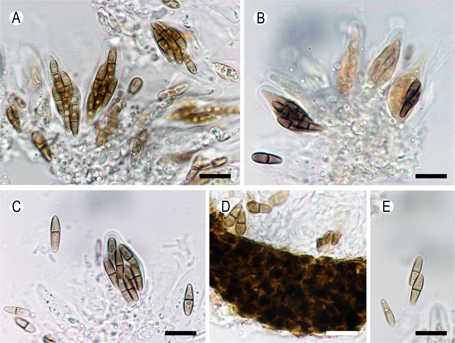
Figure 2. Long description
A multi-panel micrograph containing five sections labeled A through E.
* Panel A at the top-left shows several elongated, sac-like asci containing brown, multi-celled ascospores immersed in water.
* Panel B at the top-right displays asci treated with Lugol’s iodine solution, appearing as pale, yellowish sacs with dark brown spores inside.
* Panel C at the bottom-left shows a single ascus in water with clearly defined, septate ascospores.
* Panel D in the bottom-center shows a cross-section of the dark, thick-walled ascomatal wall with thin, thread-like pseudoparaphyses extending from it.
* Panel E at the bottom-right shows individual, two-celled brown ascospores in water.
Each panel includes a black or white scale bar in the bottom-right corner representing 10 mu m.
Ascomata perithecioid, arising singly, erumpent, black, subglobose, (35–)50–80 μm diam., not producing deformations or discoloured or necrotic patches, without visible mycelium on the host lichen thallus. Ascomatal wall brown throughout, in surface view recalling textura angularis, composed of several layers of thick-walled, radially compressed cells, 4–6.5 × 3–5.5 μm. Hamathecium composed of branched and anastomosing septate pseudoparaphyses, 1–2 μm thick, slightly swollen at the apices; hymenial gelatine I−, K/I−. Asci fissitunicate, elongate clavate to broadly clavate, shortly stalked, 8-spored, 25–45 × 6–11 μm; wall I−, endoplasma I+ brownish orange. Ascospores olive-brown, irregularly distichously arranged, narrowly ellipsoid, 1-septate, slightly constricted at the septum, cells almost equal in size, sometimes with somewhat narrower lower cell, with rounded ends, with 1–2 guttules per cell, (8–)10.2–10.4(–12.5) × (2.5–)3–3.2(–4.5) μm, l/w = (2.3–)3.3–3.5(–4.4) (n = 40); perispore amyloid, embedded in a gelatinous sheath, c. 0.5 μm thick.
Conidiomata not seen.
Etymology
The species is named in honour of Montserrat Boqueras, the prominent Spanish mycologist and lichenologist who first discovered and illustrated the fungus.
Distribution and ecology
The new species is currently known from four localities, two in the city of Kazan (Republic of Tatarstan, Russia) and two in the municipality of Deltebre (Catalonia, Spain) (Boqueras et al. Reference Boqueras, Navarro-Rosinés and Gómez-Bolea1989; Boqueras Reference Boqueras2000). In Russia, the host lichens were found growing on the bark of Acer platanoides and Populus tremula in Lebyazhe Forest Park, the largest park (32.1 km2) in Kazan. It is dominated by pine (Pinus sylvestris), broadleaved (Acer platanoides, Tilia cordata, Ulmus laevis) and mixed coniferous-broadleaved woodlands and belongs to South Taiga. The climate of Kazan is temperate (moderate) continental (Mingazova et al. Reference Mingazova, Torsueva, Latypova and Boiko2005). In Spain, P. boquerasiae was found in sunny coastal areas with high environmental humidity. There, the host lichens occurred on beach vegetation, Limoniastrum monopetalum and Tamarix gallica (Boqueras et al. Reference Boqueras, Navarro-Rosinés and Gómez-Bolea1989; Boqueras Reference Boqueras2000).
The fungus infects both the thallus and apothecia (disc and margin) of Lecania cyrtella. In the Russian material, the host lichen was also co-infected with Taeniolella delicata.
Notes
According to the modern concept of the genus, most of its representatives are host-specific, being confined to a single or closely related species. Polycoccum boquerasiae is currently the only species known to occur on Lecania. Among the known Polycoccum species, four have comparable ascospore dimensions (length between 8 and 12 μm, width between 2.5 and 4.5 μm): P. ibericum Etayo & van den Boom, P. laursenii Zhurb., P. serusiauxii and P. stictaria (Linds.) D. J. Galloway. Of these, P. ibericum is distinguished by wider, verruculose ascospores, 9.5–12.5 × 4–5(–5.5) μm, and gall induction. Polycoccum laursenii has larger ascomata (100–150 μm), longer asci ((50–)55–65(–70) × 6–8(–9) μm), and verruculose ascospores. Polycoccum serusiauxii differs in having larger ascomata (60–130 μm diam.), longer ascospores (9–15(–16) × 3–4 μm) and gall induction. Polycoccum stictaria clearly differs by the I+ deep violet reaction of the asci and its verruculose ascospores (Saccardo & Trotter Reference Saccardo and Trotter1913; Matzer Reference Matzer1996; Galloway Reference Galloway2004; Zhurbenko & Alstrup Reference Zhurbenko and Alstrup2004; van den Boom & Etayo Reference van den Boom and Etayo2014). Furthermore, all these species are confined to other host species (P. ibericum to Rinodina, P. laursenii to Cladonia, P. serusiauxii to foliicolous Byssolecania and P. stictaria to Pseudocyphellaria). However, since an I+ deep violet ascus reaction is atypical for Polycoccum, and no information regarding interascal filaments was provided in the protologue, P. stictaria may belong to a different genus.
In the specimens from Russia, the ascomata were (35–)50–70(–80) μm in diameter, the asci measured (25–)29.7–30.9(–35.5) × (6–)8.4–9(–11) μm (n = 21), and the ascospores were (9–)10.2–10.4(–12.5) × (2.5–)3–3.2(–4.5) μm. In comparison, Boqueras (Reference Boqueras2000) reported larger ascomata (70–80 μm), longer asci (30–45 × 8–10 μm) and ascospores measuring (8–)10–11 × (3–)4 μm.
Additional specimen examined
Russia: Republic of Tatarstan: city of Kazan, south shore of Glubokoe Lake, 55°50′21.5″N, 48°58′28.4″E, Lebyazhe Forest Park, Betula pendula forest with Tilia cordata and Acer platanoides undergrowth, with grasses, on thallus and apothecia of Lecania cyrtella growing on Populus tremula, 14 viii 2024, G. I. Miftakhova & V. A. Chernyshova (GSU-2481).
A key to the species of Polycoccum
Although several keys to Polycoccum are available (Vězda Reference Vězda1969; Hawksworth & Diederich Reference Hawksworth and Diederich1988; Atienza et al. Reference Atienza, Calatayud and Hawksworth2003; Calatayud Reference Calatayud, Nash, Ryan, Diederich, Gries and Bungartz2004; Ihlen & Wedin Reference Ihlen and Wedin2008; Hawksworth et al. Reference Hawksworth, Atienza and Coppins2010; Gardiennet Reference Gardiennet2012; Halıcı et al. Reference Halıcı, Akgül, Oztürk and Kiliç2013), they are currently of limited use since they include only up to 23 species, some of which have already been transferred to other genera. Here we provide a worldwide key to 62 accepted species of Polycoccum, following Atienza et al. (Reference Atienza, Hawksworth and Pérez-Ortega2021) and Diederich et al. (Reference Diederich, Lawrey and Ertz2025).

Long description
The table is structured into three columns: the identification step number, the morphological description, and the resulting species name or next step number.
Key identification pathways include:
* Step 1: Distinguishes between narrowly ellipsoid ascospores (ratio greater than 4) leading to step 2, and broadly ellipsoid ascospores (ratio less than 4) leading to step 3.
* Step 2: Identifies P. perrugosae (8-spored asci, causing galls) and P. longisporum (6-spored asci, on Pseudocyphellaria).
* Step 3: Branches based on ascospore length, with those greater than 25 micrometers leading to step 4 and those less than 25 micrometers leading to step 12.
* Step 4 to 7: Focuses on ascus spore count and hymenial gelatine reactions (I plus or I minus). Species identified include P. dzieduszyckii (2-spored), P. crassum (I plus reddish), P. marmoratum (I plus blue, with sheath), and P. opulentum (I plus blue, without sheath).
* Step 8 to 11: Differentiates species on Rhizocarpon, Placynthium, and Acarospora, including P. versisporum, P. cartilaginosum, P. diederichianum, P. acarosporicola, and P. anatolicum.
* Step 12 to 17: Branches by ascospore width (less than or equal to 5 micrometers). Includes P. stictaria, P. epiphyllum, P. serusiauxii, P. boquerasiae, P. ibericum, and P. laursenii.
* Step 18 to 24: Focuses on 4-spored asci, identifying P. sporastatiae, P. follmannii, P. crespoae, P. thallicola, P. clauzadei, and P. epizoharyi.
* Step 25 to 28: Focuses on 6-spored asci, identifying P. ochvarianum, P. decolorans, and P. islandicum.
* Step 29 to 66: A large section for 8-spored asci, further divided by spore width (greater or less than 8 micrometers), ascomata size, and host lichen. Notable species include P. vermicularium (on Thamnolia), P. trypethelioides (on Stereocaulon), P. microcarpum (on Cladonia), P. rinodinae (on Rinodina), and P. pulvinatum (on Physcia).
Measurements for asci and ascospores are provided in micrometers throughout the table, along with specific host lichen names in italics.
Acknowledgements
We warmly thank Paul Diederich, Javier Etayo and Adam Flakus for help with literature searches and for the valuable consultations. Javier Etayo and Mikhail Zhurbenko are cordially thanked for their valuable comments and suggestions.
Author ORCID
Andrei Tsurykau, 0000-0002-6309-170X.
Competing Interests
The authors declare none.



