Introduction
Septoplasty is a common otolaryngological (head and neck) surgery. Its success rate ranges from 43 to 85 per cent.Reference Samad, Stevens and Maloney1–Reference Uppal, Mistry, Nadig, Back and Coatesworth6 If a deviated nasal septum is not adequately corrected, and if nasal obstruction, headaches or other symptoms continue, revision surgery is needed.Reference Harrison and Jones7 Unfortunately, revision surgery is difficult because of scar tissue and tissue adhesions or because of deficiencies in the nasal septal cartilage or bone structure. Furthermore, in regions where the nasal septal cartilage (periosteum) is deficient, dissection may be difficult because of tissue adhesions. These issues can easily lead to perforation of the nasal septum, a drooping nose, a false hump nose or other complications. Revision surgery may even fail; therefore, deviation of the nasal septum may remain. In this study, an endoscopic revision septoplasty was explored after three years of research, which allowed simple treatment for a variety of nasal septum deformities, and satisfactory clinical outcomes were achieved.
Materials and methods
Ethics statement
This study was approved by the ethics committee of the Affiliated Zhongshan Hospital of Dalian University, Dalian, China.
Clinical data
A total of 14 patients (13 men and 1 woman; age range, 26–55 years; mean age, 40.93 ± 11.26 years) admitted to the Affiliated Zhongshan Hospital of Dalian University between September 2012 and December 2016 to undergo revision septoplasty were included in this study. These patients suffered from residual deviation of the nasal septum, headaches and nasal obstructions after undergoing septoplasty. The interval between the first and last operation was 1 to 384 months, with a mean interval of 84 months. Of the patients, one had received septoplasty twice, and another had a nasal septum fracture within one month of the first operation.
Criteria
Patients were included if they had received submucous correction for a deviated nasal septum but still suffered from nasal obstruction and headaches and required revision surgery. Patients were excluded if other diseases of the nasal septum, such as perforation of the nasal septum, were treated during revision surgery.
Examination methods
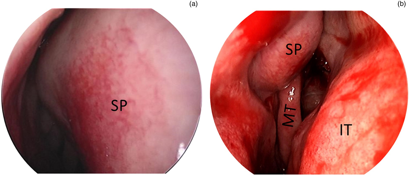
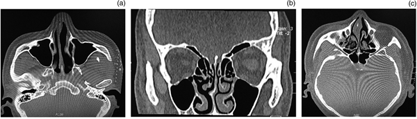
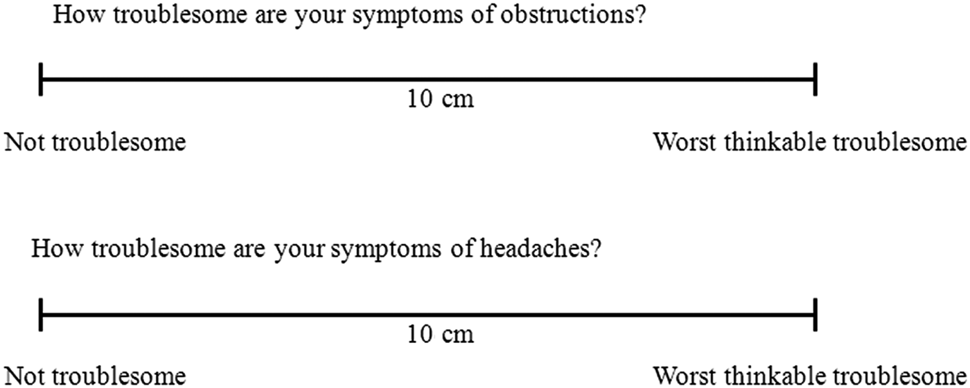
All the patients underwent pre-operative nasal endoscopy and nasal sinus computed tomography (CT) examinations (Figures 1 and 2). Therefore, diseases of the nasal cavity and sinus could be found and treated during the same surgery. These examinations revealed five cases of posterosuperior deviations, five cases of anterosuperior deviations and four cases of caudal deviations. The patients also underwent pre-operative and post-operative one-year assessments of their nasal obstructions and headaches using a visual analogue scale (VAS) with a 10-point score range, where 0 cm referred to not troublesome and 10 cm referred to the worst thinkable troublesome (Figure 3).Reference Rudmik, Mace, Ferguson and Smith8 All the patients were followed up for 30–71 months.
Nasal endoscopy images of (a) the right deviation of the upper nasal septum and (b) the left deviation of the upper nasal septum. SP = septum; MT = middle turbinate; IT = interior turbinate

Nasal sinus computed tomography (CT) images showing: (a) an axial CT scan of the right deviation of the anterior nasal septum; (b) a coronal CT scan of the right deviation of the posterior nasal septum; and (c) an axial CT scan of the right deviation of the upper nasal septum.

Visual analogue scale to measure patient scores of nasal obstructions and headaches.

Statistical analyses
The data are expressed as the mean ± standard deviation. The data were tested for normality and homogeneity of variance using the single-sample Kolmogorov–Smirnov method. As the data were non-normal, differences between sample means were analysed using Mann–Whitney U tests. When p-values were less than 0.05, differences between the sample means were considered statistically significant.
Surgical methods
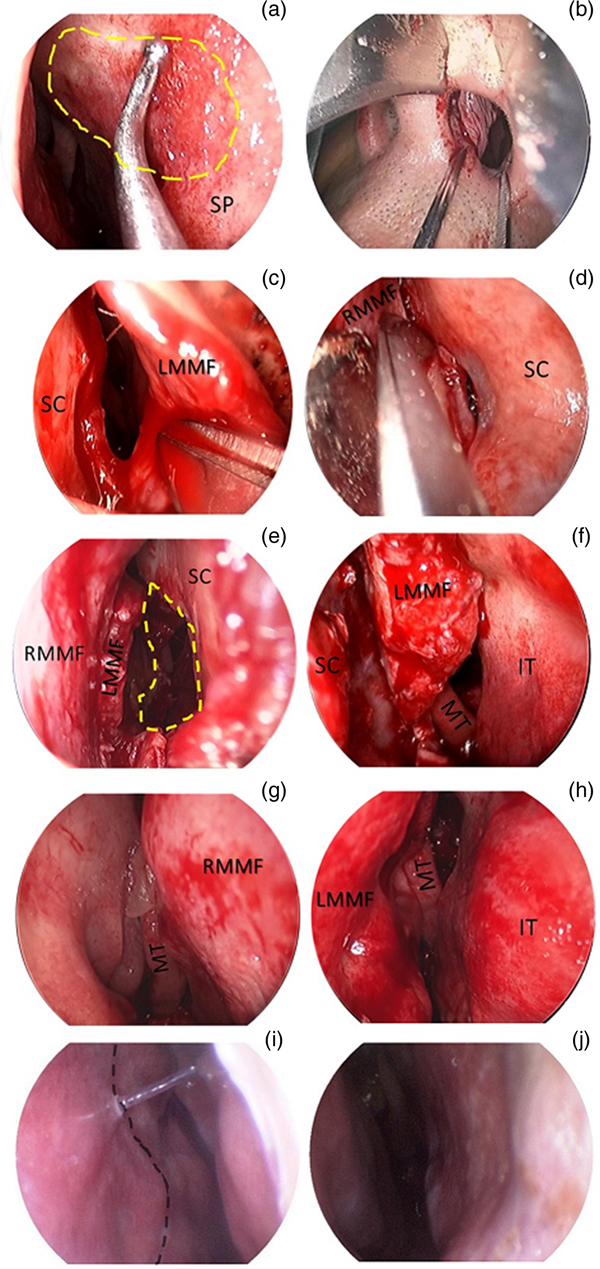
After general anaesthesia, patients underwent nasal endoscopy to allow observation of the morphology and deviation of the nasal septum as well as to explore the septal cartilage–bone defects (Figures 4a and 5a).
Endoscopic revision septoplasty showing: (a) exploration of the septal defect (the margin of the septal cartilage–bone defect); (b) the semi-penetrating straight incision in front of the caudal septum; (c) & (d) the bilateral dissection of the septal mucocartilage and mucoperiosteum along the septal cartilage–bone surface to fully expose the nasal septum; and (e) & (f) correction of the deviated cartilage and bone of the nasal septum. Nasal endoscopy (g) & (h) immediately and (i) & (j) 2 weeks post-operation showing the aligned nasal septum. The yellow line indicates the septal cartilage–bone defect. The black line indicates the healed incision. SP = septum; SC = septal cartilage; LMMF = left mucoperichondrium and mucoperiosteal flaps; RMMF = right mucoperichondrium and mucoperiosteal flaps; MT = middle turbinate; IT = inferior turbinate

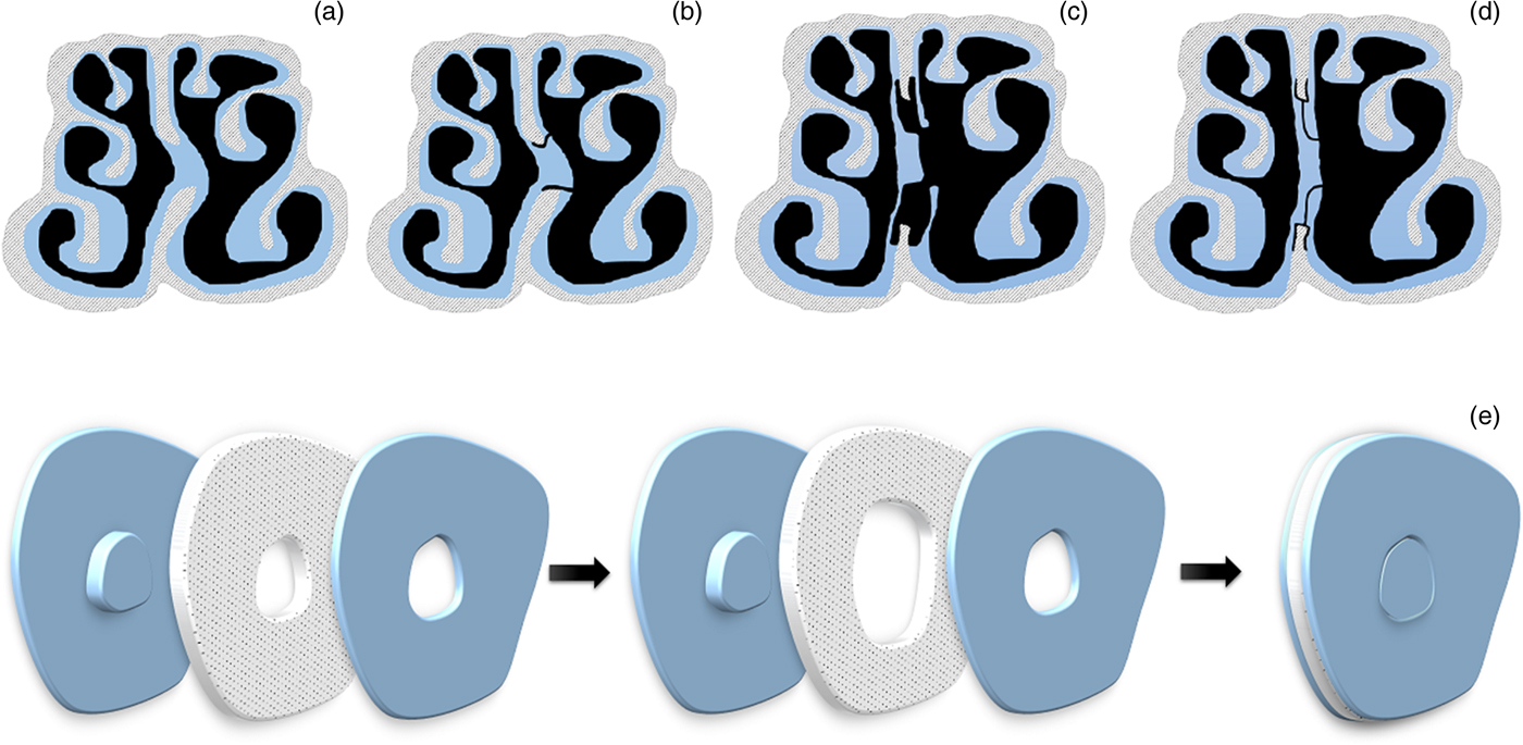
Schematic illustration of the circular incision design showing (a) nasal septum deviation, (b) dissection, (c) exposure, (d) suturation and (e) sagittal view of the circular incision.

Incision
A semi-penetrating straight incision along the left caudal septal cartilage (Figures 4b and 5b) and a semi-penetrating circular incision at the margin of the left septal cartilage–bone defect were made using an intranasal approach.
Exposure
The mucoperichondrium and mucoperiosteum were posteriorly and bilaterally dissected with a semi-penetrating straight incision at the caudal septum, along with the residual septal cartilage–bone defect of the previous operation. The whole process was performed until interlinkage with the margin of the septal cartilage–bone defect was obtained (Figures 4c and 4d). This process exposed the complete osteocartilaginous framework while keeping the right mucoperichondrium and mucoperiosteal flaps intact (Figures 4e and 4f).
The deviated cartilage and bone were removed with nasal rongeurs (Figure 5c and d). During this procedure, the cartilage (with a width of 1–1.5 cm) should be kept intact at the superior part of the cartilaginous septum, maintaining its linkage with the posterior bony septum and bony nose cone. If the residual septal cartilage presented a coronal C-shaped deviation, a horizontal scratch was marked on its concave surface. In contrast, if the septal cartilage presented a sagittal C-shaped deviation, a longitudinal scratch was marked on its concave surface.
Caudal deviations
For cases with a deviation of the caudal septal cartilage, a columellar capsule was generated with sharp dissection between the bilateral medial crura of the greater alar cartilage. The deviated cartilage was placed into the columellar capsule, followed by mattress suturing with straight needles. Other diseases, including those of the nasal cavity and sinus, were treated during the same surgery.
Results
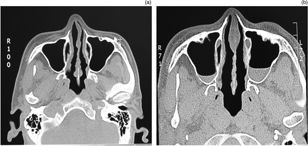
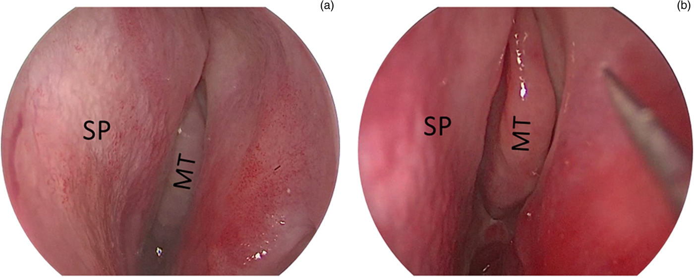
Significant improvements in the VAS scores of patients for nasal obstructions and headaches were observed (p < 0.05; Table 1). Nasal CT scans and endoscopies (Figures 6 and 7) showed aligned nasal septums and no further deviation or perforation. No patients had a saddle nose, false hump nose, contracture of the nasal columella, collapse of the nasal tip or any other complications in the external nose.
Pre-operative and post-operative VAS score differences

* Differences in pre- and post-operative nasal obstruction and headache were assessed using paired sample Mann–Whitney U tests. VAS = visual analogue scale; SD = standard deviation
Axial computed tomography scans of the nasal sinus (a) pre-operation (showing right deviation of the anterior nasal septum) and (b) post-operation (showing the aligned nasal septum).

Endoscopy images (a) pre-operation, showing right deviation of the upper nasal septum (only part of the middle turbinate is visible) and (b) post-operation, showing the aligned nasal septum (the intact front end of the middle turbinate is visible). SP = septum; MT = middle turbinate

Discussion
Deviation of the nasal septum is a common deformity of the nasal cavity. One study in the US showed that nasal septoplasty was the third highest ENT operation. Only adenotonsillectomies and myringotomies were performed more often than nasal septoplasty.Reference Manoukian, Wyatt, Leopold and Bass9 You et al. reported that deviations of the caudal, upper (above the free edge of the middle turbinate) and posterior (behind the front end of the middle turbinate) nasal septa could severely affect the normal physiological function of the nasal cavity and sinus.Reference You, Gao, Liu and Cui10 Gillman et al. suggested that patients who received inappropriate treatment for deviations of the caudal and upper nasal septa might need revision surgery.Reference Gillman, Egloff and Rivera-Serrano11 Patients who need revision septoplasty suffer from different degrees of defects in the cartilage or bone of the nasal septum and have scar tissue caused by adhesions of the bilateral mucoperichondria or mucoperiosteum from the previous operation. As such, revision surgery is generally more difficult and prone to cause nasal septal perforation, collapse of the nasal tip and other complications.
In functional reconstructive nasal surgery, the complete reconstruction of nasal function and appearance is achieved in the same surgical session. A normal structure of the nasal septum and normal physiological function of the nasal cavity are achieved by correcting all the anatomical deformities of the nasal septum.Reference Xiao, Kong, Wang, Yang and Le12 Based on this principle, semi-penetrating incisions were combined, and the deformities of the septal cartilage and bone were resected and corrected. This method easily corrected all deformities of the septal osteocartilaginous framework.
Nasal septum incision selection
A Killian incision is not a rare selection for initial surgery. All parts of the nasal septum, except for the caudal septal cartilage, are fully exposed using this method. Revision surgery using this type of incision is difficult and easily leads to septal perforation. In China, Xiao et al. applied a U-shaped incision at the front end of the nasal septum to correct the deviation of the anterior nasal septum and obtained satisfactory outcomes.Reference Xiao, Kong, Wang, Yang and Le12 However, a U-shaped incision requires the mucosa to be incised at the anterior superior part of the nasal septum, the front end of the nasal septum and the base of the cavum nasi, which will create a large incision, leading to suturing difficulties and prolonged recovery time.
Sillers et al. found that semi-penetrating incisions, penetrating incisions, Killian incisions and extranasal approaches were applicable for revision surgery, but they did not present any further descriptions.Reference Sillers, Cox and Kulbersh13 In our study, a semi-penetrating straight incision was made at the front of the caudal nasal septum, and the caudal nasal septum and nasal crest of the maxilla were fully exposed. Then, the malformation at the caudal end could be easily corrected. At the same time, this incision location avoided the incision used in the previous surgery and facilitated dissection of the mucoperichondrium and mucoperiosteal flaps. Collapse of the nasal tip and septal perforation could also be avoided due to the lowered columellar strength.
Anterior nasal septum deviation treatment
The nasal valve area, including the caudal nasal septum, lower margin of the lamina dorsi nasi (upper lateral cartilage), front end of the inferior turbinate and base of the apertura piriformis, is the narrowest part of the upper airway. Dislocation of the caudal nasal septum is the most common reason for stenosis in the nasal valve area.
Dinis et al. showed that after receiving initial surgery, 42 per cent of patients were extremely satisfied, 35 per cent were satisfied and 23 per cent were unsatisfied.Reference Dinis and Haider5 These authors also pointed out that the post-operative degree of satisfaction was closely associated with post-operative stenosis in the nasal valve area. Becker et al. found that the main reasons for revision surgery after the failure of septoplasty were the incomplete correction of stenosis in the nasal valve area and deviation of the nasal septum.Reference Becker, Dobratz, Stowell, Barker and Park14 These authors also compared the interval between the revision surgery and the initial operation and found that the average interval for patients with stenosis of the nasal valve area was 7.5 years, whereas it was 3.2 years for those without stenosis of the nasal valve area. This indicated that stenosis of the nasal valve area was often ignored by rhinologists. Grymer et al. showed that partial correction of a caudal nasal deviation was prone to present better efficacy compared with complete correction of a posterior nasal deviation because the nasal valve area is the narrowest part of the upper airway.Reference Grymer, Hilberg, Elbrønd and Pedersen15
Correction of a caudal nasal deviation is key to the revision of a deflected septum. In 1929, Metzenbaum first described the method for surgery of the causal nasal septum and pointed out that deviation of the posterior septum tended to have a smaller effect on the ventilation of the nasal cavity compared with deviation of the anterior septum.Reference Metzenbaum16 He also believed that retaining the caudal end was extremely important for supporting the nasal tip. He then applied the swinging door method and resected the caudal convex in a wedge shape to restore the caudal septum to the median line. However, while correcting the caudal septum, this method tended to weaken its strength, which was not conducive to maintaining the supporting structure of the nasal tip.
Various improvements in Metzenbaum's method have been reported, such as suturing the caudal septum to the nasal spine of the maxilla or suturing autograft materials, including nasal septum cartilage or bone, to support the caudal nasal septum cartilage.Reference Harrison and Jones7,Reference Pastorek and Becker17–Reference Giacomini, Lanciani, Di Girolamo, Ferraro and Ottaviani19 However, fixing the nasal septum at the caudal end may lead to stenosis of the nasal valve area and aggravate nasal obstruction because the nasal septum cartilage is relatively thick.Reference Constantine, Ahmad, Geissler and Rohrich20 Although the bony nasal septum, as a graft, is thinner and harder than cartilage, it often requires suturing via an open approach. This causes substantial nasal trauma and may lead to stiffness of the nasal tip. The intranasal method employed in this approach could prevent the redislocation of the caudal septum, strengthen the columella and prevent post-operative deformity of the nasal tip.
During the correction of caudal septal deviation, cartilage should be retained at the junction of the basal part of the cartilaginous nasal septum and the nasal crest area of the maxilla. This area is able to prevent the collapse of the cartilaginous nose cone, contraction of the columellar base and other complications.
Posterior nasal septum deviation treatment
During initial surgery for the correction of nasal septum deviation, it is necessary to remove the quadrilateral cartilage and part of the lamina mediana, leaving a square-shaped cartilage–bone defect. Deviation of the posterior nasal septum often occurs as a result of incomplete correction of the upper nasal septum and vomerine ridge. Xiao et al. applied a 0.5 cm longitudinal incision in the anterior deviated area to adjust the middle or upper nasal septum deviation.Reference Xiao, Kong, Wang, Yang and Le12 They applied a transverse incision at the labiogingival groove to expose the bone or cartilage in the deviated area and to enable the correction of inferior nasal septum deviation. They also ground the deviated area with an electric drill. However, this kind of incision might lead to a large injury.
In this study, a semi-penetrating circular incision was made at the margin of the septal cartilage–bone defect on the left side of the residual defect from previous surgery, and dissection occurred in various directions along the bilateral cartilage and bone surfaces. The mucoperichondrium and mucoperiosteum within the circular incision were not dissected. The defective osteocartilaginous framework was fully exposed, and the deviated parts could be removed and corrected. As the mucoperichondrium and mucoperiosteal flaps outside the septal cartilage–bone defect were mildly injured during the initial surgery (with light scarring and tissue adhesion), they could be easily dissected from the septal cartilage–bone defect during revision surgery. Thus, this method effectively exposed the deviated osteocartilaginous framework and avoided residual scar tissue and tissue adhesions from the initial surgery. Moreover, this method allowed us to freely select the incision according to the different deviation locations of the nasal septum, resulting in a relatively simple surgical procedure.
Upper nasal septum deviation treatment
Deviation of the upper nasal septum often refers to the upper deviation of the posterior cartilaginous nasal septum and the anterior lamina mediana. This type of deviation is prone to induce local pain and constriction. During surgery, the septal bone and cartilage at the deviated area were exposed fully, retaining a 1–1.5 cm ‘safe region’ at the superior part of the cartilaginous nasal septum. This was necessary in order to remove the deviated bone and cartilage and to maintain the linkage of the cartilaginous nasal septum in the safe region with the posterior bony nasal septum and nose cone. The deviated cartilaginous and bony nasal septum below the safe region could be removed. Thus, this operation was conducted outside the safe region to avoid the collapse of the nose bridge and nasal tip as well as the rotation of the cartilaginous nasal septum.
• The success rate of septoplasty ranges from 43 to 85 per cent
• Revision surgery is difficult because of scar tissue and tissue adhesions or deficiencies of the nasal septal cartilage or bone structure
• Semi-penetrating straight and circular incisions in front of the caudal septum and at the margin of the nasal septal cartilage-bone defect enabled exposure of the septal osteocartilaginous framework
• The technique allowed successful correction of the residual deviation
Limitations
This study has some limitations. First, a small number of cases were included. This study aimed to introduce a new surgical method to determine how to treat residual malformation in the anterior and posterior nasal septum after a previous operation to avoid septal perforation, a saddle nose, a false hump nose, contracture of the nasal columella or post-operative collapse of the nasal tip. After performing 14 such operations, this method was considered safe, feasible, repeatable and effective. Moreover, no control group was used in this study. This study aimed to introduce a surgical technique that was safe, feasible and repeatable for revision septoplasty surgery, rather than to prove its advantages compared with other techniques. Therefore, a self-controlled experiment was conducted instead of including a control group. Furthermore, acoustic rhinometry was not used for evaluating nasal obstruction. The placement of the nasal probe into the nasal cavity led to deformation of the caudal nasal septa with chances of false results. Finally, one patient who suffered from headaches after the operation had to undergo a sinusal CT scan, leading to additional radiation exposure. The cause of headache was acute sinusitis according to the CT scan and routine blood examination. After treatment, the patient recovered from acute sinusitis.
Conclusions
Most techniques used to correct a deflected nasal septum can easily manage mild-to-moderate deviations of the defect. However, for complex deviations, surgery is still challenging, even for experienced surgeons. This is because surgeons need to consider the structure, function and aesthetics of the nose. For patients with combined deviated nose or nasal trauma, physicians must abide by the concept of functional reconstructive nasal surgery.Reference Stupak21 They should also acquire knowledge of rhinoplasty surgery to provide the best possible outcome by repairing and reconstructing the structure, function and appearance of the nasal cavity with various graft materials.Reference Gillman, Egloff and Rivera-Serrano11 The proposed technique allowed the exposure of the septal osteocartilaginous framework and allowed a broad operational vision, thus enabling the successful correction of various deformities of the nasal septum.
Competing interests
None declared