Introduction
Angiostrongylus spp. is a nematode genus of the superfamily Metastrongyloidea, which comprehends 21 different species with worldwide distribution (Prociv et al., Reference Prociv, Spratt and Carlisle2000; Thiengo et al., Reference Thiengo, Simões, Fernandez and Júnior2013; Robles et al., Reference Robles, Kinsella, Galliari and Navone2016). Carnivores and rodents are the main definitive hosts and mollusks act as intermediate hosts (Acha and Szyfres, Reference Acha and Szyfres2003). All species are found in the pulmonary arteries and right ventricle, with the exception of A. costaricensis and A. siamensis, which inhabit the mesenteric arteries of the cecum (Robles et al., Reference Robles, Kinsella, Galliari and Navone2016). Usually, carnivores are the definitive hosts of A. vasorum, and rodents of A. cantonensis, respectively. Humans, dogs, horses, birds and other wild animals are considered accidental hosts of A. cantonensis (Lunn et al., Reference Lunn, Lee, Smaller, MacKay, King and Hunt2012).
The ability of such parasites to thrive in new areas is believed to be partially due to their wide variety of definitive and accidental hosts (Koch and Willesen, Reference Koch and Willesen2009). For instance, A. cantonensis has been diagnosed through histopathological evaluation in the North American opossum (Didelphis virginiana), grey-headed flying fox (Pteropus poliocephalus), black-and-white ruffed lemur (Varecia variegata) and in the miniature horse (Equus ferus caballus) (Reddacliff et al., Reference Reddacliff, Bellamy and Hartley1999; Costa et al., Reference Costa, McCluret, Snideirii and Stewart2000; Kim et al., Reference Kim, Stewart, Bauer and Mitchell2002); as well as through ELISA and Western blot in domestic dogs (Canis lupus familiaris) (Lunn et al., Reference Lunn, Lee, Smaller, MacKay, King and Hunt2012). Additionally, A. vasorum has been diagnosed through coproparasitological analysis in domestic cats (Felis domesticus) and in the European badger (Meles meles) (Kamenov et al., Reference Kamenov, Radev and Slateva1999; Torres et al., Reference Torres, Miquel and Motjé2004), through electron microscopy in the wolf (C. lupus) and in the crab-eating fox (Cerdocyon thous) (Segovia et al., Reference Segovia, Torres, Miquel, Llaneza and Felie2001; Duarte et al., Reference Duarte, Vieira, Louzada, Bessa and Souzalima2007; Vieira et al., Reference Vieira, Muniz-Pereira, Souzalima, Rocha and Luque2017); as well as through histopathology in the wolf (C. lupus), mustelids (Mustela erminea, Mustela nivalis, Mustela putorius), domestic dog (C. lupus familiaris), red fox (Vulpes vulpes) and in the crab-eating fox (C. thous) (Denk et al., Reference Denk, Matiasek, Just, Hermanns, Baiker, Herbach, Steinberg and Fischer2009; Eleni et al., Reference Eleni, De Liberato, Azam, Morgan and Traversa2014; Santoro et al., Reference Santoro, D'Alessio, Di Prisco, Neola, Restucci, Pagano and Veneziano2015; Simpson et al., Reference Simpson, Tomlinson, Stevenson, McLuckie, Benavides and Dagleish2016; Ferreira-Júnior et al., Reference Ferreira-Júnior, Blume, Sousa, Carvalho and Gardiner2017). Furthermore, the wide variety of definitive and accidental hosts are considered a risk to people who live in areas where such species are affected (Ma et al., Reference Ma, Dennis, Rose, Spratt and Spielman2013). In Brazil, the disease is considered emerging, and in a more recent case series, performed by Morassutti et al. (Reference Morassutti, Thiengo, Fernandez, Sawanyawisuth and Graeff-Teixeira2014), neural angiostrongyliasis has been confirmed in 34 cases. Definitive and intermediate hosts naturally infected by A. cantonensis have already been found in municipalities from north to south of Brazil, but reports of human cases are exceedingly scarce. This is probably due to the lack of knowledge of the health professionals about the infection by the parasite and its occurrence in the country (Morassutti et al., Reference Simpson, Tomlinson, Stevenson, McLuckie, Benavides and Dagleish2014). In human beings, infection by this nematode is considered an acute disease, which may be asymptomatic, and show spontaneous resolution in many cases, mainly in adults (Graeff-Teixeira et al., Reference Graeff-Teixeira, Silva and Yoshimura2009). Mortality is rare and occurs in approximately 3% of cases, mainly in children with encephalitis that may develop severe neurological signs (Li et al., Reference Li, He, Wang, Liang, Li, Men and Zhan2001; Eamsobhana and Yong, Reference Eamsobhana and Yong2009).
The objective of this study is to describe cases of angiostrongylosis in C. thous (crab-eating fox) and Lycalopex gymnocercus (Pampas fox) in the Southern region of Brazil, as well as to characterize the gross and histological findings of this infection and identify the associated Angiostrongylus spp. species through molecular analysis.
Materials and methods
Data were collected through the evaluation of necropsy reports of wild canines submitted to postmortem examination from January 2013 to December 2017. The studied animals were referred to the Sector of Veterinary Pathology by veterinarians from universities, zoos and health surveillance centres. The referred animals were originally from the following municipalities: Viamão, Canoas, Mostardas, Estrela and Cruz Alta; in some cases, however, it was not possible to determine the canid precise location, since these were found in roads in the surrounding areas of the metropolitan region of Porto Alegre. Information regarding the identification of animals, gross and histopathological findings, as well as the presumptive diagnosis present in the necropsy reports were reviewed and compiled. Cases were included in the study based on the fulfilment of one or two of the following criteria: (i) presence of eggs, larvae and adult nematodes compatible with Angiostrongylus sp. on histological slides [haematoxylin and eosin (HE)]; (ii) Angiostrongylus sp. PCR detection.
In two cases, lung impression smears were performed and stained with Romanowsky (Diff-Quik ®) stain. During necropsy procedures, tissue samples were collected, fixed in 10% formalin, routinely processed for histological examination and stained by HE. Masson's Trichrome histochemical technique was conducted in lung sections in order to identify fibrous connective tissue proliferation. In addition, sections of lung were submitted for immunohistochemistry (IHC) with mouse monoclonal antibody for smooth muscle actin (Dako ®, clone 1A4) to identify smooth muscle hypertrophy and hyperplasia in alveoli and arterioles. IHC procedure was polymer-based (MACH4); antigenic retrieval was performed with Tris-EDTA buffer, pH 9.0 in pressure cooker (120 °C, 20′), with antibody anti-smooth muscle actin (1:100) and detection was performed using 3-amino-9-ethylcarbazole chromogen (AEC).
Lung tissue samples from all canids evaluated in this study and one sample of central nervous system were submitted for polymerase chain reaction (PCR). In eight cases, fresh samples were available, and in 21 cases, formalin-fixed paraffin-embedded (FFPE) tissue blocks were evaluated. Initially, DNA was extracted from all tissue samples, following the manufacturer instructions (Quiagen). After DNA extraction, PCR for the 18S rRNA gene of Angiostrongylus spp. (SSU F: 5′-AAAGATTAAGCCATGCATG-3′ and SSUR: 5′-AGCTGGAATTACCGCGGCTG-3′) was conducted in the samples, according to the technique described by Blaxter et al. (Reference Blaxter, De Ley, Garey, Liu, Scheldeman, Vierstraete, Vanfleteren, Mackey, Dorris, Frisse, Vida and Thomas1998) and Foronda et al. (Reference Foronda, Lopez-Gonzalez, Miquel, Torres, Segovia, Abreu-Acosta, Casanova, Valladares, Mas-Coma, Bargues and Feliu2010), with modified primer annealing temperature of 62 °C. PCR products (480 bp) were evaluated through agarose gel electrophoresis (2%). In order to confirm results, sequencing was performed. PCR products purification (Purelin kit) and quantification (Qubit kit) were performed, and sequencing was conducted using an automated sequencer [BigDye® Terminator version 3.1 cycle sequencing kit in ABI PRISM 3130 Genetic Analyzer (Applied Biosystems®)]. Resulting chromatograms were evaluated with the software Staden package version 1.7.0 (Staden et al., Reference Staden, Judge, Bonfield, Krawetz and Womble2003). Then, the obtained sequences were submitted to BLAST (Basic Local Alignment Search Tool) analysis (Altschul et al., Reference Altschul, Gish, Miller, Myers and Lipman1990) in order to verify sample similarity when compared with sequences deposited in GenBank, and confirm the identity of the detected agent. Phylogenetic analysis of the obtained sequences was conducted through alignment with other sequences of Metastrongylids using Clustal/W v.1.8.1 (Thompson et al., Reference Thompson, Higgins, Gibson and Clustal1994). A maximum-likelihood phylogenetic tree was constructed applying as substitution model Tamura 3-parameter (Gamma), using Mega 7 software (Tamura, Reference Tamura1992), with 100 bootstraps replicates. Substitution model was selected using the software Mega 7 (Tamura, Reference Tamura1992) according to the lowest BIC (Bayesian Information Criterion), and sequences of the genus Dracunculus were used as outgroup.
Besides conventional PCR, DNA samples were submitted to real-time PCR using Taqman probe for A. cantonensis ITS-1 gene. The primers and probes used have been previously described by Qvarnstrom et al. (Reference Qvarnstrom, Silva, Teem, Hollingsworth, Bishop, Graeff-Teixeira and Silva2010); modifications on amplification conditions are listed: 94 °C for 5 min, followed by 50 cycles of 94 °C for 30 s, 60 °C for 30 s and 72 °C for 1 min.
Results
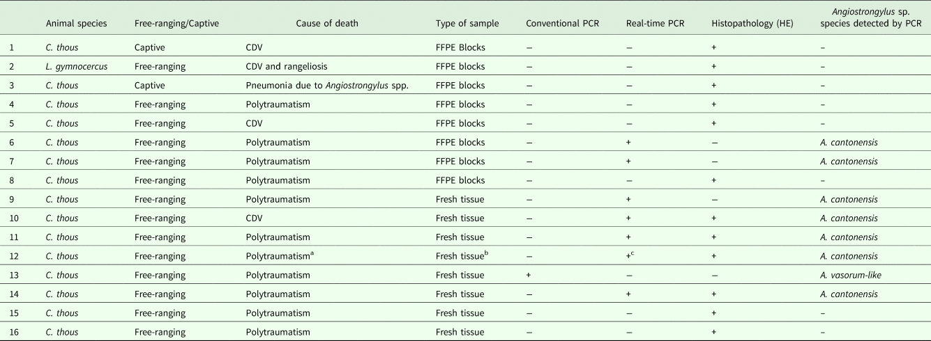
In the studied period, the necropsy of 29 wild canids (26 C. thous and 3 L. gymnocercus) was performed, 16 of which (55%) presented Angiostrongylus spp. infection. Out of these 16 animals, 15 were C. thous (93.75%) and one was L. gymnocercus (6.25%). Information regarding each individual animal is described in Table 1. Granulomatous pneumonia due to angiostrongylosis was observed without any additional evident potential causes of death in only one case. In the remaining studied wild canids, verminous pneumonia was associated with other causes of death, such as traumatism due to vehicle collision in 11 cases (56.25%), and with canine distemper virus infection in four animals (25%), among which one canid also presented rangeliosis. In one case of traumatism, besides granulomatous pneumonia, the animal presented an area of haemorrhage and malacia in the brain; however, traumatic lesions were restricted to the right hind limb.
Angiostrongylus spp. infection in C. thous and in L. gymnocercus in Southern Brazil

CDV, canine distemper virus; +, positive; –, negative.
A Cerdocyon thous presenting encephalic lesion associated with malacia and haemorrhage.
b Fresh samples of lung and brain.
c Positive results in lung and brain samples.
The following information is displayed: wild canid-infected species, free-ranging or captive, cause of death, type of sample [fresh sample or formalin-fixed paraffin-embedded (FFPE) blocks], diagnostic method and detected species of Angiostrongylus sp.
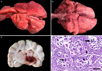
Grossly, in nine cases, lungs were not collapsed, presented multifocal dark-red areas, which were firm on palpation, displayed shiny surface (Fig. 1A), and sometimes drained serosanguinous fluid on cut surface. Multifocal small whitish firm nodules distributed in all pulmonary lobes were noted in one case (Fig. 1B). On gross examination of the encephalon, a focal area of softening and haemorrhage extending from the thalamus to the mesencephalon, and pons in the brainstem was present in one case (Fig. 1C).
Gross and histopathological findings of Angiostrongylus spp. infection in wild canids. (A) The lungs are not collapsed, and present multifocal dark-red areas of consolidation. (B) Multifocal white firm small nodules are noted in all pulmonary lobes. (C) Encephalon, a focally extensive area of softening, and haemorrhage is observed in the thalamus. (D) Lung, morulated eggs (arrowhead), larvated eggs (narrow arrow) and L1 larvae (large arrow) are observed in the pulmonary interstitium and alveoli (100 µ m bar), HE stain.
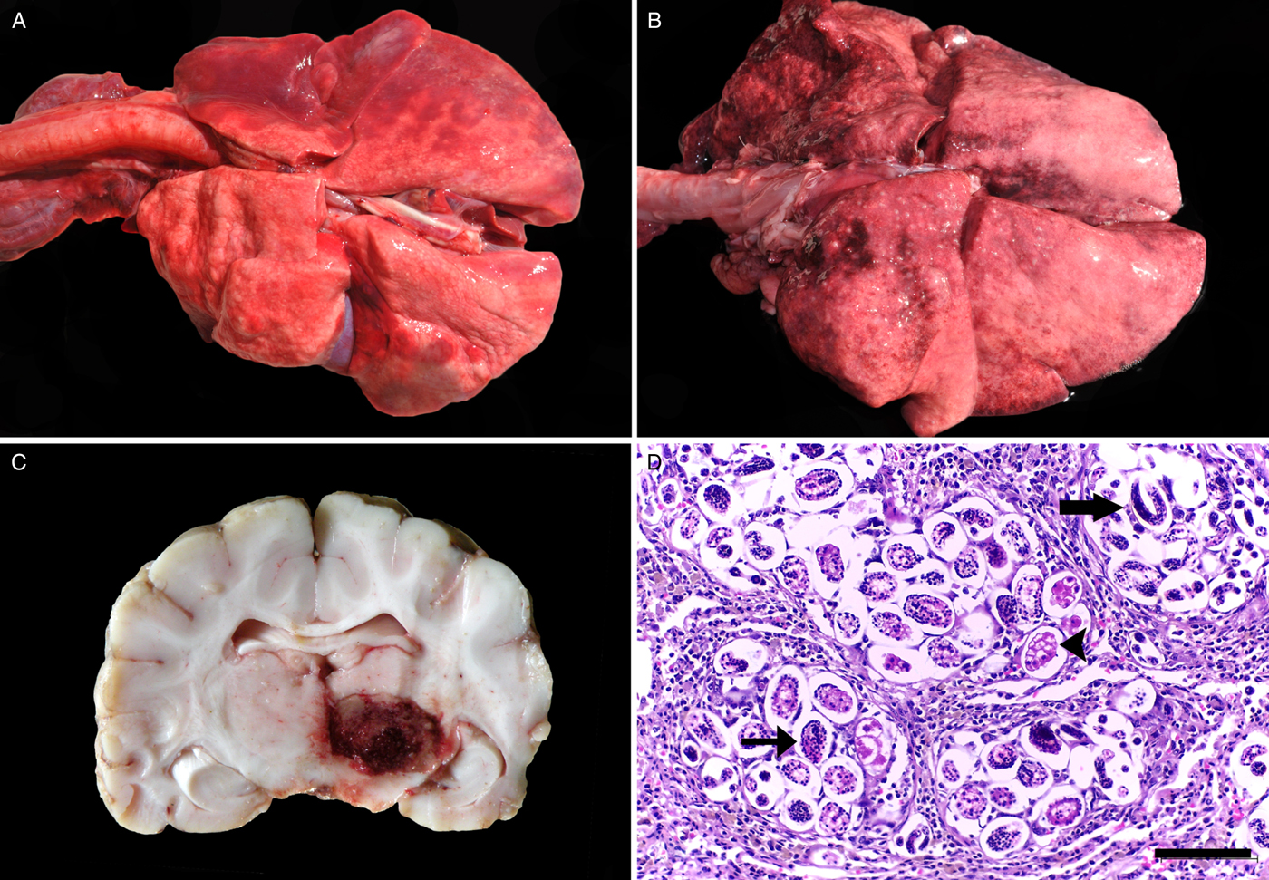
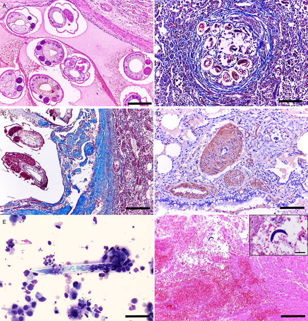
The areas presenting gross lesions in the lung parenchyma were histologically represented by granulomatous bronchopneumonia or interstitial pneumonia associated with a variable number of eggs and larvae (L1) in blood vessels, pulmonary interstitium, alveoli (Fig. 1D), and occasionally within bronchi and bronchioles, showing a multifocal distribution. In addition, adult nematodes were observed within blood vessels (Fig. 2A). Parasitic eggs were oval and thin-walled (25–40 × 30–50 µ m) and were filled with morula or larvae. Larvae were elongated with eosinophilic cuticle and contained basophilic granules (7–11 × 20–45 µ m). Larvae and eggs were surrounded by fibrous connective tissue proliferation, which was evidenced through Masson's Trichrome histochemical technique (Fig. 2B), as well as inflammatory infiltrate of lymphocytes, plasma cells, macrophages, sometimes presenting brown intracytoplasmic granules, few eosinophils, and multinucleated giant cells. Pulmonary arteries were thickened due to fibrous connective tissue deposition (Fig. 2C). Cross-sections of adult nematodes observed within blood vessels measured 220–230 × 250–420 µ m and longitudinal sections measured 190–250 × 530–840 µ m. These parasitic structures presented eosinophilic cuticle, coelomyarian/polymiarian musculature, and pseudocelom with gastrointestinal and reproductive tract. The parasites were frequently surrounded by thrombi.
Microscopic findings of Angiostrongylus spp. infection in wild canids. (A) Lung, adult nematodes are present within artery (bar 200 µ m), HE stain. (B) Lung, larvae and eggs surrounded by fibrous connective tissue proliferation (fibrosis) are evidenced. Masson's Trichrome histochemical technique (bar 100 µ m). (C) Lung, pulmonary artery wall thickening due to fibrous connective tissue proliferation. Masson's Trichrome histochemical technique (bar 200 µ m). (D) Lung, positive immunostaining for smooth muscle actin is showed, evidencing arterioles smooth muscle hyperplasia and hypertrophy. IHC, AEC (bar 100 µ m). (E) Lung, impression smear of pulmonary tissue performed during necropsy reveals larva. Romanowsky stain (Diff-Quik ®) (bar 40 µ m). (F) Encephalon, a focally extensive area of malacia, associated with neuropil rarefaction, gitter cells, haemorrhage, as well as an intralesional larva is noted (bar 200 µ m). Inset: free intralesional larva (bar 40 µ m), HE stain.
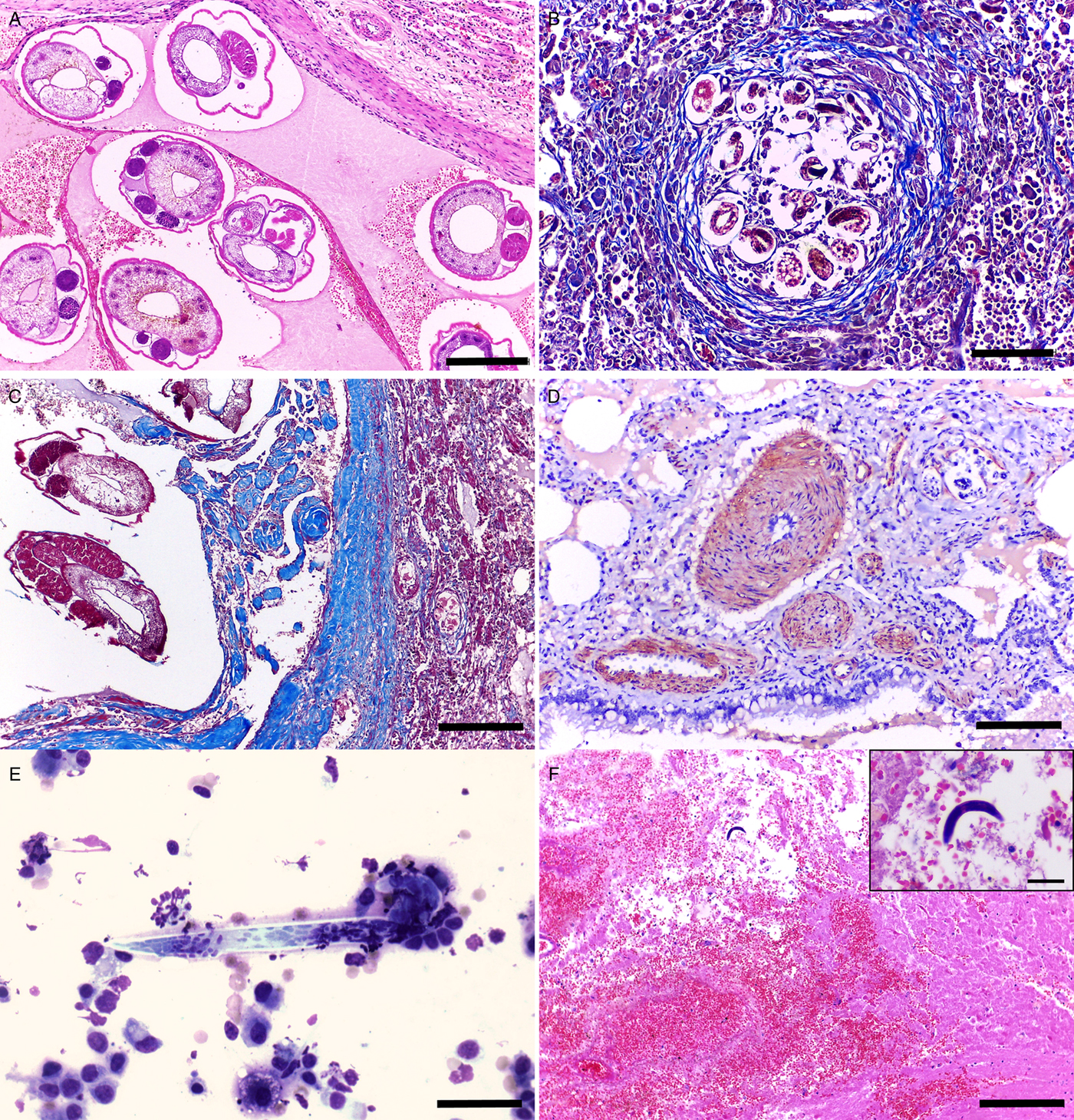
Alveolar smooth muscle hypertrophy (93.75%) and arterioles smooth muscle hypertrophy and hyperplasia (87.5%) were observed in areas adjacent to granulomas (Fig. 2D); both showed strong positive labelling for smooth muscle actin. In addition, haemorrhage in alveolar spaces, interstitium and perivascular areas (75%), thrombosis (62.5%), sometimes associated with blood vessel recanalization (43.75%) as well as alveolar oedema (43.75%) were also present. Oftentimes, the pleura was mildly thickened due to connective tissue proliferation (68.75%). Regarding the two cases in which impression smears were performed, larvae (L1) were observed in one case (Fig. 2E).
In one case, encephalon evaluation revealed a focally extensive area of malacia, characterized by neuropil rarefaction associated with gitter cells, along with haemorrhage, thrombosis and a free intralesional larva (Fig. 2F). In the remaining cases, encephalon evaluation evidenced multifocal areas of haemorrhage in 10 cases, which ranged from mild to moderate, and were located in the neuropil and around blood vessels.
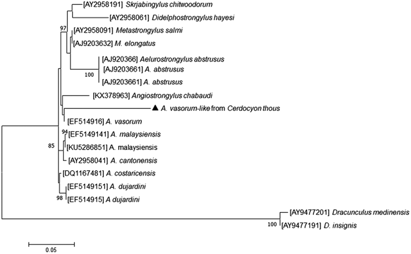
It was possible to identify in one C. thous, through conventional PCR, Angiostrongylus spp. 18S rRNA gene, generating a 480 bp amplicon. Sequencing and Blast analysis revealed sample similarity (97%) with A. vasorum; this sequence will be deposited in GenBank after publication. After sequencing, phylogenetic analysis was conducted (Fig. 3), which demonstrated greater phylogenetic proximity between the obtained sequence and A. vasorum, when compared with other Angiostrongylus species. Moreover, real-time PCR (ITS-1) yielded seven positive samples for A. cantonensis in C. thous, including the case in which encephalic lesion was observed.
Phylogenetic tree depicting genera of metastrongylids Angiostrongylus, Aelurostrongylus, Didelphostrongylus, Metastrongylus and Skrjabingylus through the sequence of 18S rRNA gene. The obtained sequence is shown in detail (triangle).

Eight positive cases of Angiostrongylus spp. were detected by PCR, and, among four of those cases, parasites were also observed on tissue sections; however, in the remaining cases, only positive PCR was available. In addition, the eight cases in which PCR yielded negative results were included in the study (six FFPE tissue blocks and two fresh samples), since parasites were seen on tissue sections, but the identification of the species of Angiostrongylus sp was not possible.
Discussion
In the present study, 55% of the analysed animals were parasitized by nematodes of the genus Angiostrongylus spp., and A. cantonensis and A. vasorum-like species were detected. The rate of infection in wild canids was 57.69% for species of C. thous and 33.33% of L. gymnocercus. These data suggest that a large number of wild canids may be infected by Angiostrongylus spp. in Southern Brazil. In Brazil, A. vasorum infection in wild canids has been reported in Lycalopex vetulus in the State of Minas Gerais (Lima et al., Reference Lima, Guimarães and Lemos1994), as well as C. thous in the States of Rio de Janeiro (Travassos, Reference Travassos1927), Minas Gerais (Duarte et al., Reference Duarte, Vieira, Louzada, Bessa and Souzalima2007; Vieira et al., Reference Vieira, Muniz-Pereira, Souzalima, Rocha and Luque2017) and the Federal District (Ferreira-Júnior et al., Reference Ferreira-Júnior, Blume, Sousa, Carvalho and Gardiner2017). Angiostrongylus cantonensis infection in C. thous or in L. gymnocercus had never been reported.
In studies conducted in Europe, the red fox (V. vulpes) has been implicated as the main host of A. vasorum. These animals show fair parasite adaptation, and are capable of enduring infection without presenting elevated mortality rates. In wild canids, infection is usually mild, and severe lesions are detected in the pulmonary parenchyma only in association with concomitant diseases or due to a massive number of lung parasites (Morgan et al., Reference Morgan, Tomlinson, Hunter, Nichols, Roberts, Fox and Taylor2008). In the present study, four animals were diagnosed with canine distemper virus infection, which may have accentuated the clinical case.
The main gross findings are restricted to the lesions in the respiratory and cardiovascular systems (Poli et al., Reference Poli, Arispici, Marroncini, Mancianti and de Monte1984; Morgan et al., Reference Morgan, Tomlinson, Hunter, Nichols, Roberts, Fox and Taylor2008). Right ventricular hypertrophy and lower heart weight/body weight ratio are findings observed during necropsy (Poli et al., Reference Poli, Arispici, Marroncini, Mancianti and de Monte1984; Jeffery et al., Reference Jeffery, Lankester, McGrath and Whitney2004; Morgan et al., Reference Morgan, Tomlinson, Hunter, Nichols, Roberts, Fox and Taylor2008); however, these lesions were not found in the present study. Respiratory system lesions were similar to what has been reported by other authors (Poli et al., Reference Poli, Arispici, Marroncini, Mancianti and de Monte1984; Jeffery et al., Reference Jeffery, Lankester, McGrath and Whitney2004; Denk et al., Reference Denk, Matiasek, Just, Hermanns, Baiker, Herbach, Steinberg and Fischer2009; Kistler et al., Reference Kistler, Brown, Allison, Nemeth and Yabsley2014) and included non-collapsed lungs; presenting multifocal red areas of consolidation, with firm encapsulated white nodules.
Pulmonary histological findings detected in the present study were similar to previously reported descriptions and consisted of multifocal to coalescing granulomatous inflammation foci in the pulmonary parenchyma; these foci showed a central area presenting multiple eggs and larvae (L1), which were surrounded by fibrosis associated with inflammatory infiltrate of lymphocytes, plasma cells, eosinophils, macrophages and occasional multinucleated giant cells. Adult nematodes may be observed within pulmonary arteries, occasionally associated with arterial thrombosis. L1 are also frequently noted within bronchi and bronchioles, associated with cellular debris (Poli et al., Reference Poli, Arispici, Marroncini, Mancianti and de Monte1984; Eleni et al., Reference Eleni, De Liberato, Azam, Morgan and Traversa2014; Kistler et al., Reference Kistler, Brown, Allison, Nemeth and Yabsley2014; Santoro et al., Reference Santoro, D'Alessio, Di Prisco, Neola, Restucci, Pagano and Veneziano2015; Robles et al., Reference Robles, Kinsella, Galliari and Navone2016). Vascular lesions may be observed, such as artery wall thickening due to severe fibrous connective tissue proliferation induced by the presence of adult nematodes in the blood vessel lumen, which may occlude artery lumen and promote thrombosis (Bourque et al., Reference Bourque, Whitney and Conboy2005).
Inflammatory infiltrate composed of eosinophils is an important component regarding host defence against parasitic diseases; although, in our study, as well as in previous studies reported in the literature, histological lesions presented minimal, if any, eosinophils, and granulomatous and chronic inflammatory components predominated (Traversa et al., Reference Traversa, Torbidone, Malatesta and Guglielmini2008; Denk et al., Reference Denk, Matiasek, Just, Hermanns, Baiker, Herbach, Steinberg and Fischer2009).
In one case, encephalon gross and histopathological evaluation revealed a focally extensive area of haemorrhage and malacia induced by A. cantonensis infection, which presented an intralesional larva; in addition, this sample presented PCR-positive results. The affected animal was involved in a vehicle accident; however, the only observed gross lesion consisted of necrosis of the right hind limb, and no fractures or additional lesions which could induce the encephalic findings of haemorrhage and malacia were noted in the skull. A similar lesion has been reported in the literature affecting miniature horse (E. ferus caballus), North American opossum (D. virginiana) and black-and-white ruffed lemur (V. variegata) (Costa et al., Reference Costa, McCluret, Snideirii and Stewart2000; Kim et al., Reference Kim, Stewart, Bauer and Mitchell2002). Lesions detected in the CNS still present a controversial explanation among researchers. According to Costa et al. (Reference Costa, McCluret, Snideirii and Stewart2000), these lesions are caused due to physical damage induced by parasite migration. However, Mason et al. (Reference Mason, Prescott, Kelly and Waddell1976) and McKenzie et al. (Reference McKenzie, Green and Wood1978) suggest that these lesions are mainly induced by the granulomatous inflammatory response against the parasite. In the present case, the best explanation for the encephalic lesion would be parasite migration, since no granulomatous inflammatory response was noted.
Even though A. vasorum cycle does not include CNS migration, this parasite may cause neurologic damage due to haemorrhages. These haemorrhages have been reported by several authors (Garosi et al., Reference Garosi, Platt, McConnell, Wray and Smith2005; Wessmann et al., Reference Wessmann, Lu, Lamb, Smyth, Mantis, Chandler, Boag, Cherubini and Cappello2006; Denk et al., Reference Denk, Matiasek, Just, Hermanns, Baiker, Herbach, Steinberg and Fischer2009), and are likely induced by a coagulopathy, and not by the presence of larva in the CNS, as observed in the infection by A. cantonensis (Wessmann et al., Reference Wessmann, Lu, Lamb, Smyth, Mantis, Chandler, Boag, Cherubini and Cappello2006). In the only PCR-positive case for A. vasorum-like detected in this study, the animal died from polytraumatism, and presented multiple fractures in the frontal, temporal and parietal cranium bones, which consequently led to haemorrhages. In the other four cases of encephalic haemorrhage, all animals were involved in vehicle accidents; thus, these lesions may have been induced by physical trauma. In one case, the animal presented canine distemper virus infection associated with rangeliosis, which may also lead to CNS haemorrhage. In the remaining four cases presenting haemorrhage, there is a possibility that the lesions were caused by angiostrongylosis.
The 18S rRNA gene sequence obtained from one C. thous infected by A. vasorum-like showed 97% homology with A. vasorum sequences and 80% with the sequences of A. cantonensis, both deposited in GenBank. When compared with A. vasorum and A. cantonensis 18 S sequences deposited in GenBank, 99% of similarity is observed, which may suggest that the sample from C. thous in Southern Brazil belongs to a new species or subspecies of A. vasorum. Previous studies evaluating fossil evidence and genetic analysis show a divergence among fox species (Vulpes spp.) in European and South American wild canids, which include C. thous and L. gymnocercus, indicating a host–parasite coevolution between wild canids and A. vasorum in South America which differ from nematodes found in European countries (Wang et al., Reference Wang, Tedford, Van Valkenburgh, Wayne, MacDonald and Sillero-Zubiri2004; Jefferies et al., Reference Jefferies, Shaw, Viney and Morgan2009).
In the present study, out of 16 wild canids infected by the nematode, only eight yielded PCR-positive results. According to Miething et al. (Reference Miething, Hering, Hanschke and Dressler2006), factors prior to tissue fixation (type and size of tissue, as well as degree of autolysis), fixation factors (pH, type of fixative and time of fixation) and factors after fixation (time of storage) may lead to degradation of nucleic acids. Furthermore, small amounts of DNA present in the studied samples, in both fresh and paraffin-embedded tissues may have led to negative results, since lesions are multifocal, samples may not present the needed amount of parasite DNA.
In conclusion, our study described the gross and histopathological findings due to Angiostrongylus spp. infection, which were characterized as granulomatous pneumonia or bronchopneumonia, as well as a case presenting encephalic lesions. Seven positive samples of A. cantonensis and one of A. vasorum-like in C. thous in the Southern region of Brazil were detected. Angiostrongylus cantonensis may represent a threat to public health, mainly to the human population living near such species, and this study is the first report of such nematode affecting wild canids in Brazil. The positive sample for A. vasorum identified in C. thous may be a new species or subspecies, since the level of similarity detected was 97% when compared with sequences deposited in GenBank; these findings require new studies regarding this topic. Angiostrongylus spp. infection in L. gymnocercus was confirmed through histopathological examination. In our study, 55% of wild canids were infected by Angiostrongylus spp., this agent was involved in cases of granulomatous pneumonia or bronchopneumonia associated with large number of parasites; however, in most cases, the infection was not related to the cause of death.
Author ORCIDs
Rafaela A. Caprioli http://orcid.org/0000-0002-9273-7450
Acknowledgments
We thank Dr Carlos Graeff-Teixeira and Professor, Drª Alessandra Loureiro Morassutti, from the Laboratory of Molecular Parasitology of the Pontifícia Universidade Católica do Rio Grande do Sul, for kindly providing the primers and probes used to perform ITS-1 real-time PCR.
Financial support
This research received no specific grant from any funding agency, commercial, or not-for-profit sectors.
Conflict of interest
None.
Ethical standards
Not applicable.