Introduction
Embryonic stem cells (ES cells) are originally isolated from the embryonic inner cell mass (ICM). Based on their capability to differentiate, ES cells are divided into ‘naïve’ (also named ‘ground-state’) or ‘primed’ states. The mouse ES cell line is the earliest established pluripotent stem cell line that satisfies standards of naïve state pluripotency, achieving germline chimeric development or tetraploid compensation (Evans and Kaufman, Reference Evans and Kaufman1981). There are 12 core genes essential to mouse pluripotency maintenance, including OCT4 (POU5f1), SOX2, NANOG, SALL4, KLF2, KLF4, ESRRB, GBX2 and TFCP2L1 (Takahashi and Yamanaka, Reference Takahashi and Yamanaka2006). Among them, Nanog, a highly differentiated protein containing a homologous domain, is a core transcription factor in pluripotent transcriptional regulatory networks (Boyer et al., Reference Boyer, Lee, Cole, Johnstone, Levine, Zucker, Guenther, Kumar, Murray, Jenner, Gifford, Melton, Jaenisch and Young2005; Loh et al., Reference Loh, Wu, Chew, Vega, Zhang, Chen, Bourque, George, Leong, Liu, Wong, Sung, Lee, Zhao, Chiu, Lipovich, Kuznetsov and Robson2006; Wang et al., Reference Wang, Rao, Chu, Shen, Levasseur, Theunissen and Orkin2006; Cole et al., Reference Cole, Johnstone, Newman, Kagey and Young2008). Nanog is critical for mouse early embryonic development (Mitsui et al., Reference Mitsui, Tokuzawa, Itoh, Segawa, Murakami, Takahashi, Maruyama, Maeda and Yamanaka2003), and is expressed in mouse pluripotent embryonic cells, ES cells, and germline cells (Chambers et al., Reference Chambers, Colby, Robertson, Nichols, Lee, Tweedie and Smith2003; Mitsui et al., Reference Mitsui, Tokuzawa, Itoh, Segawa, Murakami, Takahashi, Maruyama, Maeda and Yamanaka2003); it is an important transcription factor that maintains ES cell pluripotency and self-renewal capacity.
After the success of mouse ESC and induced pluripotent stem cells (iPSC), previous efforts to establish other mammalian ESCs and iPSCs have been made (Kues et al., Reference Kues, Nowak-Imialek, Haridoss and Niemann2010; Nowak-Imialek and Niemann, Reference Nowak-Imialek and Niemann2012; Selokar and Kues, Reference Selokar and Kues2018; Su et al., Reference Su, Zhu, Salman and Tang2020). Although a series of advances was achieved, in general, pluripotent stem cells of other species, especially farm animals, have not reached the full biological characteristics of mouse ES cells, and most of them are close to the primed state. This is also the same for attempts for pig (Beckmann and Day, Reference Beckmann and Day1993; Brevini et al., Reference Brevini, Pennarossa, Maffei and Gandolfi2012; Xue et al., Reference Xue, Li, He, Wei, Sun, Yin, Bou and Liu2016). Mouse studies indicated that ES cell lines cannot be isolated from Nanog-deficient blastocysts (Mitsui et al., Reference Mitsui, Tokuzawa, Itoh, Segawa, Murakami, Takahashi, Maruyama, Maeda and Yamanaka2003). The expression pattern of NANOG in porcine embryos was significantly later than that in mouse embryos. Therefore we tested the possibility of increasing the efficiency of forming blastocyst-derived porcine ES/pluripotent cell by overexpressing NANOG from the 4-cell stage. We conducted the current research to explore the effect of NANOG overexpression in porcine early embryos.
Materials and methods
Experimental materials
The pig ovary used in the experiment was taken from a slaughterhouse; follicular fluid containing oocytes was extracted manually. Oocytes were matured in vitro and fertilized in vitro. Boar semen for in vitro fertilization was taken from a rural pig farm affiliated to the university. All procedures related to in vitro maturation, in vitro fertilization and in vitro embryo culture were conducted as described previously in detail (Bou et al., Reference Bou, Liu, Sun, Zhu, Xue, Guo, Zhao, Qu, Weng, Wei, Lei and Liu2017). The related animal experiments observed the rules and regulations of animal experiments of Inner Mongolia Agricultural University.
Obtaining of NANOG overexpressing lentivirus
Based on the porcine NANOG mRNA sequence (XM_021092390.1) deposited in GenBank, an upstream primer 5´-ATGAGTGTGGATCCAGCTT-3´ and a downstream primer 5´-TCACATATCTTCAGGCTGTATG-3´ were designed for porcine NANOG 915-bp full-length coding sequence (CDS) cloning. PCR was performed using PrimeSTAR Max DNA polymerase. A NANOG overexpressing lentivirus was constructed using homologous recombination following the instruction of the NEBuilder® HiFi DNA Assembly Master Mix/NEBuilder HiFi DNA Assembly Cloning Kit. Following this method, we inserted the obtained porcine NANOG CDS into the lentiviral vector Lenti-EGPF bone vector, and the lentiviral overexpression vector Lenti-NANOG-EGFP was obtained subsequently. To obtain high titre lentiviruses, we performed a procedure described in our previous study (Bou et al., Reference Bou, Liu, Sun, Zhu, Xue, Guo, Zhao, Qu, Weng, Wei, Lei and Liu2017). Here, c. 50 μl of lentivirus were injected under the zona pellucida of a 4-cell stage porcine embryo using a microinjection method in order to infect the blastomeres.
Evaluation of gene expression by real-time PCR
The zona pellucida was removed from 40 embryos per group using an acidic working solution (pH 2.5), after which oocytes or embryos were washed with manipulating solution (pH 7.3). Then nude embryos or oocytes were immediately frozen in liquid nitrogen. To obtain RNA and cDNA, the Purelin Micro-To-Midi Total RNA Purification kit (Invitrogen) and High Capacity cDNA Reverse Transcription Kit (Applied Biosystems) were used individually according to their instructions. Real-time PCR was performed using SYBR Premix Ex Taq (TaKaRa). All transcripts levels were normalized against control gene GAPDH samples. Relative quantitative real-time PCR was performed using the Applied Biosystems 7500 real-time PCR system. Three to six biological replicates and three technical replicates were performed. The 2−ΔΔCt method was used for relative quantification analysis.
In vitro formation of porcine pluripotent embryonic stem cell clone from Day 5.5 blastocyst
The blastocysts were selected at Day 5.5 to form porcine pluripotent stem cell clones in vitro. At 1 day before blastocyst seeding, the feeder cells were grown overnight on 4-well plates; the basal medium was 15% FBS medium at 37°C, in 5% CO2, in a saturated humid incubator. Before seeding: (1) the blastocyst was washed twice in the manipulating solution, and the zona pellucida was scraped off using a mechanical method with a narrow pipette under a stereomicroscope; and (2) the solution in the 4-well plate was replaced with 200 μl KO medium with 10 μmol/l Y-27632 (Xue et al., Reference Xue, Li, He, Wei, Sun, Yin, Bou and Liu2016). The medium was changed every 24 h. After 1.5 days of culture, fully expanded primary clones appeared and for passage the clone was sectioned into small pieces using a glass needle.
RNA FISH
Embryos were fixed in 4% paraformaldehyde (PFA), overnight at 4°C, and stored in methanol at −20°C. In situ hybridization was performed according to Chazaud et al. (Reference Chazaud, Yamanaka, Pawson and Rossant2006) with slight modifications. RNA probes for porcine NANOG were generated by in vitro transcription of 915-bp full-length cDNA cloned in the pSPT18 plasmid. Sense and antisense probes were synthesized in vitro under the control of the T7 and SP2 promoters, respectively, and labelled with digoxigenin-UTP using the DIG RNA labelling kit (SP6/T7) (Roche). The slide was sealed with ProLong Gold antifade reagent (Invitrogen) and detected on a Leica TCS SP2 confocal microscope with a ×40 magnification oil immersion objective.
Immunofluorescence
Embryos or cells were fixed with 4% PFA in phosphate-buffered saline (PBS) for 40 min at room temperature. Then the embryos were permeabilized with 1% Triton X-100 (Sigma-Aldrich) in PBS for 5 h at 4°C, and then blocked with 1% bovine serum albumin in PBS for 1 h at room temperature. Next, samples were incubated with primary antibodies in PBS containing 0.01% Triton X-100 and 0.1% Tween 20 overnight at 4°C. Antibodies used in the current study were as follows: goat anti-SOX2 (Santacruz, sc-17320), goat anti-OCT4 (Santa Cruz, sc8628), goat anti-NANOG (Sigma, SAB2500670). After incubation, embryos were washed three times for 5 min with 0.01% Triton X-100 and 0.1% Tween 20 in PBS at room temperature, and then incubated with secondary antibodies diluted at 1:1000 in 0.01% Triton X-100 and 0.1% Tween 20 in PBS for 1 h at room temperature. Finally, clones were counterstained with Hoechst 33342 (Sigma-Aldrich), mounted on glass slides, and examined under a Nikon microscope.
Alkaline phosphatase (AP) staining
Cells were fixed in 4% PFA for 90 s at room temperature, then washed three times with DPBS. AP activity was determined using the BCIP/NBT Alkaline Phosphatase Colour Development Kit (Beyotime).
Statistics
All data from at least three replicates were analyzed using GraphPad software. Data were finally shown as mean ± standard error of the mean (SEM), a P-value < 0.05 was judged to be a significant difference.
Result
Embryonic NANOG starts to be expressed at blastocyst stage in porcine early development
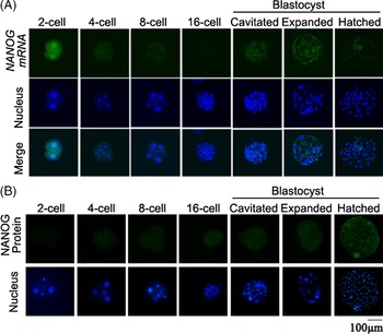
The RNA FISH experiment (Fig. 1A) clearly showed that porcine oocyte-derived NANOG mRNA was hardly sustained to the 4-cell stage and zygotic NANOG still did not start to be expressed. Zygotic NANOG transcription started in early blastocysts with small cavities. In early stage blastocysts, diffuse weak signals were present in almost all cells; in hatched large blastocysts only, stronger signals were detected, specifically in ICM cells. The IF study (Fig. 1B) to confirm showed strong protein staining in the ICM of hatched blastocysts.
NANOG expression during porcine early development. (A) RNA FISH; (B) IF.

NANOG overexpression in porcine embryos fails to promote the early development
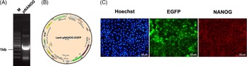
After cloning porcine NANOG full CDS (Fig. 2A), we constructed Lenti-pNANOG-EGFP incorporating virus that expressed porcine NANOG tagged by EGFP (Fig. 2B). Tests on porcine embryonic fibroblasts confirmed the high infection and expression efficiency of our packaged virus (Fig. 2C).
Lentivirus-mediated NANOG overexpression. (A) Gel image showing successful cloning of porcine NANOG CDS (961 bp). (B) Structure of Lenti-pNANOG-EGFP. (C) IF (NANOG, red) assay and virus-derived EGFP on porcine fibroblasts showing the high infection efficiency.

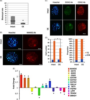
Then at Day 2 after in vitro fertilization (IVF), we injected c. 50 μl of infectious virus under the 4-cell stage embryo zona pellucida to obtain embryos with continuous overexpression of NANOG. At Day 7 after IVF, we found that, after NANOG overexpression, the blastocyst rate was significantly lower than that of the control group (Fig. 3A; P < 0.05). The NANOG IF study confirmed that Day 7 blastocysts overexpressed NANOG in all cells (Fig. 3B). We further used SOX2 (green) and CDX2 (red) co-immunoprecipitation (co-IP; Fig. 3C) to calculate the ICM and trophectoderm (TE) cell numbers for blastocysts from the two groups. There was a significant decrease in the TE proportion because of the significantly small numbers of TE cells in the NANOG overexpressing group (Fig. 3D), showing that NANOG overexpression may interfere with TE development. A further real-time PCR assay (Fig. 3E) on Day 7 blastocysts indicated that NANOG overexpression also induced significant expression changes in many development-related genes including OCT4, CDX2, SOX2, GATA4, GATA3, SALL4, TET1, FGFR2, TEAD4, and SMAD1. Among the genes we tested, only GATA6 and LIFR expression did not change.
Effect of NANOG expression on porcine early development. (A) Blastocyst rate of intact group and NANOG overexpressing group. (B) NANOG IF assay at blastocyst stage proving the successful lentivirus-mediated NANOG overexpression in porcine early embryos. (C) SOX2 and CDX2 co-IF assay could facilitate the ICM and TE calculation in porcine blastocysts. (D) Statistics on ICM and TE cell number and proportion of two groups blastocysts. (E) Real-time PCR result showing that NANOG overexpression could change the expression of many pluripotent/development-related genes and signalling. *Significantly different at P < 0.05.

NANOG overexpression is not required for obtaining embryonic stem cells from porcine blastocysts
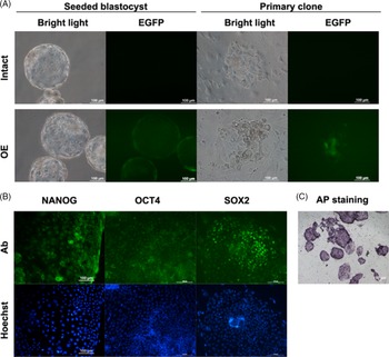
Our laboratory previously successfully established a porcine ES cell line using blastocyst seeding, and known as the KO cell line (Xue et al., Reference Xue, Li, He, Wei, Sun, Yin, Bou and Liu2016). Using the same procedures, in this experiment, we obtained Day 5.5 NANOG overexpressing blastocysts and control blastocysts for seeding (Figs 3 and 4, Table 1). During our experiments, we found that NANOG overexpressing blastocysts grew normally and initially could adhere to produce flat KO cell clones and control blastocysts. However, after a short time in culture, these cell clones began to loosen and lose their clonal morphology (Fig. 4A). Therefore, we did not obtain any cell clones that could be passaged from NANOG overexpressing blastocysts (Table 1). In contrast, using same procedure, from control blastocysts, we obtained several cell clones that expressed pluripotent genes NANOG, OCT4, and SOX2 and were AP positive (Fig. 4B).
Effect of NANOG expression on formation of pluripotent cell clone from porcine blastocysts. (A) Morphology of seeded blastocysts and primary clone after seeding for 5 days. (B) IF assays on pluripotent genes and (C) AP staining indicating the pluripotency of cell clone from the control group.

Effect of NANOG expression on cell clone formation

Discussion
Diversity in the early development of different species is accompanied by differences in gene expression, regulation and function. For instance, our study, together with other studies on different species demonstrated that the expression pattern of Nanog in early development varied among species. During mouse early development, Nanog RNA and protein are present throughout all processes, and are finally restricted in epiblasts (Chazaud et al., Reference Chazaud, Yamanaka, Pawson and Rossant2006). However, according to our study, during porcine early development, Nanog RNA and protein are strongly expressed from the early blastocyst stage and then restricted in the ICM of large hatched blastocysts. Our observation are similar to those of previous studies on human, cattle, sheep and pigs (Mitsui et al., Reference Mitsui, Tokuzawa, Itoh, Segawa, Murakami, Takahashi, Maruyama, Maeda and Yamanaka2003; Hall et al., Reference Hall, Christensen, Gao, Schmidt and Hyttel2009; du Puy et al., Reference du Puy, Lopes, Haagsman and Roelen2011; Frankenberg et al., Reference Frankenberg, Gerbe, Bessonnard, Belville, Pouchin, Bardot and Chazaud2011; Wolf et al., Reference Wolf, Serup and Hyttel2011; Habibi et al., Reference Habibi, Hosseini, Zadegan, Hajian, Ostadhosseini, Vash, Naddafpour and Nasr Esfahani2018; Simmet et al., Reference Simmet, Zakhartchenko, Philippou-Massier, Blum, Klymiuk and Wolf2018; Liu et al., Reference Liu, An, Wang, Liu, Dai, Liu, Yang and Du2019; Ortega et al., Reference Ortega, Kelleher, O’Neil, Benne, Cecil and Spencer2020). As shown, except for rodent ES cells, we did not obtain authentic ES cells from other species, especially from large animals. However, the mouse study showed that ES cell lines could not be isolated from Nanog-deficient blastocysts (Mitsui et al., Reference Mitsui, Tokuzawa, Itoh, Segawa, Murakami, Takahashi, Maruyama, Maeda and Yamanaka2003), implying that Nanog is necessary for the initial establishment of ES cell lines. Therefore, one aim of our study was also to check whether mouse-like NANOG expression patterns could benefit the development of porcine early embryos and the ES cells obtained. Consequently, we found the answer was no. At least, overexpression of NANOG alone was not enough. In terms of embryonic development, we found that the blastocyst rate and TE cell number and proportion would be lower after NANOG overexpression. The real-time PCR study explained that, despite significant upregulation of pluripotent genes such as OCT4 and SOX2, NANOG, overexpression could significantly downregulate both genes and signalling related to TE development, including CDX2, GATA4, TEAD4, SMAD1 and FGFR2 (Bou et al., Reference Bou, Liu, Sun, Zhu, Xue, Guo, Zhao, Qu, Weng, Wei, Lei and Liu2017). While, in terms of embryonic cell clone, we found that NANOG overexpression was harmful to the viability of pluripotent embryonic cells.
Recently many studies have revealed the co-operation of Nanog and epigenetic modification and chromatin structure (Lee et al., Reference Lee, Hamm, Whitworth, Spate, Park, Murphy and Prather2014; Carey et al., Reference Carey, Cao, Choi, Ganguly, Wilson, Paul and Knott2015; Liu et al., Reference Liu, An, Wang, Liu, Dai, Liu, Yang and Du2019; Veil et al., Reference Veil, Yampolsky, Grüning and Onichtchouk2019; Pálfy et al., Reference Pálfy, Schulze, Valen and Vastenhouw2020; Uh et al., Reference Uh, Ryu, Farrell, Wax and Lee2020), therefore we speculated that the effect and function of Nanog may be highly correlated with the fundamental DNA state. In addition, the effect of NANOG overexpression in our study would be different if we changed the cell state by adding some small molecules that affected DNA modification or structure.
The generation of bona fide ES and iPS cells from farm animals still remains unachieved. Notably, culture conditions and inducing systems of mouse iPS cells are inadequate to activate the endogenous pluripotent network in reprogrammed cells of farm animals, such that continuous transgene expression hinders their ability to satisfy ES/iPS requirements (Brevini et al., Reference Brevini, Pennarossa, Maffei and Gandolfi2012; Nowak-Imialek and Niemann, Reference Nowak-Imialek and Niemann2012; Chen et al., Reference Chen, Zhang, Jiang, Zhang, Wang, Zhao, Wang, Liu and Li2020; Su et al., Reference Su, Zhu, Salman and Tang2020). Our study, in an indirect way, also suggested that a thorough evaluation of upstream regulatory networks of core pluripotent genes in farm animals would provide a valuable insight into the establishment of complete reprogramming.
Funding
The study was supported by an Inner Mongolia Agricultural University Grant (NDYB2016-01).
Conflict of interest
We declare that we have no conflict of interest.