




No CrossRef data available.
Article contents
Evidence for macrofilaricidal activity of ivermectin against female Onchocerca volvulus: further analysis of a clinical trial in the Republic of Cameroon indicating two distinct killing mechanisms
Published online by Cambridge University Press: 04 November 2004
Abstract
During a 3-year trial of the effects of ivermectin (MectizanR) on adult worms and microfilariae of Onchocerca volvulus in the Republic of Cameroon, comparison was made between the percentages of calcified and uncalcified moribund (M) and dead (D) adult female worms dying following (a) the standard dose (150 μg/kg) given annually; (b) high doses (400, then 800 μg/kg) given annually; and (c and d) these same doses given at 3-monthly intervals. In the killing of adult female O. volvulus worms, the relative rôles of (a) natural causes; (b) a presumed, direct, anthelminthic, macrofilaricidal action of ivermectin; and (c) a potentially fatal pleomorphic ovarian neoplasm (PN), of which the incidence is increased by ivermectin treatment, are herein further investigated and discussed. It is concluded that ivermectin per se has a considerable direct macrofilaricidal action against female worms and that this lethal effect is supplemented by the drug's ability in some worms to increase the incidence, and the spread throughout the body of the worm, of the potentially fatal PN ovarian tumour. In moribund and dead ivermectin-treated female worms that were heavily invaded by PN, it is probable that the neoplasm was chiefly responsible for their death, but the additional direct anthelminthic action of the drug, which by itself has been responsible for the death of many other female worms, cannot be excluded as having played a supplementary lethal rôle. Similar problems as to the exact means by which adult female worms are killed may arise now that ivermectin is used in Africa for the mass treatment of lymphatic filariasis; or if and when the macrofilaricidal actions on O. volvulus of other drugs, which are closely related to ivermectin, come to be investigated.
Information
- Type
- Research Article
- Information
- Copyright
- 2004 Cambridge University Press
INTRODUCTION
In a double-blind trial, carried out in a forest-savanna mosaic area of the Republic of Cameroon, near the junction of the Mbam and Sanaga Rivers (Gardon et al. 2002) investigated the micro- and macrofilaricidal effects of (a) standard (150 μg/kg) and (b) high (400–800 μg/kg) doses of ivermectin (MectizanR), given annually or 3-monthly over 3 years to a total of 657 otherwise healthy patients, harbouring Onchocerca volvulus infections and who had not received any previous anti-filarial treatment. At the start of the trial, the highest dose permitted by Merck & Co Inc. was 400 μg/kg. Later, as the result of a safety trial in Ghana (Awadzi et al. 1995), this was increased to 800 μg/kg. Thus, the first dose on the high-dose annual regimen and the first two doses on the high-dose 3-monthly regimen were given at 400 μg/kg and the remainder at 800 μg/kg. High doses were not found to be more effectively filaricidal than the standard doses and they were liable to cause unpleasant subjective eye symptoms (Fobi et al. 2004).
Nodules, all of which had been present at the start of that trial, were excised for histological examination at 4 rounds of nodulectomy, that is (a) before treatment, in May 1994 i.e. Round 1 described by Gardon et al. (2002); (b) post-treatment early, in August/September 1997, 3 months after completing 3 years of treatment (i.e. Round 2A of Gardon et al. 2002); (c) post-treatment late (after excluding all those who had received only a single high-dose annual treatment) from one half of the remaining patients in May/June 1998, 8–9 months after finishing 3 years of treatment; and from the other half in November 1998, 14–15 months after completing 3 years of treatment (i.e. Rounds 3 and 4 of Gardon et al. (2002) combined). The post-treatment late nodules removed at Rounds 3 and 4 were pooled for examination; and all those removed at any of the post-treatment nodulectomies had been present at the start of treatment. The worms described by Gardon et al. (2002) as having been found in the relatively few nodules excised at Round 2B have been excluded from the present analysis since they all developed in nodules that first appeared subsequent to Round 1, and thus their minimum age and the duration of their exposure to ivermectin were uncertain.
Ivermectin, when given in standard doses at intervals of 3 months over 3 years, as compared with annual treatment, increased the numbers of moribund (M) and dead (D) female worms found at the early post-treatment examination by 43·75%; and the numbers of M and D female worms/nodule by 36% (Gardon et al. 2002). As well as having a direct macrofilaricidal action on adult female worms, ivermectin also induced a considerable increase in the prevalence and intensity of a pleomorphic, neoplastic tumour (PN) in the ovarian cells of many of the female worms. This tumour, described briefly by Duke, Zea-Flores & Gannon (1990) and later in greater detail (Duke et al. 2002), certainly sterilized and was often seen to become widespread and ultimately fatal, in the bodies of the M and D worms that it had attacked. The relative rôles of these two potentially lethal mechanisms of ivermectin (one directly anthelminthic and macrofilaricidal; the other neoplastic), acting on adult female O. volvulus are herein discussed.
MATERIALS AND METHODS
Excision and processing of nodules
All the nodules examined were excised surgically under local anaesthesia with full sterile precautions. Pre-treatment nodules were excised in May 1994, just before treatment began with an initial clearing dose of 150 μg/kg ivermectin, given in order to avoid severe reactions occurring after the subsequent higher doses that were to be taken later by some patients. The early post-treatment nodules were excised in August/September 1997, 3–4 months after the last (i.e. 12th) 3-monthly dose and 12–13 months after the last (i.e. 3rd) annual dose of ivermectin. The late post-treatment nodules (from Rounds 3 and 4 of nodulectomy), that were excised respectively in May/June 1998, and in November 1998 were pooled for the present assessment. Full details of the findings at each nodulectomy round and of the ivermectin treatment programme can be found in Gardon et al. (2002).
Each excised nodule was fixed in 20 or more times its volume of a mixture of pure ethyl alcohol (70 parts), glycerol (10 parts) and distilled water (20 parts) as described by Schulz-Key & Karam (1984). After 24 h, the fixative was renewed and the nodules were stored in more of the same fluid prior to sectioning. Small nodules were bisected and both halves were sectioned. Those big enough were cut at 25%, 50% and 75% of the way across the most appropriate diameter (Awadzi et al. 1997). They were then embedded in paraffin wax, sectioned at 6 μm, stained with haematoxylin and eosin, and examined independently by Dr M. Boussinesq and the present author, both of whom were experienced in this field. Any original differences of opinion were resolved by further study and discussion between the two observers, both of whom, at the time of the examinations, were aware of which nodulectomy round the slides came from but had no knowledge of the actual ivermectin dosage taken by any of the patients from whom the nodules came.
Classification of female worms
The system for classifying the female worms seen in sections of nodules was that of Duke et al. (1990). It may be summarized as follows, with the origin of the key letters herein indicated by capital italic letters.
Category F – FECUND worms, inseminated or re-inseminated, producing oocytes and embryos of all stages up to microfilariae (mfs).
Category S – potentially fertile worms, not currently inseminated, SHEDDING oocytes that are transforming into unfertilized ova and then degenerating. It includes 2 subcategories: – SF (young worms in FULL production of oocytes); and SO (OLDER worms producing reduced numbers of oocytes and sometimes also containing small numbers of degenerate mfs left over from a previous insemination).
Category E – EMPTY senescent worms with atrophic ovaries, no longer producing oocytes; sometimes still containing a few degenerate mfs.
Category M and D – MORIBUND and DEAD worms, almost or completely devoid of internal organs, with loss of turgor, collapse of the body wall, and often surrounded and/or invaded by, foreign-body giant cells. About half of these worms were calcified.
The very few worms belonging to Category I (INTERMEDIATE), i.e. those changing from Category S to F (IA) or vice versa (IZ), were placed in Category F and subcategory SO respectively.
RESULTS
Causes of death in female worms treated for 3 years with ivermectin (Table 1)
The results of the Cameroon trial (Gardon et al. 2002), and the findings concerning M and D female worms and those with PN in that trial (Duke et al. 2002), justify the following assumptions.
M and D female worms may be either calcified or uncalcified. Those that are calcified have presumably been dead for a longer period than those that are uncalcified, but it is not possible from histological observations to deduce what may have been the most likely cause of death in the calcified worms. However, as Table 1, Section C shows, the numbers of calcified M and D female worms per nodule at the early post-treatment nodulectomies were about double the numbers found at Round 1. This probably indicates that a considerable number of the calcified worms died during the earlier months of ivermectin treatment, although it was not possible to determine histologically the exact causes of their death.

PN only makes its first appearance in living worms, whose ovarian cells become neoplastic, and it only occurs in female worms that are out of microfilarial production. Its presence first sterilizes the worms by invading and destroying their ovaries and eventually it can kill them by invading the whole body. The prevalence of PN rises greatly after treatment with ivermectin. This rise is not related to the size of individual doses but rather to the frequency of dosing (Duke et al. 2002).
Table 1 separates the nos. and nos./nodule of uncalcified and calcified M and D female worms excised at each round of nodulectomy. The analysis in section B (uncalcified M and D female worms), strongly suggests that ivermectin can kill adult female O. volvulus worms in 2 ways – namely (i) indirectly (as in Section B iv), by increasing the incidence and intensity of the potentially fatal pleomorphic ovarian neoplasm (PN); and (ii) directly (as in Section B v), by an anthelminthic, macrofilaricidal action of ivermectin in its own right. However, in those uncalcified, ivermectin-treated worms that were classed as M and D and had also a heavy and widespread invasion of PN cells, it was not possible to determine the extent to which the direct macrofilaricidal action of ivermectin may have contributed to their demise.
Table 1 also gives, for the M and D female worms removed at each round of nodulectomy (pooled figures for all dosage regimens) – in Section (A) the total numbers of nodules, of female worms and of M and D female worms examined; in Section (B), which relates only to uncalcified M and D female worms, it gives the estimated total numbers, and the nos./nodule of (i) all M and D female worms; (ii) M and D female worms considered, on the basis of the Round 1 results, to be dead from natural causes other than PN; (iii) the remaining few M and D female worms estimated, on the basis of Round 1 results, to be dead from naturally-acquired PN; (iv) the M and D female worms full with PN and dead as a result thereof, supplemented by the macrofilaricidal effects of ivermectin treatment; and (v) the M and D female worms without PN which, after having eliminated all other likely causes, may be assumed to have died as a result of the direct anthelminthic, macrofilaricidal action of ivermectin.
It should also be noted that the presumed ivermectin-induced incidence of fatal PN began to fall off at the late post-treatment examinations, and that the direct macrofilaricidal action of ivermectin, detected at these examinations, included a carry-over from the early post-treatment examinations. Thus it is not possible to say to what extent, if any, these lethal actions continued for the 12–18 months after the last dosage round of the 3-year trial proper.
Table 1 also demonstrates the following additional points. (1) At the pre-treatment examination, out of 637 nodules examined, 88 uncalcified M and D female worms (0·138/nodule) were found. Of these, 80 (0·126/nodule) were considered to be dead from natural causes other than PN; and the remaining 8 (0·0126/nodule) were full of naturally-acquired PN, and were dead, presumably as a result. (2) In the nodules removed from the trial patients before treatment, 218/1422 female worms (15·3%) were classed as M and D. Many (130) of these worms were calcified, having been dead for a considerable time. In nodules, which had already been recorded at the pre-treatment examination, and which were subsequently removed from the patients at the early post-treatment examinations after 3 years of ivermectin treatment, it was found that, as compared with the pre-treatment results, the proportion of uncalcified M and D female worms rose from 0·138/nodule to 0·476/nodule i.e. by a factor of 3·45. By contrast, the proportion of all M and D worms with PN (naturally-acquired plus ivermectin-induced, i.e. Table 1Biii plus Biv) had risen from 0·0126/nodule to 0·0117+0·172=0·184/nodule, i.e. by a much higher factor of 14·6. This provides further evidence that PN itself can have a lethal action on adult female worms. (3) At the early post-treatment round of nodulectomy (which included 511 nodules), a total of 243 uncalcified M and D female worms (0·476/nodule) were found. Of them, and on the basis of the Round 1 results, again 0·126/nodule, i.e. 64 worms, were considered to have died from natural causes other than PN; 6 others (0·0117/nodule) were assumed to have died from naturally-acquired PN; and, in the remaining 88 PN-affected M and D worms (0·172/nodule), the increased prevalence and lethal effects of the neoplasm were most probably induced by ivermectin treatment, perhaps with its lethal effects in this respect being supplemented by the direct anthelminthic macrofilaricidal action of that drug. (4) The last 85 dead uncalcified M and D female worms (0·166/nodule) from the early post-treatment nodulectomies, none of which showed PN, were considered to have died from the presumed direct anthelminthic, macrofilaricidal action of ivermectin. (5) At the late post-treatment examinations, nodulectomies were carried out only on those remaining patients who had received treatment with ivermectin (a) at 150 μg/kg once a year; (b) at 150 μg/kg every 3 months; and (c) at 400–800 μg/kg every 3 months; i.e. there were no further nodulectomies done on those patients treated at 400–800 μg/kg once a year. At the late post-treatment rounds of nodulectomy, 246 nodules were removed (all of which had been present before treatment began) containing a total of 202 M and D female worms (0·821/nodule). Again, based on the pre-treatment findings 31 worms (0·126/nodule) were considered to have died from natural causes other than PN; of the 32 uncalcified females with PN, 3 (0·0126/nodule) were rated as dead from naturally-acquired PN; 29 (0·118/nodule) were assumed to have died from ivermectin-induced PN, probably combined with the drug's direct anthelminthic macrofilaricidal action; and the remaining 29 worms (0·118/nodule) were assumed to have died from the direct anthelminthic macrofilaricidal action of ivermectin.
Comparison of the increases in the prevalence of PN in SO and E worms and in M and D worms (Table 2)
Table 2 gives (A) for SO and E female worms and (B) for M and D female worms, the total numbers of worms examined at the pre-treatment and the early post-treatment rounds of nodulectomy, and expresses them as percentages of the total female worms examined at the examination concerned. The numbers of SO and E females with PN and of M and D females WITH PN are also shown and are expressed as percentages of all worms in the same class and as percentages of all female worms. The factors of increase in percentage between the pre-treatment nodulectomies and the early post-treatment nodulectomies are given for each pair of readings, being x1·40–x2·87 for SO and E females and x2·62–x14·80 for M and D females. However, over the period between the early and late post-treatment nodulectomies, there did not appear to be any further significant increase in the numbers of SO and E or of M and D worms as compared with the early post-treatment examination.
Table 2. Numbers of SO and E female worms and of M and D females at each round of nodulectomy (The indices of increase or decrease in the proportion of worms in each class as between nodulectomy examinations are also shown.)

The PN tumour was present only in live female worms of subcategory SO and Category E and, more abundantly, in M and D females. At the early post-treatment examination, its increased prevalence in SO and E worms combined (103/441 or 23·36%), as compared with the pre-treatment figures (45/395 or 11·39%), was in the ratio of 2·05[ratio ]1; whereas its prevalence in M and D worms at the early post-treatment examinations (94/456 or 20·61%) as compared with the pre-treatment figures (8/218 or 3·67%) represented a greater increase, in the ratio of 5·62[ratio ]1. These figures indicate that the prevalence of PN was considerably increased by ivermectin treatment; and, either that the incidence of PN, increased by ivermectin treatment, thereafter proved fatal to a considerable proportion of the worms in which it had spread throughout the body, or that the incidence of this neoplasm increased in worms that were dying from the direct anthelminthic, macrofilaricidal action of ivermectin.
DISCUSSION
Effects of ivermectin on the embryogenesis and vitality of female worms
At the early and late post-treatment rounds of nodulectomy, the numbers of calcified M and D female worms/nodule were shown to be over double the number found before treatment. This implies that a considerable number of calcified M and D female worms may have died as a result of the ivermectin treatment and then become calcified. However, in their calcified state it was not possible to decide whether their deaths should be attributed to natural causes; to the direct anthelminthic, macrofilaricidal effects of ivermectin; to ivermectin-induced PN; or to a combination of the latter two factors.
It was also shown that at the early and late post-treatment rounds of nodulectomy there were considerable numbers (0·172 and 0·118/nodule) of uncalcified female worms that had died with, and probably as a result of, heavy invasion by ivermectin-induced PN. Their nos./nodule were about the same as those without PN that were presumed to have died from the direct anthelminthic, macrofilaricidal action of ivermectin (i.e. 0·166 and 0·118/nodule).
Effect of ivermectin in the killing of SO and E female worms by inducing a heavy and fatal invasion of PN, thereby converting them to Class M and D
Before treatment, the prevalence of PN was low and occurred along with the shift in status of unfertilized females in the sequence from SO to E and finally to M and D. At the early post-treatment examination, after 3 years of ivermectin treatment, the number of uncalcified M and D female worms with PN/nodule had increased to about 14·80 times the pre-treatment level, while the corresponding rates in SO and E worms with PN rose only by about 2·87-fold. These observations imply that, under the influence of ivermectin, increasing numbers of SO and E worms had been killed by heavy invasion with PN and thus converted to Category M and D.
The effect of ivermectin treatment on male worms
Male worms (16–42 mm×0·125–0·200 μm) are much smaller than females (Neafie, Marty & Duke, 2000) and it is more difficult to assess their status accurately in histological sections of nodules.
The effects of drugs on male worms are better examined after their extraction from nodules that have been digested in collagenase solution (Büttner et al. 1988; Schulz-Key, 1988) but, in the present trial, which was based primarily on the histological examination of female worms in sections of nodules, it was not practical to use the collagenase method for the study of male worms. There was no evidence from the present trial that ivermectin treatment had any significant pharmaceutically induced lethal action on male worms (Gardon et al. 2002) and, in addition, the male worms were obviously not subject to the development of PN ovarian tumours.
CONCLUSIONS
The adverse systemic reactions to treatment of onchocerciasis with ivermectin at normal and at high doses, given annually or 3-monthly, have been described by Kamgno et al. (2004). High doses of ivermectin are not to be recommended as they can cause distressing visual symptoms (Fobi et al. 2005) and are no more filaricidal than standard doses. However, standard doses of 150 μg/kg, when given on a 3-monthly schedule for 3 years, certainly have a lethal action on a considerable proportion of adult female O. volvulus worms. Primarily this is caused by a direct anthelminthic macrofilaricidal action of the drug on a proportion of the adult female worms but, almost as many female worms appear to have been killed by the combined effects of the direct anthelminthic, macrofilaricidal action of ivermectin coupled with an ivermectin-induced increase in the incidence and intensity of the ovarian neoplasm (PN) which, under natural conditions, can affect a small proportion of untreated female worms, first sterilizing them and later spreading throughout the body and killing them. After prolonged treatment with ivermectin, the direct anthelminthic, macrofilaricidal effects of this drug cannot be clearly separated from the lethal effects of PN in those M and D female worms that have been attacked by the neoplasm, but it appears highly probable that the latter is chiefly responsible for the eventual death of those that are heavily loaded with tumour cells.
These findings are encouraging, and it is hoped that they may lead to further trials of 3-monthly standard-dose (150 μg/kg) ivermectin treatment in order to assess its longer-term lethal actions on adult O. volvulus. In addition, and unless previously unknown disadvantages of 3-monthly ivermectin treatment (such as selective activity only against worms of a certain genotype) are subsequently discovered, large-scale onchocerciasis control schemes in Africa, e.g. the African Programme for Onchocerciasis Control (APOC) and in Latin America, e.g. the Onchocerciasis Elimination Program in the Americas (OEPA), should surely initiate investigations into (i) the feasibility of carrying out Community-Directed Treatment with Ivermectin (CDTI) using the standard dose administered 3-monthly; and (ii) the effect of such 3-monthly treatment in reducing transmission by different Simulium vector species. Likewise, if and when other compounds that are closely related to ivermectin (e.g. moxidectin) come to be investigated for the treatment of onchocerciasis, similar problems as to their mode of action may arise. It would also be most interesting to determine whether or not ivermectin has similar effects on the adult worms of other human filarial parasites, especially Wuchereria bancrofti in Africa, where ivermectin plays a major rôle in the Programme for the Elimination of Lymphatic Filariasis (PELF).
This trial received ethical clearance from the Cameroon Ministry of Public Health and from Merck & Co. Inc. The author wishes to thank Merck & Co. Inc, especially Dr P. Gaxotte, for supplying the specially formulated gellules of ivermectin and their placebo controls; Drs J. Gardon, M. Boussinesq, N. Gardon-Wendel, J. Kamgno and Demanga-Ngangue, who carried out the trial in the field; Drs J. Millan and J. M. V. Martin, former Directors of the Centre Pasteur, Yaounde, Cameroon, and Dr Michel Santiago for undertaking the histological preparation of the nodules; Dr Aileen Marty and Dr Daniel Connor for assistance and advice in examining slides of PN; and the River Blindness Foundation (especially its founders, Mr and Mrs John Moores) for financing the greater part of these long-term investigations.
References
REFERENCES
AWADZI, K.,
OPOKU, N. O.,
ADDY, E. T. &
QUARTEY, B. T. (
1995).
The chemotherapy of onchocerciasis XIX: the clinical and laboratory tolerance of high dose ivermectin.
Tropical Medicine and Parasitology 46,
131–137.Google Scholar
AWADZI, K.,
OPOKU, N. O.,
ATTAH, S. K.,
ADDY, E. T.,
DUKE, B. O. L.,
NYAME, P. K. &
KSHIRSAGAR, N. A. (
1997).
The safety and efficacy of amocarzine in African onchocerciasis and the influence of ivermectin on the clinical and parasitological response to treatment.
Annals of Tropical Medicine and Parasitology 91,
281–296.CrossRefGoogle Scholar
BÜTTNER, D. W.,
ALBIEZ, E. J.,
VON ESSEN, J. &
ERICHSEN, J. (
1988).
Histological examination of adult Onchocerca volvulus and comparison with the collagenase technique.
Tropical Medicine and Parasitology 39,
390–417.Google Scholar
DUKE, B. O. L.,
MARTY, A. M.,
PEET, D. L.,
GARDON, J.,
PION, S. D. S.,
KAMGNO, J. &
BOUSSINESQ, M. (
2002).
Neoplastic change in Onchocerca volvulus and its relation to ivermectin treatment.
Parasitology 125,
431–444.CrossRefGoogle Scholar
DUKE, B. O. L.,
ZEA-FLORES, G. &
GANNON, R. T. (
1990).
On the reproductive activity of the female Onchocerca volvulus.
Tropical Medicine and Parasitology 41,
387–402.CrossRefGoogle Scholar
FOBI, G.,
GARDON, J.,
KAMGNO, J.,
AIMARD-FAVENAC, L.,
LAFLEUR, C.,
GARDON-WENDEL, N.,
DUKE, B. O. L. &
BOUSSINESQ, M. (
2005).
A randomised, double-blind controlled trial of the effects of ivermectin at normal and high doses, given annually or 3-monthly, against Onchocerca volvulus: ophthalmological results.
Transactions of the Royal Society of Tropical Medicine and Hygiene 99 (in the Press).CrossRefGoogle Scholar
GARDON, J.,
BOUSSINESQ, M.,
KAMGNO, J.,
GARDON-WENDEL, N.,
DEMANGA-NGANGUE &
DUKE, B. O. L. (
2002).
Effects of standard and high doses of ivermectin on adult worms of Onchocerca volvulus: a randomised controlled trial.
Lancet 360,
203–210.CrossRefGoogle Scholar
KAMGNO, J.,
GARDON, J.,
GARDON-WENDEL, N.,
DEMANGA-NGANGUE,
DUKE, B. O. L. &
BOUSSINESQ, M. (
2004).
Adverse systemic reactions to treatment of onchocerciasis with ivermectin at normal and high doses given annually or three-monthly.
Transactions of the Royal Society of Tropical Medicine and Hygiene 98,
496–504.CrossRefGoogle Scholar
NEAFIE, R. C.,
MARTY, A. M. &
DUKE, B. O. L. (
2000).
Onchocerciasis. In
Pathology of Infectious Diseases I, Helminthiases (
ed. Meyers, W. M.), pp.
287–306. Armed Forces Institute of Pathology, American Registry of Pathology, Washington D.C.
SCHULZ-KEY, H. (
1988).
The collagenase technique: how to isolate and examine adult Onchocerca volvulus for the evaluation of drug effects.
Tropical Medicine and Parasitology 39,
423–440.Google Scholar
SCHULZ-KEY, H. &
KARAM, M. (
1984).
Quantitative assessment of microfilariae and adults of Onchocerca volvulus in ethanol-fixed biopsies and nodules.
Transactions of the Royal Society of Tropical Medicine and Hygiene 78,
157–159.CrossRefGoogle Scholar

Table 1. Numbers (and nos./nodule) of uncalcified and calcified M and D female worms found at each round of nodulectomy, and the presumed reasons for their deaths
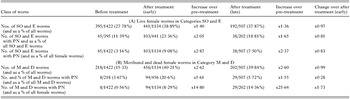
Table 2. Numbers of SO and E female worms and of M and D females at each round of nodulectomy
 You have
Access
- 29
- Cited by