Introduction
When observing a normal tympanic membrane, two common clinical phenomena are frequently noticed: a convex contour of the eardrum (seen mainly in infants), and (in some ears) a temporary inward movement during swallowing. Convexity indicates the existence of positive pressure inside the middle-ear cavity. Inward movement during swallowing indicates active evacuation (of gas) from the middle ear through the eustachian tube.
These two well known phenomena do not support the common concept of the ventilation mechanism of the middle ear, which postulates that gas is supplied by the eustachian tube.Reference Aoki, Mitani, Tuji, Hamada, Utahashi and Horiyama1–Reference Takahashi, Hayashi, Sato and Honjo3 Due to these two clinical observations, we speculated that gas may be produced by the middle ear and evacuated via the eustachian tube during swallowing together with mucus, in contrary to the presently accepted concept.
Few published reports support a mechanism of active gas production in the middle ear.Reference Tideholm, Carlborg, Jonsson and Bylander-Groth4 Buckingham et al. Reference Buckingham and Ferrer5, Reference Buckingham, Stuart, Girgis, Geik and McGee6 observed that gas was generated in the middle ear, creating a slightly positive pressure within it. They studied ears with otitis media with effusion, and observed inward movement of the tympanic membrane during swallowing. They found gas absorption in 9 per cent of 70 ears, but a positive pressure in 37 per cent. They also found an increase in middle-ear pressure during hypoventilation.Reference Buckingham, Stuart, Girgis, Geik and McGee6 They concluded that further experiments were needed. Ostfeld et al. Reference Ostfeld, Blonder, Crispin and Szeinberg7 and DoyleReference Doyle8 reported that the composition of middle-ear gas resembled that of venous blood rather than air.
These findings, along with our own clinical observations, led us to study the mechanism of middle-ear ventilation and clearance, and to attempt to replicate Buckingham and colleagues' findings.
Patients and methods
We began our observations on patients suffering from sudden hearing loss but with otherwise normal ears. These patients were treated with intra-tympanic steroid solution (dexamethasone).
First, a thorough, otologically relevant clinical history was taken and a standard audiometric evaluation was performed (along with tympanometry in some patients).
After signing an informed consent form, dexamethasone (Dexacort; Lumenis, Yokneam, Israel) was injected into the middle ear through a laser myringotomy. All patients were examined and treated in the recumbent position using an operating microscope. The tympanic cavity was filled with the steroid solution. Any excess fluid that escaped was suctioned (not from the myringotomy site), leaving the outer surface of the tympanic membrane clean.
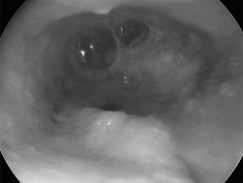
After injecting the steroid solution, the myringotomy site was inspected to observe the dynamics of the middle-ear gas. Fluid and air bubbles emerged continuously from the myringotomy as long as the observation continued, or until the ear emptied (up to 50 minutes of observation). The steroid solution usually covered the myringotomy opening, forming a fluid film and then a bubble (Figure 1).
Operating microscope photograph showing gas bubbling through a tympanic membrane myringotomy in a non-inflamed right ear treated with intra-tympanic steroid solution for sudden hearing loss. The myringotomy diameter is approximately 0.5 mm. Thin arrow = umbo; thick arrow = laser myringotomy.

After inspecting about 20 patients with sudden hearing loss as above, we decided to also include in our observations patients with traumatic tympanic membrane perforation caused by blast injuries (due to local terrorist attacks), and other patients with traumatic perforation who required cleaning of particles from their ears. After signing an informed consent form, these patients' ears were cleaned by sterile normal saline irrigation with suction. A clinical history was taken from these patients to verify that there was no history of middle-ear pathology.
Two patients with Ménière's disease (treated intra-tympanically with aminoglycoside solution) were also included.
Initially, all the patients were asked not to swallow, and the fluid surface at the tympanic membrane perforation or myringotomy was observed for inward or outward movement at rest. (It was easier to observe the fluid movement after suctioning, leaving a fluid film.) After 30–90 seconds of continuous observation, patients were asked to swallow while being observed for fluid film movement (Figure 1). The first 20 ears (from the normal ears) were observed and video-recorded for up to 50 minutes. Those who needed the steroid treatment were inspected during the whole treatment session (about 30 minutes). Patients from other groups were examined for few minutes as part of simple otoscopy (diagnostic or for ear suction treatment.). The same physician made all observations during one observation session, but several physicians made observations in different sessions when repeated treatment was necessary.
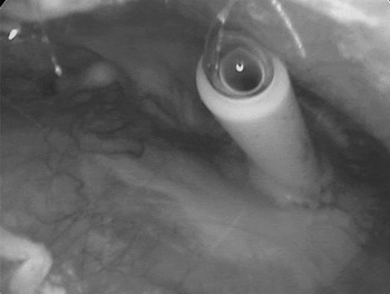
After observing 30 normal ears and identifying a common pattern of observations, we decided to also include patients with otitis media of various aetiologies and involving various inflammatory states in the “otitis media” group all the patients with chronic otitis media had perforated tympanic membrane. In the “otitis media with effusion” group the patients had either a tympanostomy tube who needed cleaning or post tympanostomy tube residual perforation. In ‘dry’ and ‘wet’ ears, the perforation was covered either with a thin film of mucus from the ear, or with 0.1–0.21 ml of sterile normal saline to create a fluid film. The mucous or fluid film was observed for movement at rest and during swallowing, as above (Figures 2 and 3). (Saline was also used to soften and clean crusts.)
Operating microscope photograph showing acute otitis media in the left ear, with pus and bubbles due to spontaneous perforation.

Operating microscope photograph showing gas bubbling from a tympanostomy tube covered with saline, inserted into the tympanic membrane of the right ear.

Movement of the fluid film (indicating the gas production or evacuation rate) differed between ears – the rate of gas production varied, as did the degree of fluid film movement (neither of which could be measured). For simplicity of data analysis, we recorded only the major types of fluid film behaviour observed at rest and swallowing, as follows.
At rest, the observer noted whether bubbling was present (i.e. bubbles forming and emerging via the perforation) or absent.
During swallowing (the patient was instructed to swallow while the observer held their hand on the patient's larynx, to feel the timing of swallowing, while observing the middle ear), three modes of fluid film movement were noted: inward (i.e. the fluid film moved inward towards the tympanic cavity); outward (i.e. the fluid film ballooned outwards); and no motion. In most ears, there was a sinusoidal movement, that is, the fluid film moved in and then out, returning to its previous position.
In addition to noting bubbling and fluid film movement, observers also noted the inflammatory condition of the ear and the perforation size. We analyzed the impact of the perforation size on the bubbling and film movement and found it statistically non-significant. The inflammatory condition of the ear was recorded as ‘dry’ (i.e. normal, thin, non-inflamed mucosa), ‘wet’ (i.e. various grades of inflammation but without purulent discharge; for simplicity, these inflammation grades were considered as one group) and purulent (i.e. purulent discharge filling the middle ear). To enable fluid film observations, large perforations were partially covered with a small piece of thin paper affixed to the tympanic membrane with mucus or normal saline, leaving a small opening which was then covered with a film of liquid.
Data on bubbling, fluid film movement, inflammatory condition and perforation size were recorded for each observation session, representing the state of that ear at that particular time point.
We also recorded patients' exact ear pathology diagnoses.
Statistical analysis indicated that they reacted similarly in their main group (Every main group included subgroups, such as: ears with otitis media with effusion included subgroups of ‘with tympanostomy tube’ and with ‘residual perforation’ without a tube in the ear. They reacted similarly.). Therefore, all patients were divided into three main groups, for simplicity: (1) those with non-inflamed ears (i.e. sudden hearing loss, blast injury tympanic perforation and other traumatic perforation); (2) otitis media with effusion included those having tympanostomy tubes (with crusts softened and cleaned by saline solution), acute otitis media with rupture or myringotomy and those shortly after a laser myringotomy for otitis media with effusion; and (3) those with chronic otitis media (including such cases with permanently perforated eardrums from any cause).
Statistical analysis
Patients who were treated intra-tympanically (i.e. the sudden deafness and Ménière's groups), and some patients from other groups, were observed up to 15 times per ear. Most of these ears were identical throughout repeated observation sessions; however, some ears changed (e.g. different observation sessions noted differing fluid film movement or gas production), especially in the otitis media with effusion group. In order to reduce the statistical impact of many identical observations from repeatedly examined ears, compared with single observations from ears examined only once, and in order to accurately express the degree of variety observed within the same ear over time, we conflated identical responses from the same ear. Thus, when observations for the four ear parameters (i.e. bubbling, fluid film movement, inflammatory condition (dry, wet or purulent) and perforation size) were identical in a number of sessions, all these observation sets were considered for statistical purposes to constitute one ‘observation type’. When observations for the four ear parameters varied in different observation sessions, the observation sets were conflated into two or more observation types, each type representing all occasions on which that particular combination of parameter results had been observed in that particular ear. These observation types were used for statistical analysis.
Data were entered into an Excel spreadsheet (Microsoft, Redmond, Washington, USA). Data analysis was performed using Epi Info 3.4.3 software (CDC, Atlanta, Georgia, USA). We applied the non-paired, two-tailed Student's t-test and chi-square test, or Fisher's exact test, as required. A p value of 0.05 was set as significant.
Results
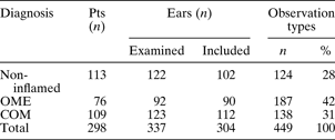
Basic patient data are shown in Table I. Thirty-three ears were excluded due to incomplete data, leaving 304 ears. We recorded 912 observations regarding gas bubbling, fluid film movement, inflammatory condition and perforation size for these 304 ears (an average of three observations per ear). Analysis of these observations indicated that different ears showed different patterns of combined observations (referred to as ‘observation types’, e.g. bubbling with fluid movement in one session and bubbling without it in another session), and that these observation types sometimes varied over time. Thus, some ears demonstrated more than one observation type (see explanation in Statistical Analysis section, above). Overall, we detected 449 different observation types within the whole study group of 304 ears. Except when otherwise specified to enable further analysis, the study group ears were also divided into the following categories: non-inflamed, otitis media with effusion and chronic otitis media (Table I).
Basic patient data
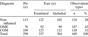
Pts = patients; OME = otitis media with effusion; COM = chronic otitis media
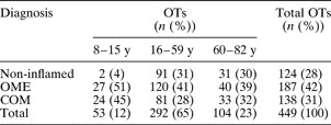
Patients' ages ranged from eight to 82 years (mean 48.6 ± standard deviation 18.4 years; Table II).
Observation types in various ear diagnoses, by patient age

See text for explanation of observation types (OTs). Y = years; OME = otitis media with effusion; COM = chronic otitis media
Of the 449 observation types recorded, 235 (52 per cent) included a dry middle ear, 160 (36 per cent) a wet middle ear and 54 (12 per cent) a purulent middle ear (Table III).

In order to verify that middle-ear gas was not the result of diffusion of room air through the fluid film, we filled the bony (i.e. medial) part of the external ear canal with sterile dexamethasone solution during the intra-tympanic treatments. This created a 5–10 mm fluid layer, reducing the possibility of gas diffusion. Even so, bubbles formed in these ears continuously. This was not due to fluid entering the middle ear, as we had already filled the middle ear at the beginning of the treatment, and we observed that the fluid level in the external canal rose due to expulsion (due to gas production in the middle ear there was increase of the fluid/gas volume and overflow of the fluid level towards the outside) (Figure 1, Table III). Detailed data on gas escape are presented in Tables III, IV, VII. Gas bubbles were observed much more often in non-inflamed ears than in inflamed ones. Acute otitis media occurred mainly in adults without a history of otitis media (i.e. with no previous mucosal pathology) (Figures 2 and 3; Table III – in the pus group, and IV – included in OME group).
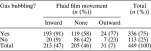
Observation types in various ear diagnoses, by gas bubbling and fluid film movement*

See text for explanation of observation types.
* During swallowing. OME = otitis media with effusion; COM = chronic otitis media
During swallowing, the fluid films of some ears demonstrated various inward–outward movements.
Inward fluid film movement (i.e. movement toward the eustachian tube) was a component of 74 per cent of the observation of normal ear types, but only 32–41 per cent of the observation types recorded for otitis media ears and video-recorded in the first 20 patients. The normal ears' types evacuated in 74 per cent compared to 50 per cent of the otitis media with effusion types (P < 0.001) and to 59 per cent of the chronic otitis media types (p < 0.001), (Table IV). When comparing the impact of the inflammatory condition on the evacuation toward the Eustachian tube, 70 per cent of the non-inflamed ears evacuated in comparison to 24 per cent of the inflamed ones (p < 0.001) and in comparison to 7 per cent of the purulent ones (p < 0.001) (Table III). In ears that were repeatedly observed, we noticed that this inward movement appeared during most but not all observed swallows. Even in the normal ears an inward movement does not happen in every swallowing but in most of them (74 per cent, Table IV). In otitis media it was found to be significantly less. This observed inward fluid film movement indicates that the normal mechanism during swallowing would appear to be clearance from the middle ear towards the eustachian tube, rather than in the reverse direction.
Outward movement of the fluid film during swallowing was a component of only one observation type recorded for normal ears (of 124; 1 per cent), but was a component of 17 observation types (of 187; 9 per cent) recorded for otitis media with effusion ears and 13 observation types (of 138; 9 per cent) recorded for chronic otitis media ears (Tables III to IV). Most of the observation types which included gas bubbling as a component also included inward fluid film movement (Table VII).
We found no difference in ear behaviour or inflammatory condition (i.e. The ears in the various age groups responded alike accordings to their inflammatory condition and their diagnostic groups), comparing various patient age groups.
There was no association between perforation diameter and fluid film movement. The size of the perforation was measured as big (5–2.5 mm), medium – (2.5–1 mm) and small – smaller than 1 mm. However we were aware that this method might bear inaccuracy. Technically it was impossible to create a film when there was a perforation larger than 5 mm.
Discussion
In the middle-ear cavity, there are only two dynamic processes affecting ventilation and drainage: gas supply and mucus evacuation. While mucus drainage via the eustachian tube is understandable, the mechanism of gas supply requires further clarification. The aim of the present study was to investigate the source of middle-ear gas, and its clearance.
According to the current concept, gas is supplied to the middle ear via the eustachian tube. However, it is not clear from the current literature whether such gas is supplied simply by passive air entry from the nasopharynx into the middle ear via the eustachian tube (when the latter opens), or by an active pump action. Current theories on the mechanism of middle-ear ventilation involve three factors: mastoid cell volume (the reservoir theory),Reference Doyle8–Reference Magnuson10 mucosal diseaseReference Kawabata, Nomura and Dohi11–Reference Tanabe, Takahashi, Honjo and Hasebe14 and eustachian tube malfunction.Reference Aoki, Mitani, Tuji, Hamada, Utahashi and Horiyama1–Reference Takahashi, Hayashi, Sato and Honjo3 The composition of middle-ear gasReference Sade and Ar2, Reference Ostfeld, Blonder, Crispin and Szeinberg7, Reference Doyle8 resembles that of venous blood. A few recent publications have shown that CO2 is produced by the middle ear,Reference Tideholm, Carlborg, Jonsson and Bylander-Groth4 supporting the findings of Buckingham and colleaguesReference Buckingham and Ferrer5, Reference Buckingham, Stuart, Girgis, Geik and McGee6 and also MagnusonReference Magnuson10 and Hergils and Magnuson.Reference Hergils and Magnuson15 We searched the literature for studies involving direct measurement of gas supply via the eustachian tube to the middle-ear cavity. We found only speculation based on indirect measurements of eustachian tube functions (e.g. pressure equilibration of artificial high or low pressure applied to the middle ear, or sonotubometry possibly indicating opening or partial opening). However, we found no studies reporting the measurement of air entry into the middle ear via the eustachian tube at normal atmospheric pressure.
To the best of our knowledge, the current paper is the first to observe middle-ear gas production in non-inflamed ears directly via a tympanic membrane perforation, and to compare this with observations in ears with otitis media. We found evidence for gas production in the middle ear, when the patient did not swallow, especially in non-inflamed ears. Such gas production would explain the convexity commonly seen in normal, intact tympanic membranes, especially in infants. Further support for our findings is provided by Luntz and Sade's observation that atelectatic ears become inflated after a night's sleep.Reference Luntz and Sade16 In this study, the observed pressure increment in the ears was explained by congestion and increased venous volume in the mastoid mucosa, which increased the middle ear pressure by 25 mm H2O.Reference Sade and Ar2, Reference Luntz and Sade16 However, we find it difficult to accept that venous congestion alone could cause such an increase in the volume of the middle-ear veins that increases the pressure. This volume increase can be better explained by a rise in CO2 venous pressure occurring during hypoventilation, as observed by Buckingham et al. in a canine ear model: these authors reported that ‘… raising the pCO2 and lowering the pO2 by hypoventilation caused positive pressure’.Reference Buckingham, Stuart, Girgis, Geik and McGee6 The difference in gas bubbling between non-inflamed and inflamed ears may be explained by pathology in the gas-generating mechanism in the latter group. A large mastoid mucosal surface area, as found in ears with well developed mastoids, enables better gas production, and this may explain the connection between well developed mastoids and normal ears. Our gas bubble observations agree with Buckingham and Ferrer's report of continuous gas generation in the middle ear (they reported that ‘… often before even one minute elapsed we found that a pulsatile bubble was forming and enlarging’);Reference Buckingham and Ferrer5 Buckingham et al. also stated in another paper that ‘… the human middle ear is aerated from the blood stream’.Reference Buckingham, Stuart, Girgis, Geik and McGee6
It seems that the eustachian tube may function in two modes: as an active opening with suction towards the nasopharynx, and possibly in a passive mode. In the latter mode, it may be the case that a gas bubble's surface tension is too weak to push the bubble towards the eustachian tube, and therefore the bubble shows no motion. We observed retrograde movement (i.e. regurgitation) from the eustachian tube into the middle ear (causing outward movement of the fluid film) in 9 per cent of the diseased ears, types but in only one normal ear; we believe this difference suggests that such retrograde movement is pathological. Buckingham and Ferrer have reported:
Often in the presence of a bubble… swallowing either had no effect or caused a deflation of the bubble. If deflation is caused by opening of the tube, then the function of the Eustachian tube in these ears is release of air into the nasopharynx rather than the reverse.Reference Buckingham and Ferrer5
It seems that the eustachian tube clears gas together with mucus from normal, non-inflamed ears, but fails to do so from otitis media ears. Failure to clear secretions indicates eustachian tube dysfunction, which may explain some of the pathophysiology of otitis media.
In the human body, peristaltic movements are well known to occur in various hollow organs as a clearance mechanism which also prevents stasis and infection (e.g. in the gall bladder, ureters, intestines and salivary glands). However, retrograde movement does not normally occur in any such organ. Retrograde movement (or regurgitation) towards a ‘cul de sac’ organ (such as the middle ear) may, in our opinion, increase the risk of infection and is theoretically illogical. Our current findings support the hypothesis that the eustachian tube generate subtle, episodic suction to clear mucus and gas from the middle ear.
Our findings, together with the known correlation between well developed mastoids and healthy middle ears, point to the importance of mastoid cellularity, not only as a volume reservoir but also as an active gas-producing organ with a wide mucosal surface.
Our findings may have clinical implications regarding: questioning the rationale of clearing all mastoid cells in mastoidectomies; explaining the low rate of tympanostomy tube infections due to swimming (Goldstein et al.);Reference Goldstein, Mandel, Kurs-Lasky, Rockette and Casselbrant17 and elucidating the occurrence of acute otitis media in bottle-fed infants. The existence of an evacuation function of the eustachian tube would explain the tympanic membrane retraction observed in some cases of cholesteatoma and otitis media with effusion, in which the middle ear fails to produce gas but the eustachian tube continues to evacuate gas.
• According to the current concept, the eustachian tube supplies gas to the middle ear
• This study found evidence to support the theory that the middle ear produces gas, especially under normal conditions
• Middle-ear gas and mucus are evacuated via the eustachian tube
• Evidence for a retrograde (regurgitation) movement via the eustachian tube into the middle ear was found only in diseased ears
• Inflamed middle ears showed impairment of both middle-ear gas production and evacuation mechanisms
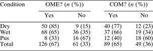
We could not answer the question of whether eustachian tube malfunction causes otitis media or vice versa. We did not find any statistical difference in the ear parameters observed, comparing the otitis media with effusion and chronic otitis media groups (see Tables IV to VII).
Observation types including differing ear inflammatory conditions, for otitis media with effusion and chronic otitis media ears
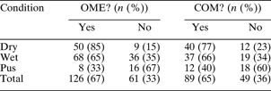
See text for explanation of observation types. OME = otitis media with effusion; COM = chronic otitis media
Observation types including differing ear inflammatory conditions, for ears with otitis media with effusion and chronic otitis media, by fluid film movement*

See text for explanation of observation types.
* During swallowing. FFM = fluid film movement; OME = otitis media with effusion; COM = chronic otitis media
Observation types including and not including gas bubbling, by fluid film movement*
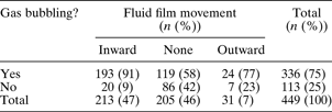
See text for explanation of observation types.
* During swallowing.
Disordered gas production in diseased ears may possibly be explained by the role of biofilms in the diseased middle-ear mucosa.Reference Vlastarakos, Nikolopoulos, Maragoudakis, Tzagaroulakis and Ferekidis18–Reference Jacobs, Dagan, Appelbaum and Burch21 In the inflamed middle ear, biofilms are believed to obstruct the mucosal surface.
In this study, we recorded our clinical observations but could not assess the underlying mechanisms involved.
Our findings may help to determine the timing of myringoplasty surgery in difficult cases; however, further studies are needed to explore their implications for pre-surgical evaluation.
In this study, we repeated Buckingham and colleagues' investigations,Reference Buckingham and Ferrer5, Reference Buckingham, Stuart, Girgis, Geik and McGee6 and supported their findings with a large number of clinical observations. We found clinical evidence for continuous gas generation within the middle ear, and for active evacuation of middle-ear contents by the eustachian tube. These findings suggest the presence of a finely tuned system in which middle-ear gas and mucus production is balanced by evacuation via the eustachian tube. In diseased ears, this system may be disturbed.
Conclusion
In the normal middle ear, gas is produced by the middle-ear mucosa. This gas is evacuated, together with mucus, by the eustachian tube, probably by a peristaltic mechanism during swallowing as well as by ciliary clearance. We found movement of gas from the nasopharynx via the eustachian tube into the middle ear in only 1 per cent of non-inflamed ears but in 9 per cent of otitis media ears. We consider this to represent abnormal regurgitation. In diseased ears, our findings indicated less middle-ear gas production and less eustachian tube evacuation. In this respect, we could find no difference between ears with otitis media with effusion and those with chronic otitis media.