Introduction
Chronic sinonasal diseases are among the most common conditions in otorhinolaryngology that require surgical intervention.Reference Zacharek, Han, Allen, Weissman and Hwang1 Today, endoscopic sinus surgery (ESS) is the most widely used surgical approach for treatment.Reference Luong and Marple2 Therefore, to prevent serious post-operative complications, it is essential to have a thorough understanding of the anatomical variations in this region.Reference Sarı, Yıldırım, Önol, Ozdemır, Arslanoglu and Gurpınar3
Nasal septal deviation is one of the most frequently encountered anatomical abnormalities of the nasal cavity by otorhinolaryngologists. A marked deviation of part or all of the nasal septum may contribute to the development of disease in the paranasal sinuses.Reference Aktas, Kalcioglu, Kutlu, Ozturan and Oncel4 Various studies have demonstrated that a deviated septum can result in osteomeatal complex disease, compensatory turbinate hypertrophy and an enlarged ethmoid bulla.Reference Elahi, Frenkiel and Fageeh5 However, only a limited number of studies have evaluated the relationship between septal deviation and the depth of the skull base.
Paranasal sinus computed tomography (PNS CT) is a widely used imaging modality for evaluating the nasal cavity, paranasal sinuses, nasopharynx and surrounding bony structures.Reference Karatay and Avcı6 The Keros classification is commonly used to assess the depth between the skull base and the ethmoid roof.Reference Sarı, Yıldırım, Önol, Ozdemır, Arslanoglu and Gurpınar3 In the Keros classification, the depth of the olfactory fossa (OF) is categorised into three types: type 1 (1–3 mm), type 2 (4–7 mm) and type 3 (8–16 mm).Reference Sarı, Yıldırım, Önol, Ozdemır, Arslanoglu and Gurpınar3
In this study, we aim to investigate whether a severe unilateral septal deviation, characterised by the mucosal contact point (MCP) with the lateral nasal wall structures such as the turbinates and also additional angle of mucosal contact points (MCPA), affect the depth of the skull base and other related anatomical structures. This will be evaluated through PNS CT imaging by comparing patients diagnosed with septal deviation and those without any septal deviation.
Materials and methods
This retrospective study was conducted in accordance with the guidelines outlined in the Declaration of Helsinki at the Departments of Otorhinolaryngology and Radiology, Faculty of Medicine, Kırıkkale University. PNS CT scans were accessed from the database of the Department of Radiology, Faculty of Medicine, Kırıkkale University. Ethical approval for the study was obtained from the Non-Interventional Research Ethics Committee of Kırıkkale University on 29 June 2022 with reference number 2022/11.
The data for this study were derived from the first author’s medical specialty thesis.Reference Şimşek7
Patients
The study was conducted by forming two groups: 60 patients with septal deviation and mucosal contact point on the right side, and 60 control subjects without septal deviation, all of whom underwent PNS CT at Kırıkkale University Faculty of Medicine Hospital between January 2014 and June 2022. In this study, patients’ PNS CT data were retrospectively reviewed via the Picture Archiving and Communication System (PACS) system and recorded in patient data forms.
All patients were between 18 and 75 years of age. None of the participants had previously undergone surgery involving the paranasal region. Subsequently, the patients were divided into two groups: (1) patients with a mucosal contact point on the right side (N = 60) or (2) patients without septal deviation (N = 60).
The minimum number of individuals required to determine the statistically significant change between the groups was found to be 102 in total, with 51 in each group (α = 0.05, 1-β = 0.80), to conduct statistical analysis using Gpower3.1 version
Radiological measurements
The CT images of the patients included in the study were obtained at the Department of Radiology, Faculty of Medicine, Kırıkkale University, using a 64-slice multidetector computed tomography (MSCT) scanner (Brilliance 64, Philips Medical Systems, Best, The Netherlands). All scans were performed using the following parameters: tube voltage = 120 kV, effective mAs = 350, slice thickness = 1.00 mm, field of view (FOV) = 180 mm and image matrix = 768 × 768.
The evaluation of the OF was conducted in a horizontal plane at the intersection point of a horizontal line with the infraorbital nerves. Standard anatomical landmarks were identified and used in all measurements. These included the medial ethmoid roof (the point where the medial aspect of the ethmoid roof joins the lateral lamella) and the cribriform plate (CP). OF depth was measured as the vertical distance from the medial ethmoid roof to the horizontal plane of the CP (referred to as medial ethmoid roof height). Keros classified OF depth into three types based on the height of the lateral lamella: type 1 (1–3 mm), type 2 (4–7 mm) and type 3 (8–16 mm) (Figure 1).
Measurement of the olfactory fossa (Keros type 3).

In the coronal plane, the height and width of the crista galli (CG) were measured at its most prominent point (Figure 2), using the outer bony contours for reference. Pneumatisation of the CG was categorised as follows: none (0), less than 50 per cent pneumatisation (1), more than 50 per cent pneumatisation (2) and full pneumatisation (3). Evaluation of the CP width in the coronal plane was performed by measuring the distance between the inferior point of the lateral lamellae and the CG (Figure 3).
Crista galli height.

Cribriform plate width.

MCPA was defined as the angle between two lines: one drawn from the midpoint of the CP to the base of the septum, and the other drawn from the base of the septum to the most lateral point of septal deviation contacting the concha (Figure 4). The septal deviation angle was defined as the angle between a line drawn from the midpoint of the CP to the base of the septum and a second line drawn from the same midpoint to the most lateral point of the septal deviation (Figure 5).
Mucosal contact point angle.

Septal deviation angle.

Anatomical variations observed in our study were classified as follows: aplastic frontal sinus, silent maxillary sinus, concha bullosa (Figure 6), Onodi cell, Haller cell, paradoxical middle turbinate, pneumatised pterygoid process (Figure 7) and pneumatised clinoid process.
Left middle concha bullosa.

Pneumatisation of the pterygoid process.

Statistical analysis
The patient data collected in this study were analysed using the IBM Statistical Package for the Social Sciences (SPSS) for macOS version 29.0 (IBM Corp., Armonk, NY). Descriptive statistics were presented as frequencies and percentages for categorical variables, and as mean, standard deviation, median, minimum and maximum for continuous variables. The normality of the distribution of continuous variables was assessed using the Kolmogorov–Smirnov test.
For comparisons between groups, the Independent Samples t-test was used for normally distributed variables in two-group comparisons, while the Mann–Whitney U test was used for non-normally distributed variables. For comparisons involving more than two groups, one-way analysis of variance (ANOVA) was applied for normally distributed variables, and the Kruskal–Wallis H test was used for non-normally distributed variables. The chi-squared test or Fisher’s exact test was applied for the comparison of categorical variables. When significant differences were found among more than two groups, the post-hoc Bonferroni test was used to determine which specific groups contributed to the difference.
The relationships between continuous variables were evaluated using Pearson or Spearman correlation analysis, depending on the distribution. A p-value of less than 0.05 was considered statistically significant.
Results
The study included 60 patients in the control group and 60 patients in the patient group. The control group consisted of 33 females and 27 males, while the patient group comprised 30 females and 30 males. The ages of patients in the control group ranged from 18 to 70 years, with a mean age of 32 plus or minus 10 years. In the patient group, ages ranged from 18 to 69 years, with a mean age of 30 plus or minus 10 years.
Mucosal contact point
In the patient group, 68.3 per cent (n = 41) of the septal deviations were in contact with the inferior turbinate, while 31.7 per cent (n = 19) were in contact with the middle turbinate. The mucosal contact point was located in the cartilaginous septum in 50 per cent (n = 30) and in the bony septum in the remaining 50 per cent (n = 30) of the cases.
Keros classification
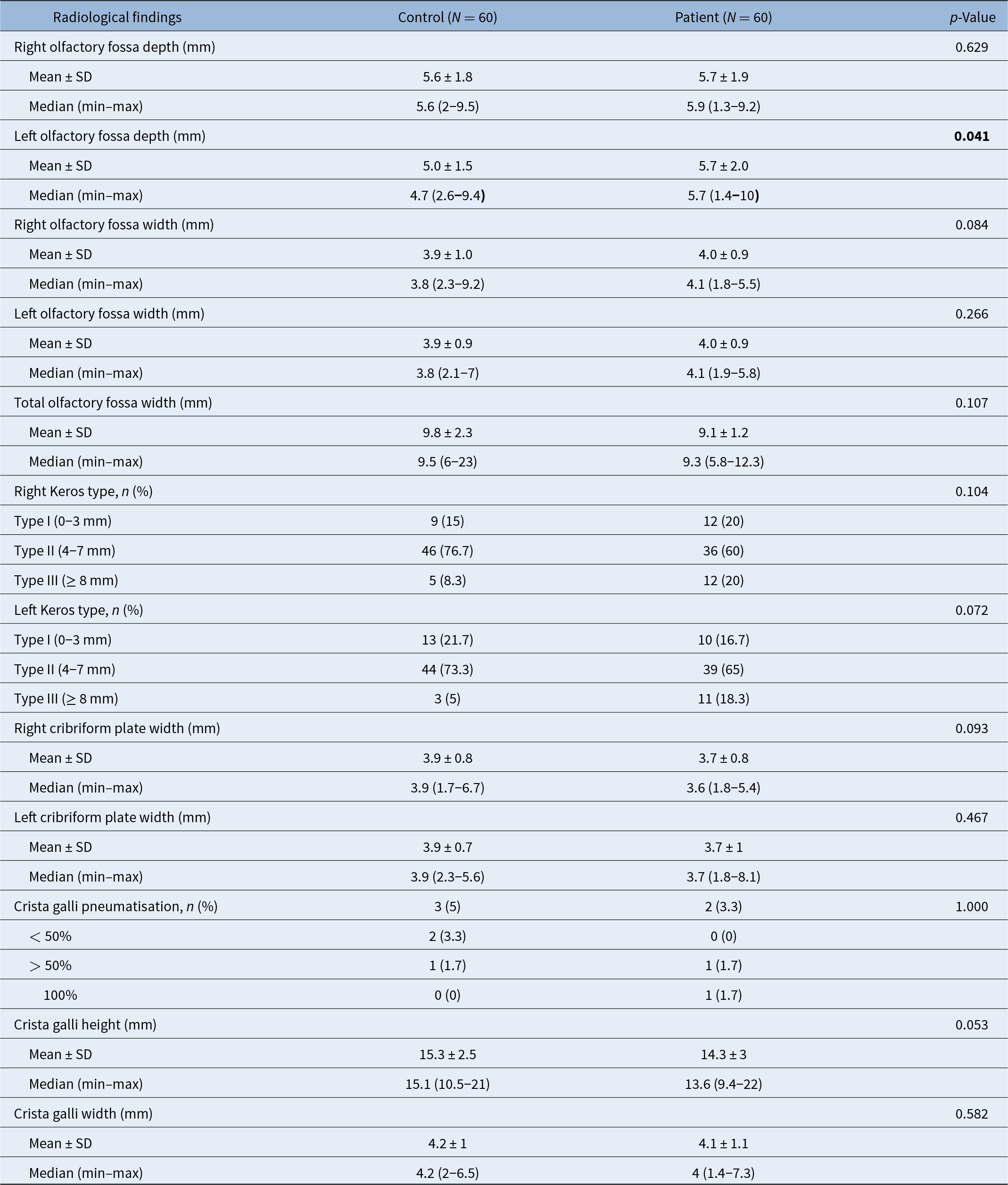
In terms of Keros classification on the right side, types 1, 2 and 3 were observed in 15, 76.7 and 8.3 per cent of participants, respectively. On the left side, type 2 was the most frequent (69.2 per cent), followed by type 1 (19.2 per cent) and type 3 (11.7 per cent). The widths of the CP on the right and left sides were both measured as 3.8 plus or minus 0.8 mm and 3.8 plus or minus 0.9 mm, respectively. Pneumatisation of the CG was observed in 4.2 per cent of cases. The mean CG height and width were 14.8 plus or minus 2.8 mm and 4.1 plus or minus 1.1 mm, respectively (Table 1).
Distribution of radiological findings among participants

* Only assessed in the patient group.
Olfactory fossa depths and widths
The OF depths on the right and left sides were 5.6 plus or minus 1.8 mm and 5.0 plus or minus 1.5 mm, respectively, in the control group, and 5.7 plus or minus 1.9 mm and 5.7 plus or minus 2 mm, respectively, in the patient group (Table 2). The left OF depth was significantly greater in the patient group compared to the control group (p < 0.05).
Distribution of radiological findings in patient and control groups

Bold in the p-value shows that p<0.05, there is a statistically significant difference.
The OF widths on the right and left sides were 3.9 plus or minus 1.0 mm and 3.9 plus or minus 0.9 mm, respectively, in the control group, and 4.0 plus or minus 0.9 mm and 4.0 plus or minus 0.9 mm, respectively, in the patient group (Table 2). No statistically significant differences were found between the patient and control groups in terms of OF width, CP width, CG width or CG height (p > 0.05).
Anatomical variations
When examining the anatomical variations among participants, at least one anatomical variation was observed in 72.5 per cent of cases. The most common variation was concha bullosa, seen in 37.5 per cent of participants. The distribution of anatomical variations is presented in Table 3. When evaluated by group, bilateral pneumatisation of the pterygoid process was observed in 35 per cent of the patient group and 15 per cent of the control group (Table 4).
Distribution of anatomical variation findings among participants

Distribution of anatomical variation findings in patient and control groups

Analysis according to the Keros type
When analysing the distribution of radiological and anatomical findings according to right-sided Keros type, CG width was measured as 3.5 plus or minus 1.0 mm in type 1, 4.2 plus or minus 1.0 mm in type 2 and 4.4 plus or minus 1.0 mm in type 3. An increase in CG width was observed with increasing Keros type, and this difference was statistically significant (p < 0.05) (Table 5).
Distribution of radiological and anatomical variation findings according to right-sided Keros classification

* Post-hoc Bonferroni test.
0–3 mm1 shows Keros type 1
4–7 mm2 shows Keros type 2
8 mm and more3 shows Keros type 3
Bold in the p-value shows that p<0.05, there is a statistically significant difference.
Analysis according to the age groups and gender
When patients were grouped by age (18–25 and ≥26 years), no significant differences were found in any of the measured parameters (p > 0.05). Similarly, no significant differences were observed between male and female patients (p > 0.05).
Location of the deviation and Keros classification
Finally, the location of the deviation (i.e., whether it contacted the inferior or middle turbinate), or whether it was located in the cartilaginous or bony portion of the septum, as well as the septal deviation angle and MCPA, were not found to affect Keros classification(p > 0.05).
Discussion
High-resolution CT is particularly useful in the anatomical assessment of the nasal septum and skull base due to its ability to distinguish between soft tissue and bone through contrast differences. Measurements are taken using bone window settings.Reference Vemuri, Karanam, Manchikanti, Dandamudi, Puvvada and Vemuri8 The Keros classification is an important parameter for estimating skull base depth in ESS.Reference Mafee9 One of the most important complications following trauma involving the anterior skull base is cerebrospinal fluid (CSF) leakage. A connection exists at the junction of the thick orbital rim of the frontal bone and the thin lateral lamella of the CP, extending posteriorly to the frontal recess. This region lies in close proximity to the anterior skull base and is the most common site for CSF leakage due to surgical penetration.Reference Hatipoğlu, Cetin and Yüksel10 Therefore, detailed pre-operative evaluation of these structures is essential before performing ESS.
In a CT study of 120 patients by Skorek et al.,Reference Skorek, Tretiakow, Szmuda and Przewozny11 Keros types 1, 2 and 3 were reported in 9.2, 75.8 and 15 per cent of cases, respectively. They found no significant differences in Keros classification related to patient age or sex. Similarly, studies by Arıkan et al.Reference Arikan, Unal, Kazkayasi and Koc12 and May et al.Reference May and O’Daniel13 identified minor asymmetries between right and left Keros grades, though these were not statistically significant. Erdem et al.Reference Erdem, Erdem, Miman and Ozturan14 also reported no significant differences in OF depth between the right and left sides. In a study by Çufalı et al.,Reference Çufalı, Çetin, Kum and Karakurt15 the mean depth of the right OF was found to be 5.75 plus or minus 2.15 mm and the left OF 5.51 plus or minus 1.99 mm. That same study concluded that deviation in the cartilaginous septum did not significantly affect Keros classification or OF depth; however, bony septal deviations significantly altered both OF depth and Keros type (p < 0.05). In contrast, Bayrak et al.Reference Bayrak, Aktuna Belgin and Orhan16 reported no statistically significant relationship between septal deviation and OF depth. Saylısoy et al.Reference Saylisoy, Acar, San, Karabag, Bayar Muluk and Cingi17 found that OF depth was greater on the contralateral side of the deviation (5.22 ± 1.58 mm) compared to the ipsilateral side (4.9 ± 1.56 mm), and this difference was statistically significant (p < 0.05).
In our study, right-sided Keros types 1, 2 and 3 were found in 15, 76.7 and 8.3 per cent of cases, respectively. On the left, they were observed in 19.2, 69.2 and 11.7 per cent, respectively, with type 2 being the most common and type 3 the least frequent. We did not find any statistically significant differences in OF depth or width based on age or sex in either the control or patient groups (p > 0.05). In the control group, right-sided OF depth was significantly greater than the left (p < 0.05), while in patients with MCP, no significant difference was found between the sides (p > 0.05). Right–left asymmetry of the ethmoid roof/lateral lamella is common; in some cohorts the right side has been reported deeper, which alone may yield a greater right OF depth even without overt septal deviation.Reference Erdogan, Keskin, Topdag, Ozturk, Sari and Mutlu18
Among patients with MCP, the contralateral OF depth was significantly greater than that in patients without septal deviation (p < 0.05). In contrast to the study by Çufalı et al.,Reference Çufalı, Çetin, Kum and Karakurt15 we found that whether the deviation was located in the cartilaginous or bony part of the septum did not significantly affect OF depth (p > 0.05).
Şahan et al.Reference Şahan, Inal, Muluk and Şimşek19 and Asal et al.Reference Asal, Bayar Muluk, Inal, Şahan, Doğan and Arikan20 found a statistically significant increase in OF width on the contralateral side compared to the ipsilateral side in patients with septal deviation (p < 0.05). In our study, we observed similar OF widths on both the ipsilateral and contralateral sides in patients with MCP.
Saylısoy et al.Reference Saylisoy, Acar, San, Karabag, Bayar Muluk and Cingi17 found no significant differences in CP width between ipsilateral and contralateral sides of the deviation. Consistent with this, we also found no significant differences in CP width between sides. However, when we stratified the groups by sex, we found that men without septal deviation had significantly greater bilateral CP width compared to women (p < 0.05).
Komut et al.Reference Komut and Golpinar21 reported mean CG height as 14.22 plus or minus 3.11 mm and width as 3.57 plus or minus 1.48 mm, with a CG pneumatisation rate of 2.3 per cent. In our study, CG height was 14.8 plus or minus 2.8 mm, width was 4.1 plus or minus 1.1 mm and the pneumatisation rate was 4.2 per cent. Although CG height was greater in patients with MCP compared to those without septal deviation, this difference was not statistically significant (p > 0.05). When evaluating the relationship between CG width and Keros types, we found that CG width increased with increasing Keros type—measured as 3.5 plus or minus 1 mm in type 1, 4.2 plus or minus 1 mm in type 2 and 4.4 plus or minus 1 mm in type 3—which was statistically significant (p < 0.05).
In the study by Kim et al.,Reference Kim, Rha, Weissman, Hwang and Most22 the mean septal deviation angle was found to be 13° on the right and 12° on the left in 25 patients. Kubat et al.Reference Orhan Kubat and Ö23 reported a mean septal deviation angle of 11.53 plus or minus 4.72°. Asal et al.Reference Asal, Bayar Muluk, Inal, Şahan, Doğan and Arikan20 found no significant relationship between septal deviation angle and Keros classification (p > 0.05). Similarly, we found no statistically significant relationship between septal deviation angle and Keros classification, anatomical variations, CG height or width or whether the MCP was located in the bony or cartilaginous septum (p > 0.05).
In the present study, we investigated whether a severe unilateral septal deviation, characterised by the MCP with the lateral nasal wall structures such as the turbinates and also additional MCPA, affect the depth of the skull base and other related anatomical structures. A novel contribution of our study to the literature is the definition of the MCPA, measured as the angle between a line drawn from the midpoint of the CP to the base of the septum, and a second line from the septal base to the most lateral contact point of the deviation with the turbinate. In our study, the mean MCPA was 27.9 plus or minus 6°. When the deviation contacted the inferior turbinate, the MCPA was 30 plus or minus 5.4°, and when it contacted the middle turbinate, it was 23.2 plus or minus 4.1°, a statistically significant difference (p < 0.05). This difference is expected given the higher anatomical position of the middle turbinate.
We found that MCPA did not vary significantly based on whether the contact point was in the cartilaginous or bony part of the septum, CG pneumatisation status, anatomical variations or Keros types (p > 0.05).
In a study by Haerian et al.Reference Haerian, Ghaffari and Azari24 of 192 patients with frontal or maxillary sinus hypoplasia/aplasia, those with frontal sinus hypoplasia/aplasia had significantly shorter skull base height (p < 0.05). In our study, bilateral pneumatisation of the pterygoid process was more common in patients with MCP, and this difference was statistically significant (p < 0.05). No other anatomical variations differed significantly between groups (p > 0.05). However, only one patient in our study had frontal sinus aplasia, limiting interpretation of this variation.
The nasal cycle is a physiological, cyclical fluctuation in nasal airway resistance. It has been reported to be present in approximately 80 per cent of healthy individuals. During the cycle, one side of the nose undergoes congestion while the contralateral side undergoes decongestion. On the decongested (i.e., active) side, the cross-sectional area of the nasal cavity increases; conversely, on the congested (i.e., passive) side, the cross-sectional area decreases.Reference Eccles25
In patients with septal deviation, this cycle would be expected to function less evenly. When the cavity contralateral to the deviated side enters the passive phase, bilateral obstruction may occur. Accordingly, the non-deviated, more patent side should still exhibit alternating increases and decreases in airflow, but with lower amplitude than that observed in healthy controls. Thus, the nasal cycle in deviated septum is presumed to operate differently from normal.Reference Letzel, Darbinjan and Hummel26
In their study, Letzel et al.Reference Letzel, Darbinjan and Hummel26 reported that the nasal cycle became more balanced after septoplasty. This finding suggests that patients with septal deviation have an autonomic neural innervation that is organised differently from that of healthy subjects. Before surgery, because the narrow side contributes little to the cycle, the autonomic nervous system may compensate by increasing the frequency of nasal cycle phases on the non-deviated side. Following septoplasty, as both sides can again undergo congestion and decongestion of the erectile tissue, this compensation is no longer required, leading to a reduction in phase frequency on the previously more open side and a more even bilateral distribution of cycle phases.Reference Letzel, Darbinjan and Hummel26
A thorough understanding of the anatomy and endoscopic technique is extremely important, particularly regarding adjacent structures such as the CP and other components of the anterior skull base.Reference Socher, Santos, Correa and da Silva27 In such cases, the use of 30°–45° angled endoscopes and shorter, fine-tipped instruments may improve visualisation and reduce the upward force applied to the skull base.
The presence of significant septal deviation may increase the difficulty of surgery by reducing the surgeon’s manoeuvreability within the nasal cavity, narrowing the FOV and hindering the identification of anatomical landmarks. When the OF depth is asymmetrical, it may be safer to limit superior dissection near the skull base until the anatomic boundaries are confirmed by intra-operative navigation.
In the study published by O’BrienReference WT, Hamelin and Weitzel28 and colleagues, the authors emphasised common anatomic variations of the paranasal sinuses that may place patients at risk for surgical complications and identified critical regions summarised by the mnemonic “CLOSE”: Cribriform plate, Lamina papyracea, Onodi cell, sphenoid sinus pneumatisation and (anterior) ethmoidal artery. This mnemonic-based approach provides a practical framework for recalling and documenting key anatomic variants that should be carefully reviewed in pre-operative imaging reports. These findings highlight that individualised surgical planning, guided by detailed pre-operative CT assessment, can optimise access, enhance visualisation and reduce the risk of intracranial penetration or iatrogenic injury.
Although numerous studies have examined the anatomy and features of the paranasal sinuses, our study not only assessed these structures but also compared patients with MCP and those without septal deviation, both within and between groups. We believe our findings contribute to the literature by providing anatomical insights for surgeons, particularly in settings where pre-operative CT imaging is unavailable or intra-operative recall of these structures is necessary.
• Endoscopic sinus surgery (ESS) is the most widely used surgical approach for treatment
• In patients with mucosal contact points (MCPs), olfactory fossa (OF) depth on the contralateral side of the deviation was greater compared to patients without septal deviation
• An increase in crista galli height was associated with a higher Keros classification
• Increased OF depth and greater crista galli height, the risk of intracranial penetration increased
• The presence and differences of MCP should alert surgeons both before and during the surgical procedure
Conclusion
In patients with mucosal contact points, the OF depths on the right and left sides were found to be similar. However, the OF depth on the contralateral side of the deviation was greater compared to patients without septal deviation. Additionally, an increase in CG height was associated with a higher Keros classification, particularly on the right side. In patients with increased OF depth and greater CG height, the risk of intracranial penetration increased, underscoring the need for heightened caution during ESS procedures.
Availability of data and materials
All data for this study is are presented in this paper.
Competing interests
There are no financial disclosures of the authors. The authors declare that they have no conflicts of interest. There are no funds for this article.
Ethics committee approval
This study is retrospective. Ethics committee approval was obtained from Kırıkkale University Non-invasive Research Ethics Committee (Date: 29.06.2022, Number: 2022/11). There is no need to take informed consent, because the data were evaluated retrospectively.
Author contributions
Bugra SIMSEK: Planning, designing, data collection, literature survey, interpretation of the results, active intellectual support, writing. Nuray BAYAR MULUK: Planning, designing, literature survey, interpretation of the results, active intellectual support, writing, submission. Yunus Emre TOKYUREK: Planning, designing, data collection, literature survey, active intellectual support. Mikail INAL: Planning, designing, data collection, literature survey, active intellectual support. Burak Mustafa TAS: Planning, designing, literature survey, active intellectual support. Ziya SENCAN: Planning, designing, literature survey, active intellectual support. Ela COMERT: Planning, designing, literature survey, active intellectual support.
Acknowledgements
None.
Consent for publication
All authors give consent for publication.