Intrauterine growth retardation/restriction (IUGR) is a fetal growth disorder that may have a negative impact on some birth somatic parameters such as birth weight, birth length, and head and abdominal circumference. Its prevalence is estimated at ~3–15% of all newborns in developed countries and reaches 20–30% in developing countries.Reference Ego, Subtil and Grange 1 – Reference Black, Victora and Walker 3 Usually, intrauterine growth restriction leads to a birth weight that is below the 10th percentile for gestational age, classified by the American Academy of Pediatrics as small-for-gestational age. 4 Nevertheless, 1–1.5% of the small-for-gestational age newborns is constitutionally small and do not experience intrauterine growth restriction.Reference Ananth and Vintzileos 5 – Reference Mamelle, Boniol and Riviere 7 At the same time, some of the fetuses affected by transient intrauterine growth restriction – as observed in subsequent fetal ultrasound examinations – are born as appropriate-for-gestational age.
Intrauterine growth restriction is an important issue, not only during pregnancy and the perinatal period but also in the following years of life. According to metabolic programming theory, a fetus affected by intrauterine growth restriction, in order to protect crucial organs, adapts to an insufficient amount of oxygen, glucose, and amino acids through changes in metabolic rate, hormone concentration, tissue sensitivity to those hormones, as well as by blood re-distribution. As a result, metabolic, structural, and functional changes take place, which subsequently can lead to the frequent occurrence of cardiovascular diseases and metabolic disorders, including insulin resistance, diabetes mellitus, obesity, and the metabolic syndrome.Reference Barker and Martyn 8 , Reference Barker 9
Fetal stress resulting from homoeostasis disturbance in utero, as well as altered function of the hypothalamic–pituitary–adrenal axis, can lead to impaired development of the autonomic nervous system.Reference Phillips 10 It is proved that autonomic disorders may contribute to ischaemic heart disease,Reference Voss, Schroeder and Vallverdu 11 insulin resistance,Reference Flanagan, Vaile and Petley 12 diabetes mellitus,Reference Salvi, Hingorani, Ramasamy and Kothari 13 obesity,Reference Vanderlei, Pastre, Freitas Junior and Godoy 14 and hypertension.Reference Ojeda, Johnson, Dwyer and Alexander 15 There is much evidence that increased renal sympathetic stimulation is one of the risk factors for hypertension.Reference Mizuno, Siddique, Baum and Smith 16 In order to answer the question whether in small-for-gestational age patients there are autonomic abnormalities, which may lead to more frequent causes of cardiovascular diseases, different methods of autonomic nervous system assessment are used.Reference IJzerman, Stehouwer, de Geus, van Weissenbruch, Delemarre-van de Waal and Boomsma 17 – Reference Aziz, Schlindwein, Wailoo, Biala and Rocha 19
Heart rate variability is a recognised, non-invasive, quantitative method that assesses the activity of the autonomic nervous system and sympathetic–parasympathetic balance based on the constant electrocardiographic evaluation. Assessment of heart rate variability consists of time and frequency domain analysis. Parameters of the time domain analysis are obtained from the calculation of mean normal-to-normal (N-N) intervals during the entire recording and statistical measures of the variance between N-N intervals. 20 In the literature, there are only a few studies assessing autonomic function using time domain heart rate variability in the paediatric population and young adults born small-for-gestational age, but the results are inconsistent. Some of them confirm lower heart rate variability measurements and/or relative sympathetic advantage among small-for-gestational age childrenReference Aziz, Schlindwein, Wailoo, Biala and Rocha 19 , Reference Rakow, Katz-Salamon, Ericson, Edner and Vanpée 21 and infants,Reference Galland, Taylor, Bolton and Sayers 22 whereas other studies conducted on newborns do not conclude on statistically significant differences in heart rate variability analysis between small- and appropriate-for-gestational age neonates.Reference Schäffer, Burkhardt and Müller-Vizentini 23 Moreover, in the group of young adults with low birth weight, higher values of time domain parameters are observed.Reference Weitz, Bonnemeier, Süfke, Wellhöner, Lehnert and Dodt 24
The aims of this study were to compare time domain parameters of heart rate variability in children born as small-for-gestational age and appropriate-for-gestational age and to assess the correlation of the postnatal and current somatic parameters with the time domain parameters.
Materials and methods
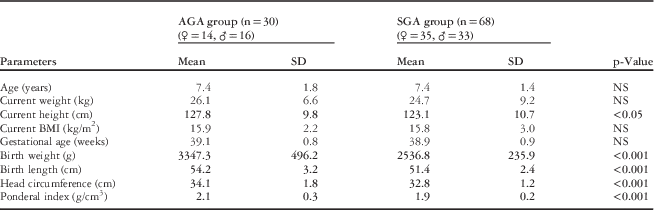
This prospective study was carried out between 2010 and 2013 in the Department of Children’s Cardiology and Rheumatology of the 2nd Chair of Pediatrics at the Medical University of Lodz in Poland. Perinatal data on children classified as small-for-gestational age were obtained from the neonatal ward. This group of patients was invited to participate in the prospective study including cardiovascular tests. In total, 68 children aged 5–10 years were included in the small-for-gestational age group. All of them were born at term, and their birth weights were below the 10th percentile for sex and gestational age.Reference Malinowski and Chlebna- Sokół 25 In the control group, 30 term-born neonates who were appropriate-for-gestational age were included. They were sex- and age-matched with the study group. Children in the control group were mainly healthy volunteers. A few patients were enrolled into the study during their short hospitalisation period as they had various symptoms included in the interview – for example, musculosceletal pain. After all medical tests, patients were found to be healthy, and they did not suffer from any ailments at the time of the study. In order to provide similar study environment, all the patients remained in the hospital for 24 hours. Informed consent was obtained from all parents, and the study was approved by the ethics committee. Perinatal history including delivery, gestational age, birth weight, birth length, head circumference, Ponderal Index, and Apgar scores was obtained from all patients. Subsequently, physical examination with measurements of weight, height, and body mass index was conducted (Table 1). Blood pressure measurement, electrocardiographic and echocardiographic studies, and 24-hour echocardiographic recording were also performed. Patients with any diseases that could affect heart rate variability, such as infections, CHDs, arrhythmias, vasovagal syncope, or any chronic diseases, were excluded from the study. None of patients felt any pain, such as headache, musculoskeletal pain, abdominal pain etc., during echocardiographic monitoring.
Characteristics of the group.

AGA=appropriate-for-gestational age; BMI=body mass index; SGA=small-for-gestational age; ♀=girls; ♂=boys
Ponderal index [PI=birth weight (g)×100/birth length3 (cm3)]
BMI=weight (kg)/height2 (m2)
The time domain analysis of heart rate variability was based on 24-hour echocardiographic recording. All the patients were advised to stick to their normal daily routine during recording. They did not have excessive physical and mental activity. No medical procedures were carried out at this time. Most of the patients stayed on the ward with parents or other relatives, who helped in maintaining a diary of all activities including sleep and wake timings.
The 24-hour, 12-lead echocardiographic recordings were obtained using Oxford DMS 300–12 recorder with a sample rate of 1000/128 Hz/channel and analysed using Oxford Cardioscan 12 software. All the recordings were manually verified and examined for artefacts, which did not constitute >10% of the recordings. Next, the Holter system checked for different types of beats – supraventricular or ventricular beats accounted for <1% of the recording – further analysing only normal R-R intervals. Prepared data were used for the time domain heart rate variability analysis. According to the standards of the Task Force of the European Society of Cardiology and the North American Society of Pacing and Electrophysiology, 20 the following time domain parameters were calculated: SDNN, standard deviation of all N-N intervals, SDNN index, the mean of the standard deviations of all N-N intervals for all 5-minute segments of the entire recording, and SDANN index, the mean of the standard deviations of the averages of N-N intervals in all 5-minute segments of the entire recording. The parameters reflect total activity of the autonomic nervous system, both sympathetic and parasympathetic components. Moreover, the parameters rMSSD, the square root of the mean of the sum of the squares of differences between adjacent N-N intervals, and pNN50, number of pairs of adjacent N-N intervals differing by >50 ms in the entire recording divided by the total number of all N-N intervals, were assessed, as they describe the difference between the N-N intervals and are considered as a good reflection of the parasympathetic activity.Reference Dąbrowska, Dąbrowski and Piotrowicz 26 The 24-hour average heart rate was also calculated.
Statistical analysis
Descriptive statistical analyses were carried out by computing the means and standard deviations for scale variables or frequencies for nominal variables. The significance level was computed for the differences between variables in both study and control groups. To evaluate the differences between the two groups, a parametric Student’s t-test and a non-parametric Mann–Whitney test were performed. The normal distribution of scale variables was verified using the Shapiro–Wilk test. All tests were two sided and performed at 0.05 significance level. Pearson’s or Spearman’s correlation coefficients were computed to evaluate the degree of association between selected variables either for the appropriate-for-gestational age or the small-for-gestational age group.
Results
Statistically significant differences between the small-for-gestational age and appropriate-for-gestational age groups were found in the distribution of birth weight, birth length, head circumference, and Ponderal Index. There was no statistically significant difference in early postnatal outcome according to Apgar scores. None of the children included in the study were born with low Apgar scores (0–3); nine patients from the small-for-gestational age group and one child from the appropriate-for-gestational age group received intermediate Apgar scores, 4 – Reference Mamelle, Boniol and Riviere 7 p=NS. The remaining children were born with normal Apgar scores.Reference Barker and Martyn 8 – Reference Phillips 10 At the time of the study, children from the small-for-gestational age group were characterised by statistically significant lower height than peers from the appropriate-for-gestational age group. Other anthropometric parameters such as current weight and current body mass index did not differ significantly between groups (Table 1).
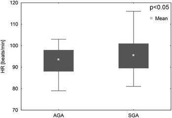
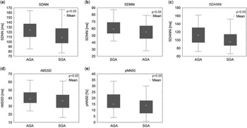
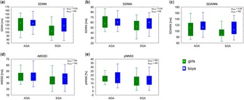
On the basis of the 24-hour echocardiographic monitoring, higher average heart rate was observed in the small-for-gestational age group than in the appropriate-for-gestational age group (Fig 1). In the heart rate variability analysis, it was found that all time domain parameters were lower in the small-for-gestational age group than in the appropriate-for-gestational age group (Fig 2a–e). Moreover, a sex-specific analysis of the time domain parameters conducted in both groups showed that among small-for-gestational age patients, girls had lower values of SDNN, SDNNi, and SDANNi than boys. At the same time, in the small-for-gestational age group, parameters such as RMSSD and pNN50 did not differ significantly between girls and boys. In the appropriate-for-gestational age group, none of the time domain parameters differed significantly according to sex (Fig 3a–e).
The average heart rate (HR) in the 24-hour echocardiograph recording. AGA=appropriate-for-gestational age; SGA=small-for-gestational age.

Time domain heart rate variability parameters ( a ) SDNN – standard deviation of all N-N intervals ( b ) SDNN index – the mean of the standard deviations of all N-N intervals for all 5-minute segments of the entire recording ( c ) SDANN index – the mean of the standard deviations of the averages of N-N intervals in all 5-minute segments of the entire recording ( d ) rMSSD – the square root of the mean of the sum of the squares of differences between adjacent intervals ( e ) pNN50 – number of pairs of adjacent N-N intervals differing by more than 50 ms in the entire recording divided by the total number of all N-N intervals. AGA=appropriate-for-gestational age; SGA=small-for-gestational age.

Time domain heart rate variability parameters in small-for-gestational age and appropriate-for-gestational age group according to sex ( a ) SDNN – standard deviation of all N-N intervals ( b ) SDNNi – the mean of the standard deviations of all N-N intervals for all 5-minute segments of the entire recording ( c ) SDANNi – the mean of the standard deviations of the averages of N-N intervals in all 5-minute segments of the entire recording ( d ) rMSSD – the square root of the mean of the sum of the squares of differences between adjacent intervals ( e ) pNN50 – number of pairs of adjacent N-N intervals differing by >50 ms in the entire recording divided by the total number of all N-N intervals. AGA=appropriate-for-gestational age; SGA=small-for-gestational age.

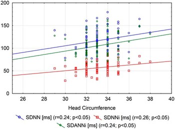
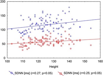
In both groups, a positive correlation between SDNN, SDNNi, SDANNi, and head circumference was observed (Fig 4). None of the time domain parameters correlated significantly with other perinatal parameters such as birth weight, length, and Ponderal Index (data not shown). Among the current measurements, only height correlated positively with SDNN and SDNNi (Fig 5). There was no significant correlation between time domain parameters and current weight or body mass index (data not shown).
Correlation between SDNN, SDNNi, SDANNi, and head circumference.

Correlation between SDNN, SDNNi, and current height.

Discussion
Our study confirmed that Holter echocardiographic monitoring with heart rate variability analysis could be a useful method to assess malfunction of the autonomic nervous system in small-for-gestational age children. The comparison of basic echocardiographic parameters revealed higher average heart rate in the small-for-gestational age group; however, a statistically significant difference in heart rate of 3 beats/minute is not clinically significant and can prove slight sympathetic dominance in this group of patients. Our findings are consistent with many other publications that relate to resting heart rate. In a large Japanese study that included 12- to 13-year-old boys, the authors observed higher pulse rate in children born lighter, shorter, and with smaller head circumference.Reference Abe, Minami, Ohrui, Ishimitsu and Matsuoka 27 Likewise, Johansson et alReference Johansson, Norman, Legnevall, Dalmaz, Lagercrantz and Vanpée 28 found that 9- to 10-year-old children born as small-for-gestational age had higher heart rate than their appropriate-for-gestational age peers. Higher heart rate, which suggests sympathetic dominance, is a well-known risk factor for cardiovascular disease including hypertension.Reference Inoue, Iseki, Iseki, Kinjo, Ohya and Takishita 29 Time domain analysis of heart rate variability allows for a precise assessment of the autonomous system balance.
Results of our study indicate that all the time domain parameters are lower in small-for-gestational age patients. This is true for SDNN, SDNNi, and SDANNi, which reflect both sympathetic and parasympathetic components, and for rMSSD and pNN50, which represent mainly parasympathetic influence. Thus, decreased heart rate variability with sympathetic predominance could be an explanation for increased risk of cardiovascular disease in adults born as small-for-gestational age. In the literature, there are only a few studies using the heart rate variability method for autonomic nervous system assessment in small-for-gestational age paediatric populations. Results of this study are mostly consistent with a Swedish study,Reference Rakow, Katz-Salamon, Ericson, Edner and Vanpée 21 which was conducted among 86 children aged ~9 years, of whom 31 were born premature, 27 were born at term as small-for-gestational age (birth weight below 2 SD), and the remaining children were born as appropriate-for-gestational age. Only SDNNi and the mean R-R interval were measured. Both parameters appeared to be lower in the small-for-gestational age and premature group than in the appropriate-for-gestational age group. This finding indicates that low birth weight, which is a result of either prematurity or intrauterine growth retardation, is a predictive factor for incorrect autonomic response to external stimuli. This might be a consequence of the intrauterine disorders that persist in subsequent years of life.
Our observations could be compared with the results obtained by Aziz et al. The authors examined 37 children aged 9–10 years, born at term, with intrauterine growth retardation, based on ultrasonographic measurements during pregnancy and/or percentiles after birth.Reference Aziz, Schlindwein, Wailoo, Biala and Rocha 19 In this study, no statistically significant differences between the study and control groups were found; however, after selecting 20 children with birth weight below 2.5 kg (so-called IUGR-1), significantly lower heart rate variability parameters were observed in comparison with both healthy peers and intrauterine growth-restricted children with birth weight above 2.5 kg (so-called IUGR-2). Among time domain parameters, lower values were noted for SDNN, SDANN, RMSSD, and pNN50, similar to the present study. This allows researchers to conclude that children born with low birth weight (below 2.5 kg) have impaired autonomic system function. It is worth mentioning that in the cited publications, as in this study, heart rate variability analysis was performed on the basis of 24-hour echocardiographic recording during normal activity documented in the patient’s diary.
In the literature, there are a number of studies evaluating heart rate variability in newborns and neonates. Galland et al investigated neonates born at term with birth weight below the 10th percentile in the first and then in the 3rd month of life. Evaluation of the autonomic nervous system was conducted using heart rate variability methods in short echocardiographic fragments. Moreover, the authors assessed cardiac reflexes in response to change in body position. Echocardiographic recording was performed during active and quiet sleep in the prone and supine positions. The findings indicate reduced autonomic activity and cardiac reflexes with higher sympathetic component in small-for-gestational age infants. This could suggest the immaturity in autonomic control of the cardiovascular system in small-for-gestational age patients compared with appropriate-for-gestational age individuals.Reference Galland, Taylor, Bolton and Sayers 22
Most of the studies assessing heart rate variability in the paediatric population have obtained results similar to our study. These publications confirm the hypothesis about intrauterine disturbance in autonomic nervous system development, probably due to intrauterine growth restriction and insufficiency of nutrition and oxygen supply. Decreased time domain parameters of heart rate variability in the small-for-gestational age group can confirm autonomic dysfunction and impaired adaptation to environmental factors. Moreover, higher heart rate suggests prevalence of sympathetic nervous system in this group of patients.
The correlation of heart rate variability with gender remains a matter of interest to many scientists; however, existing findings covering the paediatric population are limited and are inconsistent. In a large cohort study including healthy patients aged 10–99 years, time domain parameters were lower in female patients under 30 years of age. The observed difference decreased in the group aged 30–50 years and disappeared in the group including adults aged above 50 years.Reference Umetani, Singer, McCraty and Atkinson 30 It is important to indicate that in the presented study there was no difference depending on gender in the appropriate-for-gestational age group. At the same time, in the small-for-gestational age group, most of the time domain parameters (SDNN, SDNNi, SDANNi) were significantly lower among girls. Results obtained by Aziz et al were opposite. The authors did not reveal any significant difference in heart rate variability analysis between boys and girls born with intrauterine growth restriction. In the control group, only SDNN was lower in females.Reference Aziz, Schlindwein, Wailoo, Biala and Rocha 19 Sex-dependent differences are difficult to explain and remain in the area of hypothesis. The differences found in our study may have been due to smaller numbers in the appropriate-for-gestational age group.
In our study, time domain parameters were correlated with available perinatal parameters such as birth weight, length, head circumference, and the Ponderal Index, irrespective of the group. The significant positive correlation was observed only between head circumference and SDNN, SDNNi, and SDANNi. It seems interesting that there was no significant correlation between heart rate variability parameters and birth weight, length, or Ponderal Index. According to the authors, this is the first study with such a detailed analysis of correlations between heart rate variability measurements and perinatal parameters defining intrauterine growth restriction. Only a few researchers have evaluated the relationship between postnatal parameters and resting heart rate.Reference Abe, Minami, Ohrui, Ishimitsu and Matsuoka 27 , Reference Phillips and Barker 31
The low final height of small-for-gestational age children was not correlated with the shift in autonomic balance but with the global decline in autonomic nervous system activity, as indicated by interdependence of actual height with SDNN and SDNNi, which was found in our study; however, owing to the lack of access to medical documentation from the infantile and early childhood period, it was impossible to investigate the rate of growth. Probably, such data would help associate heart rate variability parameters with occurrence of “a catch-up growth”. It is known from the literature that low final height concerns 10–15% of small-for-gestational age patients, especially the ones without catch-up growth.Reference Clayton, Cianfarani, Czernichow, Johannsson, Rapaport and Rogol 32 On the other hand, not adjusting for height in the statistical analysis could be a confounder because the size of the child itself can potentially affect heart rate variability, especially parasympathetic components (rMSSD, pNN50). These did not correlate with height in our study. Current weight and body mass index did not correlate with any of the heart rate variability measurements. In the available literature, correlations of the somatic parameters with heart rate variability measurements were not investigated.
The authors are aware of the limitations of this study. A major limitation is the lack of antenatal history, especially the results of fetal ultrasonographic examinations, which would have enabled us to more clearly define the occurrence of intrauterine growth disorders in both groups of patients. There was insufficient information about the pregnancy period, including preeclampsia or infection, and about postnatal health conditions of newborns. Information was obtained only from the interview with the mother and it was incomplete. It seems that evaluation of catch-up growth as well in the study population would be an important complement to this study. Another limitation is the relatively small group of patients, especially the control group; however, this was a single-centre study, which could constitute the beginning of a wider research.
Conclusions
Children born as small-for-gestational age have impaired function of the autonomic nervous system. All time domain parameters are lower in children born small-for-gestational age in comparison with appropriate-for-gestational age peers. Moreover, in small-for-gestational age children, autonomic balance is shifted towards the sympathetic component, which is evidenced by higher heart rate. The 24-hour echocardiographic recording with time domain heart rate variability analysis is a useful method to assess malfunction of the autonomic nervous system. Children with faster heart rates and lower heart rate variability parameters may be at risk of cardiovascular disease. They should be covered by particular prevention, health education, and regular cardiologic control.
Acknowledgements
None.
Financial Support
The study was supported by the Medical University of Lodz grant number 501-03/8-000-01/501-64-004.
Conflicts of Interest
None.
Ethical Standards
The authors assert that all procedures contributing to this work comply with the ethical standards of the relevant national guidelines and have been approved by the research ethics committee Medical University in Lodz.