Congenital long QT syndrome refers to a genetical and clinical paradigm, mainly defined by the prolongation of corrected QT interval on surface electrocardiogram, recurrent syncope, and even a propensity to sudden cardiac death.Reference Schwartz, Crotti and Insolia 1 Since the first genetic discovery in the early 1990s, 15 different long QT syndrome-causing genes have been reported.Reference Mizusawa, Horie and Wilde 2 Among them, the “loss-of-function” mutations in the KCNQ1-encoded Kv7.1 α subunit accounts for ~70% of all long QT syndromes.Reference Abriel and Zaklyazminskaya 3 Extensive genotype–phenotype studies have shown that the severity of long QT syndrome could be influenced by the genetic substrate. In addition, mutation location and type in the causative KCNQ1 gene are associated with the variability of clinical symptoms and the different response to β-blocker therapy. For type 1 long QT syndrome patients, KCNQ1 mutations in the transmembrane domain of the Kv7.1 channel are much more malignant than those in other domains,Reference Moss, Shimizu and Wilde 4 whereas those with KCNQ1 mutations in the C-loop domain also exhibit a high risk for life-threatening events and derive a pronounced benefit from β-blocker therapy.Reference Barsheshet, Goldenberg and O-Uchi 5 Nevertheless, it is still unknown whether the Chinese long QT syndrome patients have similar characteristics of clinical manifestations and susceptibility to β-blockers.
Overall, genetic testing provides important implications regarding risk assessment and management of long QT syndrome patients. To date, five causative genes – KCNQ1, KCNH2, SCN5A, KCNE1, and KCNJ5 – have been reported in Chinese long QT syndrome patients; however, there are limited data about genetic-based clinical manifestation and β-blocker treatment in Chinese type 1 long QT syndrome patients. We performed the candidate gene testing to identify long QT syndrome-causing mutations, characterised the severity of individual mutations, and we have discussed the clinical response to β-blockers in Chinese long QT syndrome patients.
Materials and methods
Clinical investigation
The proband and her first-degree relatives were recruited from the local hospital. All the patients underwent clinical evaluation, including medical and family history, physical examination, 12-lead electrocardiogram, and echocardiography examinations. The clinical diagnosis of congenital long QT syndrome can be made when the patient has a corrected QT interval >480 ms in the absence of a secondary cause for QT prolongation.Reference Schwartz and Crotti 6
Genetic screening
Genomic DNA was extracted from the peripheral blood lymphocytes. Primers of the candidate genes – KCNQ1, KCNH2, and SCN5A – were designed using Primer 5.0 Software. All exons and exon–intron boundaries of the candidate genes were amplified by polymerase chain reaction. The amplicons were purified with SAP-EXON I purification method, directly sequenced in both strands using a Bigdye Terminator Mix® (Applied Biosystems, Carlsbad, California, United States of America), and then analysed by cycle sequencing using an ABI PRISM 3130XL Automated DNA Sequencer (Applied Biosystems). In addition, 400 healthy control individuals from the same ethnic background were sequenced by the same method.
Site-directed mutagenesis and transfection
The primers for site-directed mutagenesis were designed from the published sequence of the complementary cDNA fragments of human KCNQ1 (GenBank number NM_000218.2). Wild-type KCNQ1 cDNAs were amplified by polymerase chain reaction and cloned into pcDNA3.1+ vector (Invitrogen, Carlsbad, California, United States of America). Site-directed mutagenesis was performed to introduce the variant nucleotide using the GeneTailor Site-Directed Mutagenesis System (Invitrogen Corp., California, United States of America). The clones were sequenced completely on both strands to ensure the presence of the mutation and the absence of other substitutions introduced by the polymerase chain reaction. The wild type or mutant KCNQ1 constructs were transfected into COS-7 cells using Lipofectamine and Plus reagent (Invitrogen) following the manufacturer’s instructions. In order to achieve equal transfection efficiency and avoid biases from transient transfection, the tool cells were derived from a stable COS-7 cell line separately.
Electrophysiological analysis
The wild type or mutant KCNQ1 constructs were heterologously expressed in COS-7 cells. Membrane currents were measured by whole-cell voltage-clamp recordings using EPC 10 amplifier (HEKA Instruments Inc., Bellmore, New York, United States of America). Recordings were made in symmetrical Ringer solution at room temperature.
The internal pipette solution for the Kv7.1 channel consisted of (in mM) K-aspartate 100, KCl 45, EGTA 5, HEPES 5, CaCl2 1, ATP-Mg 5, and phosphocreatine-disodium 5 (pH 7.3 with KOH). The extracellular solution contained (in mM) NaCl 145, KCl 5.4, HEPES 5, MgCl2 0.5, NaH2PO4 0.33, CaCl2 1.8, and Glucose 5.5 (pH 7.3 with NaOH). Voltage-dependent potassium currents were recorded at various membrane potentials from −80 to +80 mV in 10 mV increments for 2 seconds, holding at a potential of −80 mV. Steady-state activation was estimated by measuring the peak potassium currents. Conductance G(v) was calculated using the following equation: G(v)=I/(Vm−Erev) – I: peak currents, Vm: membrane potentials, Erev: the measured reversal potentials. Steady-state inactivation was estimated using a pre-pulse protocol. Data for steady-state activation and steady-state inactivation were fitted with the Boltzmann equation.
Data acquisition and statistical analysis
All signals were acquired at 20 kHz with PatchMaster software (HEKA Instruments Inc.). The data were analysed using Fitmaster, SigmaPlot 10.0, and Excel data handling. Results are presented as mean±standard error of the mean. The mean values and SEM were compared by the t-test, and values of p<0.05 were considered to be statistically significant.
Results
Clinical findings
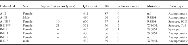
We characterised two unrelated long QT syndrome families and screened three candidate genes including KCNQ1, KCNH2, and SCN5A. The results showed that two disease-causing mutations, R380S (family A) and W305L (family B), were present in the KCNQ1 gene in both the long QT syndrome families. No mutations were found in the KCNH2 and SCN5A genes. All clinical findings are presented in Table 1.
Clinical and genetic characteristics of two unrelated Chinese LQT1 families.

HR=heart rate in beats per minute; n.f=not found; QTc=corrected QT interval; SCD=sudden cardiac death; TdP=torsade de pointes
* The proband with mutation R380S(c.1248G>T) who finally suffered SCD without medication
** The proband with mutation W305L(c.1022G>T) remained asymptomatic (no syncope episodes) during the 3-year follow-up visit after administration of β-blockers (metoprolol; 50 mg, Tid)
Family A
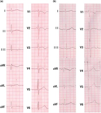
Family A comprised of four members – two males and two females – of three generations (I1, I2, II1, and III1 in Fig 1a). The proband (III1) defined by an Schwartz risk score of 3 points was a 11-year-old girl with a history of recurrent syncope attacks since the age of 10 years. The syncope often elicited during sports or emotional stress and was spontaneously terminated, and she finally suffered sudden cardiac death at home without any medication. She suffered from a pathological corrected QT interval of 660 ms and broad T wave with a heart rate of 77 beats/minute during exercise (Fig 3a). Compared with the normal resting heart rate, the peak heart rate during exercise was much lower; however, the proband’s father (II1) with the same mutation R380S was asymptomatic and had normal corrected QT interval even during exercise. None of the patients had structural heart disease as assessed by echocardiography. There was no history of cardiac events and sudden cardiac death in the family as well.
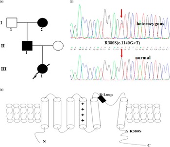
The location, pedigree, and screening results of the proband with mutation R380S. ( a ) The R380S mutation located in the C-terminal region of the KCNQ1 gene. ( b ) The pedigree of the proband with mutation R380S. ( c ) Electropherogram showing a G to T substitution at nucleotide 1248 of KCNQ1(c.1248G>T), resulting in the substitution of Arg by Ser at codon site 380 (R380S).

Family B
Family B included seven members – two males and five females – and they were clinically evaluated (I1, I2, II1, II2, III1, III2, III3 in Fig 2a). The corrected QT interval was between 450 and 480 ms in the proband (II1), and normal electrocardiograms were found in six individuals (I1, I2, I2, III1, III2, III3) even during Holter electrocardiogram. Of the seven family members, only two individuals were found to be symptomatic (II1 and II2). The proband in this family could be easily diagnosed with a Schwartz long QT syndrome risk score of 5 points, whereas “latent” long QT syndrome with normal corrected QT at rest was found in the proband’s younger sister (II2) who showed a non-diagnostic risk score of 1 point but suffered an unexplained history of syncope attacks. Of these mutation carriers, the proband’s elder daughter (III1) and son (III3) were asymptomatic. There was no other history of cardiac events and sudden cardiac death in the family. No abnormalities were found during the echocardiography examination.
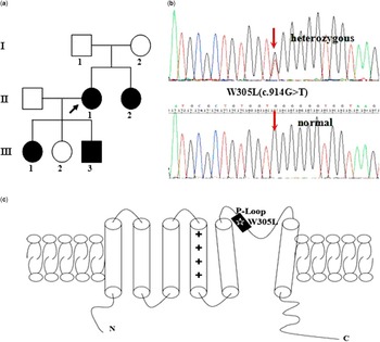
The location, pedigree, and screening results of the proband with mutation W305L. ( a ) The W305L mutation located in the P-loop region of the KCNQ1 gene. ( b ) The pedigree of the proband with mutation W305L. ( c ) Electropherogram showing a G to T substitution at nucleotide 1022 of KCNQ1(c.1022G>T), resulting in the substitution of Trp by Leu at codon site 305 (W305L).

The proband in this family was a 36-year-old woman who experienced her first cardiac event, a syncope episode, at the age of 21 years without any secondary causes of corrected QT prolongation. The 12-lead electrocardiogram at rest showed a prolonged corrected QT interval of 572 ms with a heart rate of 70 beats/minute (Fig 3b). Immediately after her syncope attack, she had a documented electrocardiogram with frequent episodes of R-on-T and torsade de pointes. Cardiac arrhythmias were most frequently observed during the day even with no adrenergic drives. After performing electrical defibrillation and injecting xylocaine, the episodes of R-on-T and torsade de pointes disappeared and the corrected QT interval was shortened to 480 ms. After oral intake of β-blockers (metoprolol, 50 mg, Tid), this proband has had no syncope episodes up to the 3-year follow-up visit, and continuous electrocardiogram monitoring recorded normalised corrected QT interval.
Genetic screening
Direct sequencing of the KCNQ1 gene in the forward and reverse directions in two probands with type 1 long QT syndrome revealed two KCNQ1 missense mutations – R380S and W305L (Figs 1b and 2b). The mutation R380S showed G to T transition at nucleotide 1248 of the KCNQ1 gene (c.1248G>T), resulting in the substitution of Arg by Ser at codon site 380, and was reported in a study by Tester et alReference Tester, Will, Haglund and Ackerman 7 in 2005, but was not detected in Chinese type 1 long QT syndrome patients. This mutation was also identified in the proband’s father in family A. The mutation W305L showed G to T transition at nucleotide 1022 of the KCNQ1 gene (c.1022G>T), resulting in the substitution of Trp by Leu at codon site 305, and was a novel heterozygous mutation. This mutation was also identified in the proband’s family members (family B), including her younger sister, her son, and elder daughter. These two mutations were absent in the control group and in “1000 Genomes” (http://browser.1000genomes.org/). Protein sequence alignment of KCNQ1 orthologs showed that the arginine (R) and tryptophan (W) residues are highly conserved through evolution (http://genome.ucsc.edu/) .
12-lead electrocardiogram recordings of the two probands: ( a ) the baseline electrocardiogram of the proband with mutation R380S showing a markedly prolonged QTc interval (660 ms); ( b ) the baseline electrocardiogram of the proband with mutation W305L showing a prolonged QTc interval (572 ms).

Electrophysiological analysis
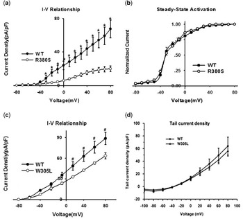
The slowly activating component of the delayed rectifier potassium current was conducted from the COS-7 cells transiently transfected with wild type or mutant KCNQ1 cDNA using the whole-cell patch clamp technique. The peak current density of R380S-Kv7.1 was decreased significantly compared with that of the wild-type Kv7.1 channel (67.46±10.51 versus 20.04±3.29 pA/pF, n=10, p<0.01, Fig 4a); however, the steady-state inactivation of potassium current was unchanged between wild type and R380S-Kv7.1 (Fig 4b). Similarly, the peak current density of W305L-Kv7.1 was reduced compared with that of the wild-type Kv7.1 channel (88.6±8.66 versus 63.38±4.26 pA/pF, n=10, p<0.05, Fig 4c). The tail current density curves showed no significant difference between wild type and W305L-Kv7.1 (Fig 4d). In addition, compared with the wild-type channel, the half-maximal activation of the delayed rectifier potassium current (V1/2) of both R380S- and W305L-Kv7.1 channel was unchanged. Data demonstrated that both R380S and W305L mutants indicated “loss-of-function” effects on the Kv7.1 channel.
( a ) Current–voltage relationships for wild type and the R380S mutation. ( b ) Voltage dependence of peak conductance for wild type and R380S mutation, the normalised peak conductance was plotted against membrane potentials. ( c ) Current–voltage relationships for wild type and the W305L mutation. ( d ) The tail current density for wild type and W305L mutation. *p<0.01 compared with wild type, #p<0.05 compared with wild type.

Discussion
Mutations in the KCNQ1 gene are the most frequent subtype of all long QT syndrome referred as type 1 long QT syndrome. Our present study showed two heterozygous missense mutations – R380S and W305L – in the KCNQ1 gene, which led to “loss-of-function” effects on the Kv7.1 channel in two unrelated Chinese pedigrees with long QT syndrome. These two mutations were located in the C-terminus and P-loop domian of the Kv7.1 channel, respectively (Figs 1c and 2c). Mutation R380S was detected in a large cohort of 541 consecutive unrelated long QT syndrome patients previously,Reference Tester, Will, Haglund and Ackerman 7 but was first reported in Chinese type 1 long QT syndrome patients, whereas mutation W305L was a novel heterozygous mutation in one pedigree.
Clinically, the diagnosis of type 1 long QT syndrome is complicated by variable phenotypic expressivity, whereby an estimated 25% of genotype-positive individuals present with “latent” type 1 long QT syndrome phenotype with normal corrected QT interval at rest.Reference Giudicessi and Ackerman 8 Our study revealed different clinical phenotypes – asymptomatic, syncope, and sudden cardiac death – in the family members carrying the same familial mutation as the proband. In other words, Chinese type 1 long QT syndrome patients showed incomplete penetrance with variable expressivity. Typical cases present no diagnostic difficulty for physicians aware of long QT syndrome, as the typical symptom and prolongation of corrected QT interval are too striking to be missed. On the other hand, it is difficult to unveil the “latent” patients with atypical clinical symptoms using traditional experience and methods.Reference Schwartz, Crotti and Insolia 1 Nevertheless, those “latent” patients have a certain level of risk for developing life-threatening arrhythmias later. Therefore, it is much better to provide a “reasonable suspicion”, which needs to be confirmed or not missed by genetic testing. In the present study, the younger sister of the proband with mutation W305L, who had episodes of an unexplained syncope but a normal corrected QT interval of 425 ms, was conformed as a “latent” type 1 long QT syndrome patient by genetic testing. It is of vital importance for those persons to improve compliance to prophylactic β-blocker therapy and avoid the use of a corrected QT-prolonging drugs. Such “latent” cases of type 1 long QT syndrome can also be recapitulated by cellular electrophysiological studies, in order to provide further insights into its mechanisms and help guide the future management. As previously noted, a KCNQ1 mutation (I235N) contributes to the “latent” type 1 long QT syndrome case by limiting the upregulation of the delayed rectifier potassium current associated with protein kinase A phosphorylation.Reference Bartos, Giudicessi and Tester 9 In addition, three genetically affected members were “silent” mutation carriers in two unrelated Chinese pedigrees with long QT syndrome. Accordingly, genetic testing will help in identifying pathogenic mutations in the “latent” individuals and “silent” mutation carriers, which lays the theoretical foundation for clinical risk assessment and genetic counselling, as well as guides potentially life-saving pharmaco-genomic therapy.
The mutation R380S has been reported previously, but further functional analysis has not been carried out so far. Our electrophysiological study showed that both the R380S and W305L mutations significantly disrupted upregulation of the delayed rectifier potassium current, resulting in a delayed re-polarisation of ventricular cells, and manifested on the 12-lead electrocardiogram as prolongation of the corrected QT interval. Importantly, the delayed rectifier potassium current is the adrenergic sensitive potassium current, and the cardiac symptoms of type 1 long QT syndrome patients are often initiated by specific triggers – for example, emotional stress, physical exertion, diving, and swimming. Consequently, a “loss-of-function” missense mutation of R380S in the KCNQ1-encoded Kv7.1 channel matched the proband’s symptoms with recurrent syncope attacks caused by sports or emotional stress in this study.
The Kv7.1 α subunit is composed of six transmembrane segments (S1–S6), which are partitioned into distinct functional modules including a voltage-sensing module (S1–S4) and an ion-conducting module (S5–S6). Mutations in the transmembrane domain cause more arrhythmogenic phenotypes than mutations in the C-/N-terminus of the Kv7.1 channel;Reference Abriel and Zaklyazminskaya 3 however, several studies also show that mutations in the N-/C-terminus of the Kv7.1 channel impair the delayed rectifier potassium current in various heterologous expression systems.Reference Donger, Denjoy and Berthet 10 , Reference Schmitt, Calloe and Nielsen 11 In the present study, mutation R380S located in the C-terminus manifests with severe clinical phenotypes, and shows “loss-of-function” effects on the Kv7.1 channel. As such, we deduce that certain mutation located in the C-terminus of the Kv7.1 channel may also be associated with an increased risk for cardiac events, in agreement with the type 1 long QT syndrome phenotype of C-terminal KCNQ1 mutation R555CReference Donger, Denjoy and Berthet 10 and M520R.Reference Schmitt, Calloe and Nielsen 11 Such mutations in the C-terminus may cause defects in Kv7.1 channel assembly,Reference Donger, Denjoy and Berthet 10 or impair protein-trafficking deficiency.Reference Schmitt, Calloe and Nielsen 11 As previously reported, the syncope attacks do not occur in type 1 long QT syndrome patients with C-terminal domain mutations until the age of 6–9 years,Reference Yasuda, Hayashi, Horie, Taketani and Yamaguchi 12 which favoured the proband with mutation R380S who suffered recurrent syncope attacks since the age of 10 years. In addition, mutation W305L is located in a putative pore helix selectivity filter segment (P-loop) that connects S5 and S6 of the Kv7.1 α subunit. Compared with type 1 long QT syndrome patients with non-P-loop mutations, patients with P-loop missense mutations display higher incidence of sudden cardiac death.Reference Schwartz, Crotti and Insolia 1 In general, patients with mutations at the transmembrane domain of the Kv7.1 channel are at higher risk for long QT syndrome-related cardiac events and have greater sensitivity to sympathetic stimulation; however, we made a hypothesis in our study that the different effects of β-adrenergic stimulation could also be related to the specific location of the mutation in the P-loop. As to the mutation R380S, tryptophan at position 305 may have an important role in maintaining the properties of the Kv7.1 α subunit, similar to W305S,Reference Neyroud, Denjoy and Donger 13 W305X,Reference Schwartz, Stramba-Badiale and Crotti 14 and W305R.Reference Kapplinger, Tester and Salisbury 15
Not all β-blockers act similarly in the management of symptomatic type 1 long QT syndrome patients, and metoprolol should not be recommended for symptomatic type 1 long QT syndrome patients.Reference Chockalingam, Crotti and Girardengo 16 In the present study, the proband with mutation W305L who rejected to accept device implantation had to be treated with metoprolol. A more exciting thing is that she benefited from the full dosage intake of metoprolol (50 mg, Tid). Administration of metoprolol is associated with a reduction in the cardiovascular responses to sympathetic stimulation in non-adrenalectomised pithed rats.Reference Vila and Badia 17 , Reference Guimaraes and Moura 18 We, therefore, assume that the protective effects of metoprolol may be associated with attenuation of sympathetic stimulation to maintain the normalised corrected QT interval during the 3-year follow-up visit. Thus, aggressive medical intervention – for example, device implantation and sympathetic denervation – may not be needed in all the affected individuals as β-blocker therapy is generally effective in some patients with KCNQ1 mutations. The Registry’s retrospective study indicated that the efficacy of different β-blockers – atenolol, metoprolol, propranolol, or nadolol – differed by genotype in the treatment of long QT syndrome – for example, nadolol was the only β-blocker associated with a significant risk reduction in patients with type 2 long QT syndrome.Reference Abu-Zeitone, Peterson, Polonsky, McNitt and Moss 19 It is still unknown whether metoprolol is effective in type 1 long QT syndrome patients or even superior to other β-blockers.Reference Wilde and Ackerman 20 Therefore, we should further extend our study and try to discover the effects of metoprolol in our Chinese type 1 long QT syndrome patients.
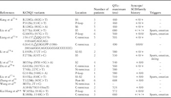
In order to better understand the genetic characteristics of Chinese type 1 long QT syndrome pedigrees, we reviewed all related published studies regarding genotype–phenotype correlations.Reference Kang, Yang and Zhang 21 – Reference Wang, Li and Zhang 29 The corrected QT interval of all type 1 long QT syndrome probands ranged from 490 to 680 ms (568±13 ms, n=18). The longest corrected QT of 680 ms was reported in one proband with mutation S277L located in the S5 region of the Kv7.1 channel. Sports and emotional stress were the main triggering factors for cardiac events in Chinese type 1 long QT syndrome patients. Overall, 19 putative pathogenic mutations were found in KCNQ1 gene, most of which were missense mutations (15/19[79%]). As shown in Table 2, three missense KCNQ1 mutations were found in the P-loop domain of the Kv7.1 channel, whereas four KCNQ1 mutations were located in the C-terminus, including one missense mutations, two deletion mutations, and one insertion mutation. Of these C-terminal mutations, three probands suffered from recurrent syncope and two probands’ family members had a history of syncope, consistent with our findings. Although some of those nine Chinese studies (e.g., Liu et alReference Liu, Yang and Hu 22 ) revealed a family history of sudden cardiac death, none of them presented any proband with C-terminal mutation suffering sudden cardiac death finally, or with P-loop domain mutation who benefited from metoprolol. Our study, therefore, further filled a gap in the gene mutations database regarding Chinese type 1 long QT syndrome patients and might be of great benefit for the early prevention and development of individual therapeutics.
Summary of Chinese LQT1-associated KCNQ1 variants.

n.a=not available; QTc=corrected QT interval; SCD=sudden cardiac death
* The reference was retrieved from SinoMed of China (http://www.sinomed.ac.cn/)
** The data are reported in the present study
Conclusions
There is a large possible association between mutation W305L in P-loop domain of Kv7.1 channel and clinical response to β-blocker therapy. The mutation R380S located in the C-terminus may also be associated with a high risk of sudden cardiac death. Genetic testing can be helpful in identifying the “silent” family members at risk of sudden cardiac death. These findings expand the KCNQ1 mutation spectrum in Chinese type 1 long QT syndrome patients and further emphasise the important role of genetic testing in identifying long QT syndrome patients at risk, screening specific drugs, and optimising the effective treatments.
Acknowledgements
The authors acknowledge Prof. Ali J Marian (Center for Cardiovascular Genetics at The University of Texas Health Science Center-Houston and Texas Heart Institute, Houston, Texas, United States of America) and Dr Li Zhang (Center for Clinical Cardiology, Lankenau Institute for Medical Research, Jefferson Medical College, Philadelphia, Pennsylvania, United States of America) for their kind help and valuable suggestions for this manuscript. The authors thank the patients and their family members for taking part in this study and also thank Xiuxia-Liu and Xia-Yan for carrying out DNA extraction.
Financial Support
This work was supported by the National Basic Research Program of China (973 Program: 2013CB531103), the National Natural Science Foundation of China (81160023, 81370288), and the Ministry of Chinese Education Innovation Team Development Plan (IRT1141, HK).
Conflicts of Interest
None.
Ethical Standards
This study was conducted in accordance with national law and the Code of Ethics of the World Medical Association (Declaration of Helsinki), and was approved by the Hospital Review Board of the Second Affiliated Hospital of Nanchang University. Written informed consents were obtained from all study patients or their guardians in line with the standards established by local institutional review board.