INTRODUCTION
The differential effects of unilateral brain insult on language are well established. Lesions to the perisylvian region of the left hemisphere in adulthood typically render individuals permanently dysphasic or aphasic. In contrast, prenatal or infant-onset left or right hemisphere lesions often result in little or no permanent language disturbance, with language continuing to develop through childhood and adolescence (Bates et al., 2001). Although subtle long-term effects of early lesions on language have been reported (see Reilly et al., 1998; Vargha-Khadem et al., 1985), the effects are diminutive compared with the robust impairments following left hemisphere lesions in adulthood. The relative sparing of language function following early injury is believed to reflect plasticity afforded especially to the developing brain.
Findings from intracarotid sodium amobarbital procedure (IAP), used to determine hemispheric language dominance prior to epilepsy surgery, provide strong evidence for functional reorganization in early development. Increased atypical (right or bilateral) language representation associated with early left hemisphere insult has been well documented (Brazdil et al., 2003; Helmstaedter et al., 1997; Rasmussen & Milner, 1977; Saltzman-Benaiah et al., 2003; Satz et al., 1988). Collectively, findings indicate a rapidly decreasing potential for language to reorganize from the left to the right hemisphere after the age of 5 years (see also, Vargha-Khadem et al., 1985).
The neuroimaging data relating age of seizure onset to language laterality generally support IAP findings. Positron emission tomography (PET) and magnetoencephalography (MEG) have shown increased right hemisphere activity relative to baseline during language tasks among individuals with seizure onset in early childhood (Duncan et al., 1997; Muller et al., 1998), and increased right hemisphere activity compared with individuals with later seizure onset (Muller et al., 1999; Pataraia et al., 2004). Similarly, increased atypical language representation associated with early insult has been demonstrated with functional magnetic resonance imaging (fMRI, Springer et al., 1999), although findings have been mixed (c.f., Liegeois et al., 2004). In a recent fMRI study, Brown et al. (2005) described normative changes in language representation across early development and into young adulthood (subjects 7–32 years of age): healthy children demonstrated widespread and bilateral language representation, whereas adults demonstrated language representation focused in frontal and parietal regions of the left hemisphere. Atypical language representation observed in children with early lesions may reflect diffuse neural processes normally associated with early developmental stages. If so, language may develop within atypical regions (relative to adult norms) in the context of injury to classical language areas of the left hemisphere.
Interestingly, neuroimaging studies have also revealed atypical activations for language in adults with late-onset injury, in the absence of preserved language function. Increased right hemisphere activation on PET was reported in adults with left hemisphere tumors (Thiel et al., 2001) and strokes involving Broca's area (Blank et al., 2003; see also, Rosen et al., 2000). Although left hemisphere injury engenders right hemisphere activation for language tasks (see also, Berl et al., 2005), many participants remain dysphasic. Although de novo activation suggests reorganization of the language cortex in adulthood, the atypical representation does not predict preserved or restored language ability to the extent observed in children. These findings suggest there may be developmental limits to plasticity for this functional domain.
While the bulk of research on language plasticity focuses on gross hemispheric shifts in language representation, researchers have acknowledged the possibility of intrahemispheric reorganization. Using intraoperative stimulation mapping in adults with epilepsy, Penfield and Roberts (1959) first reported language interference at sites both within and beyond neuroanatomical regions described by Broca (1961) and Wernicke (1874). Others have made similar observations following stimulation mapping (Ojemann, 1979; Ojemann et al., 1989; Rasmussen & Milner, 1977), although the extent to which findings represent plastic responses to lesions or seizures versus normal variability is unknown. Of interest, functional neuroimaging in adult stroke patients suggests that better recovery may be associated with activation of perilesional areas in the dominant hemisphere, in contrast to activation of homologous regions contralaterally (Cao et al., 1999; Fernandez et al., 2004; Rosen et al., 2000). Thus patterns of ipsilateral connectivity may confer an advantage on intrahemispheric reorganization following brain insult in adulthood.
There is increasing, although inconclusive, evidence for the occurrence of intrahemispheric reorganization in children with left hemisphere injury. DeVos et al. (1995) used IAP to study language dominance in children with tumors proximal to or encroaching on classical language areas and failed to observe reorganization to the right hemisphere in most cases. Tumor resections failed to produce lasting aphasias, suggesting accommodation within the left hemisphere.
Duchowny et al. (1996) used extraoperative stimulation mapping (ESM) with subdurally implanted electrode grids in children undergoing neurosurgery for epilepsy and reported interhemispheric shifts in language only when acquired lesions encroached classical language areas. In contrast, children with developmental lesions of the left hemisphere apparently did not experience language reorganization to the right. Because Duchowny et al. did not confirm laterality independently with another method such as IAP or fMRI, and because electrode grid arrays varied in coverage, the nature of language representation in this group could not be conclusively determined. Duchowny et al. acknowledge the possibility of intrahemispheric reorganization in participants who were negative for ESM for language in the left hemisphere, yet were unable to observe language sites beyond classical language regions due to limited electrode grid coverage (and a paucity of independent measures of language laterality).
Interestingly, Ojemann et al. (2003) reported that variability of language sites within the dominant hemisphere increases with age (cp Ojemann et al., 1989). Adults undergoing intraoperative and extraoperative stimulation mapping demonstrated naming errors at broader cortical regions than individuals 4 to 16 years of age. Within the child/adolescent group, older participants demonstrated language at broader cortical regions than the younger children. It is unknown whether the increased areas of cortical representation associated with advancing age reflects increased intrahemispheric reorganization in the context of brain injury or normal maturational processes. Indeed, the findings of Ojemann et al. (2003) are consistent with previous reports of variability in language maps of adults with epilepsy (e.g., Bhatnagar et al., 2000; Ojemann, 1979; see also Devinsky et al., 2000, and Schwartz et al., 1998, for predictors of variability in anterior temporal naming and reading sites), although they are inconsistent with recent neuroimaging studies of changing representation across early development (e.g., Brown et al., 2005).
The purpose of the present study was to assess and quantify subtle intrahemispheric reorganizing effects of early epilepsy on language representation. We report on nine children with refractory epilepsy who underwent ESM prior to neurosurgery; all demonstrated language interruptions from left hemisphere stimulations. The electrode grid arrays used provide extensive cortical coverage, permitting detection of language at sites beyond classical language areas (i.e., permitting detection of possible intrahemispheric reorganization). To facilitate comparison of findings across participants, we developed a novel coregistration technique whereby ESM data are represented on a model brain and described in standard space (Talairach & Tournoux, 1988). The occurrence of language-positive sites in noncanonical areas may serve to support the notion that language can reorganize intrahemispherically following early insult.
METHODS
Participants
Nine children and adolescents (six male, three female; 5.6–17.7 years of age) with medically intractable epilepsy were studied. They represent a subset of all patients who underwent ESM in the left hemisphere for language prior to epilepsy surgery at the Hospital for Sick Children (Toronto, Canada) from 1996 through 2004. The participants studied include all individuals who met the following criteria: left hemisphere epileptogenic foci, thin-slice MR images amenable to standardization and 3D rendering, intraoperative digital images that clearly indicate cortical morphology and electrode placement, and positive language mapping (observed language interruptions from stimulation) of the left hemisphere. This study was approved by the Hospital's Research Ethic's Board.
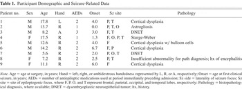
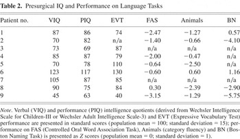
The presurgical evaluations have been described previously (Minassian et al., 1999; Snead, 2001), and included video electroencephalography (EEG) monitoring, structural MRI, and neuropsychological assessment; in several cases, patients also underwent MEG and PET. Subsets underwent IAP (n = 6) and fMRI (n = 7) for determination of hemispheric dominance for language. All participants received surgical excisions, with tissue samples studied to determine pathology. Seven received multiple subpial transections (MSTs) in addition to resections. Demographic and seizure-related data are presented in Table 1. Select neuropsychological findings are presented in Table 2.
Participant Demographic and Seizure-Related Data

Presurgical IQ and Performance on Language Tasks

Procedure
Subdural electrode grids
Participants underwent craniotomy for subdural electrode grid array implants for localization of epileptogenic foci (electrocorticography, ECoG) and ESM. Antiepileptic medications were withdrawn for ECoG and ESM. Protocols for surgical implantation, ECoG, and ESM have been reported previously (Chitoku et al., 2001; Onal et al., 2003), and are described only briefly below.
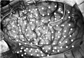
Custom subdural electrode grid arrays (Ad-Tech Medical Instrument Co., Racine, WI) were designed for each patient based on seizure semiology, scalp EEG recordings (ictal and interictal), and in some cases MEG data, in combination with 3D MRI brain-volume modeling. Each grid consisted of 70–116 platinum electrodes (mean = 95), 5 mm in diameter, spaced 10–13 mm apart (measured center-to-center), and embedded in a Silastic sheet. Typical grid coverage is depicted in Figure 1.

Photograph of the lateral surface of the left hemisphere (anterior brain at left) of Participant #6 immediately prior to grid implantation. White dots represent electrode placement and indicate typical grid coverage (Participant #6 received a 98-electrode grid implant); white lines indicate location of sylvian fissure and central sulcus (confirmed with electrocorticography/extraoperative stimulation mapping for motor and somatosensory function).
A large craniotomy was performed in each case, and a large dural opening was fashioned. Subdural grids were first positioned on the cortex, then sutured to the dura to prevent displacement. Digital photographs were taken prior to and following grid placement to document cortical morphology and electrode positioning (Rutka et al., 1999). Multiple digital photographs of the lateral surface of the brain, with a field of view centered over and perpendicular to the inferior rolandic region, allowed for precise and direct mapping of the cortical contact points for each electrode. Duraplasty was performed, and the bone flaps were hinged loosely superiorly following grid placement. Cables were routed through the scalp away from the incision, and secured with sutures. Plain skull X rays and head computed tomography verified electrode grid placement.
Electrocorticography
ECoG consisted of split-screen time-locked video EEG (BMSI 5000; Nicolet, Madison, WI) for comparison of electrocortical activity and semiology. The digitized EEG data (200 samples/s per channel) were stored and reviewed; events were identified by alarm button triggers and automated seizure and spike detection programs. ECoG data were assessed to determine epileptogenic foci.
Extraoperative stimulation mapping
Eloquent cortex was mapped in one to three sessions, taking place over 1 or 2 days. First stimulation mapping sessions occurred between the second and fourth days following implantation. Motor cortex mapping always preceded somatosensory and language mapping.
Stimulation thresholds established at motor cortex mapping were used in somatosensory and language mapping. Stimulations were delivered in 50 Hz biphasic pulses (duration = 0.2 ms) for up to 25 seconds per train. Initial stimulations were delivered at 2 mA; current was increased in 1–2 mA increments until motor responses or afterdischarges (rhythmic high-frequency spike and sharp waves that follow applications of electrical stimulation) were observed. Stimulation was not delivered beyond 20 mA. The distance reference technique of Lesser et al. (1984) was used: stimulation occurred between the target electrode and an electrode at the periphery of the array. EEG activity was continuously recorded and monitored. Stimulation was paused when afterdischarges were detected. For some patients, afterdischarge thresholds exceeded the maximal stimulation levels and were not observed at any stimulation site (see, Chitoku et al., 2001, 2003).
Classic language areas and the surrounding cortex, as well as epileptogenic regions (which may or may not reside within canonical language anatomy) and surrounding cortex were mapped for language. For each patient, baseline language task performance was established prior to implantation. Tasks included counting, reciting the alphabet, color naming (using an array of colored squares), and picture (object) naming. The pictures were colored drawings of common objects familiar to young children and were selected from a variety of standardized naming tasks and from commercially available flash cards designed for use with young children. Items or entire tasks for which correct responses were not achieved at baseline testing were excluded from the stimulation battery. To confirm understanding of task requirements and sufficient visual and auditory acuity and attention at the time of stimulation mapping, electrical currents were withheld on each trial until several initial items were responded to correctly. Whenever possible, picture (object) naming was the first test of choice because naming has long been the standard for establishing cortical language areas (Ojemann, 1983). Color naming, reciting the alphabet, and counting were used when children's cooperation with naming was lacking, when swelling around the eyes (from the implant) reduced vision, and/or when IAP or fMRI had identified tasks that were especially sensitive to the language results obtained with those procedures.
To test cortical sites for language, stimulations were applied while the child carried out the various language tasks. If the child produced incorrect responses, incoherent or inappropriate speech, or total or subtotal speech arrest in the absence of motor arrest (apraxia), the site would be flagged as a possible language site and would later be retested. Likewise, inconclusive testing warranted retest. Motor arrest in language (apraxia) was distinguished from language interference through additional motor stimulation testing (e.g., application of stimulation while observing oral–motor activity) and by instructing the child to engage in automatic speech (e.g., counting or reciting the alphabet). Repetition of simple phrases, or recitation of well-learned leading phrases such as “this is a _____” in picture naming, were also useful in distinguishing motor effects from language processing effects in expressive errors. A site was determined necessary for language if errors were observed at retest, in the absence of afterdischarge, clinical seizure, and motor arrest. In the absence of errors at retest without afterdischarge, the site would be assessed a third time at higher stimulation intensity (+1–2 mA), exceeding the threshold established through motor-strip testing. The site was deemed necessary for language only if interruptions were observed during two separate stimulation periods.
Assessing ESM findings in standard space
Preoperative structural MR images were obtained for each patient using a GE 1.5-T Signa Advantage 5.6 unit (GE Medical Systems, Milwaukee, WI). For inclusion in the current study, volumes for each participant necessarily lacked motion artifact that would preclude normalization and 3D transformation with retained sulcation/gyration.
All coregistration processing of ESM and structural MRI data were completed using freeware applications running on a Microsoft Windows 98 Intel Pentium III PC, processing at 1.7 GHz with 256 Mb of RAM, and visualized on a 17-inch flat screen monitor (24-bit true color, 1024 × 768 pixels). Each T1-weighted MR brain volume was mounted and preprocessed (cropped, oriented, and converted from DICOM to Analyze format) using MRIcro v. 1.36 software (Rorden & Brett, 2000). Volumes were then spatially normalized in SMP96 for Windows (Friston et al., 1995), to a supplied T1 template within an MNI bounding-box using only linear iterations, 7 × 8 × 7 (X × Y × Z) basis functions, re-sliced at 1 × 1 × 1 voxels. Brain volumes were then remounted in MRIcro and skull-stripped using the Brain Extraction Tool plug-in (Smith, 2002) and 3D-rendered for ESM–MRI coregistration. A composite of surface- and volume-rendered images provided visible cortical morphology at default values.
The normalized 3D-rendered brain scans were oriented to approximate the orientation and field of view of the respective digital photographs. Two independent raters compared the ESM maps (as printouts from labeled digital photographs) to 3D-rendered brain images. Guided by the electrode grid placement and the cortical morphology available on ESM maps, raters freely navigated the corresponding 3D renderings and independently determined stereotactic coordinates (provided by MRIcro) for each language site in the standard space of Talairach and Tournoux (1988).
Assessing proximity of lesions/epileptogenic regions to classical language areas
Due to varied findings from stimulation mapping, inconsistent descriptions, and multiple nomenclatures (i.e., gyral–sulcal landmarks vs. Brodmann's areas), it is difficult to relate classical language areas to specific neuroanatomical structures (Poeppel & Hickok, 2004). In the absence of consensus in describing canonical language regions by structure, adopting operational definitions of Broca's and Wernicke's areas poses a significant yet necessary challenge.
A review of the literature indicates that the frontal operculum (pars opercularis) emerges as the most consistent structural center for Broca's area (Blank et al., 2003; Quinones-Hinojosa et al., 2003; Tomaiuolo et al., 1999; see also Mohr et al., 1978); the anterior portion of Brodmann area 44 (BA 44) is represented in the superficial cortical component of the operculum (Petrides & Pandya, 1994, in Tomaiuolo et al., 1999). The frontal operculum occupies the third convolution of the inferior frontal gyrus and is bordered rostrally by the vertical ramus of the sylvian fissure (distinguishing pars opercularis and pars triangularis) and caudally by the precentral sulcus (Tomaiuolo et al., 1999). Trained independent raters assessed digital photographs merged with ECoG findings (by electrode site) to determine whether epileptogenic zones encroached on the operculum.
Operationally defining the anatomical boundaries of Wernicke's area is additionally challenging. Bogen and Bogen (1976) discuss the multitude of early anatomical descriptions of Wernicke's area: conservative neuroscientists identify a small region of the posterior superior temporal gyrus as Wernicke's area, while others identify larger regions that extend ventrally within the temporal lobe (i.e., to middle and inferior temporal gyri) and dorsally and caudally to regions surrounding the temporal–parietal junction (i.e., angular and supramarginal gyri). For the current project, raters studied the maps of the posterior receptive language area of Penfield and Roberts (1959), compiled from stimulation mapping investigations, and decided whether or not lesions/epileptogenic regions encroached on Wernicke's area (i.e., the posterior superior temporal lobe and area surrounding the temporal–parietal junction).
Intracarotid sodium amobarbital procedure for language laterality
The IAP protocol used at the Hospital for Sick Children has been previously documented (see Fernandes & Smith, 2000; Saltzman-Benaiah et al., 2003) and is described only briefly here. Participants underwent neuropsychological testing prior to IAP; language performance at pre-IAP assessment served as baseline. Items and/or procedures that were not negotiated successfully at baseline testing were excluded from the IAP battery. Patients' internal carotid arteries were catheterized via the femoral artery. Hemisphere-selective injections of sodium amobarbital (Amytal) were administered at 1.5 mg/kg (by body weight). Brain activity was monitored by scalp EEG to verify drug effects; paralysis of limbs contralateral to the barbiturate injections confirmed fronto-central drug perfusion. Language testing commenced immediately following verification of drug effects. Testing typically included counting, reciting days of the week or months of the year or the alphabet, spelling, reading, naming pictures, and occasionally naming small objects or naming colors. Errors (relative to baseline) and/or speech arrest were taken as evidence for language in the perfused hemisphere.
If only left-sided or only right-sided injections produced speech arrest and/or speech errors, the individual was deemed to possess lateralized speech (left or right hemisphere, respectively). If language was conclusively interrupted following injections to both hemispheres, or if neither left- nor right-sided injections produced errors or speech arrest, language representation was classified as bilateral.
fMRI for language dominance
A minimum of two of four established covert language tasks were used, including verb generation, picture and color naming, phonemic fluency, and word repetition. A flexible battery allowed procedures to be tailored to each participant's skill and cooperation level. Of the multiple tasks used, covert verb generation and covert naming to confrontation with line drawings were preferred, and attempted in most cases.
An experienced neurologist judged language representation after visually assessing activation across each patient's whole cerebrum on a case-by-case basis. Recently, Gaillard et al. (2002) demonstrated that expert judgment of language laterality is comparable to quantitative region of interest analyses for determining language dominance, as verified by IAP. Activation data are categorized as left, right, or bilateral.
RESULTS
Lesion/Epileptogenic Encroachment Status of Classical Language Areas
Raters' judgments of lesion/epileptogenic region proximity to Broca's area (the opercular portion of BA 44 in the left hemisphere) were in perfect agreement, as were raters' judgments of whether or not lesions/epileptogenic regions encroached on classical Wernicke's area. Participants #3, #5, #6, and #9 had lesions/epileptogenic regions that encroached on Broca's area. Participants #1, #2, #3, #4, #7, and #8 had lesions/epileptogenic regions that encroached on Wernicke's area.
Coregistration of 2D ESM Data with 3D Brain Renderings
A total of 28 language sites were analyzed for rater agreement: 15 frontal-lobe sites (across 8 participants), and 13 extra-frontal sites (across 6 participants) were assessed. The average difference (in three dimensions) between raters' estimates of the 28 language sites was 3.9 mm (SD = 2.0). Frontal-lobe sites were more consistently mapped (mean difference = 3.2 mm, SD = 1.3, n = 15) than extra-frontal language sites (mean difference = 4.8 mm, SD = 2.3, n = 13) [t(27) = 2.35, p < .05]. Because the x-plane was automatically selected by the MRIcro software (snapped to surface of rendering), two-dimensional (y- and z-plane) point-to-point differences better reflect rater agreement. The average difference (in two dimensions) between raters' estimates of language was 3.5 mm (SD = 1.9). Given that electrodes were 5.0 mm in diameter, interrater reliability was determined to be excellent. Only coordinates from one rater's estimates were analyzed for the sections that follow.
ESM Language Findings in Stereotactic Space
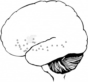
ESM identified language sites within and outside of classical language areas. Positively mapped language sites across the group are presented in Figure 2.

Language-positive sites from extraoperative stimulation mapping for all participants studied in standard space (n = 9). Language sites are collapsed across the x-plane and depicted on the lateral surface of the left hemisphere (in the diagram, left is anterior, right is posterior). Diamonds represent language sites stimulation-mapped in the participants' frontal lobes; triangles represent language sites simulation-mapped outside the frontal lobe. The shaded region represents the cortical portion of the frontal operculum, or Broca's area.
Stimulation of the third frontal convolution of the inferior frontal gyrus interrupted articulation (not accepted as a language error), naming, and fluency; stimulation of the posterior region of the superior temporal gyrus interrupted naming and elicited confusion (not accepted as a language error). Stimulations of mid-temporal and anterior superior temporal regions interrupted naming, as expected (see Penfield & Roberts, 1959; Schwartz et al., 1998). Language sites where stimulation affected articulation, naming, and fluency were also observed in regions anterior to classical Broca's area.
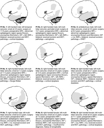
Possible relationships between demographic and seizure variables (e.g., age at seizure onset) and language representation could not be assessed using inferential statistics due to the small sample size. However, demographic and seizure-related data, as well as IAP and fMRI data (where available), for these individuals are presented alongside plots of ESM findings in Figure 3.

Diagrams represent language-positive sites from extraoperative stimulation mapping for each participant. Diamonds indicate language sites stimulation-mapped in participants' frontal lobes; triangles indicate language sites stimulation-mapped outside the frontal lobe. Epileptogenic regions are lightly shaded; where epileptogenic regions overlap eloquent cortex (identified through extraoperative stimulation mapping), sites were either spared or received multiple subpial transections. The darker-shaded regions indicate the cortical portion of the frontal operculum, or Broca's area.
Language Laterality
Seven participants underwent either IAP or fMRI for language lateralization. In all cases, IAP and fMRI findings indicated that the left hemisphere supported language (in some cases, both the left and right hemispheres supported language; predominantly right hemisphere language representation was not observed in this sample). These findings are concordant with positive ESM for language in the left hemisphere.
Six patients underwent both IAP and fMRI investigations. The IAP and fMRI findings were in perfect agreement for one patient (#6). Patients #1, #2, #4, and #5 had IAP indicating left hemisphere dominance, whereas fMRI findings suggested bilateral language representation. Patient #8 demonstrated bilateral language representation on IAP and left hemisphere representation on fMRI. The tendency for fMRI to suggest bilateral language representation more frequently than IAP is not uncommon; others have reported right hemisphere activation even when predominant activation is lateralized to the left hemisphere (see Vingerhoets et al., 2004). The discordance of IAP and fMRI for language across the participants suggests nonequivalence of these procedures; different language tasks were used in the two procedures, and activation in fMRI does not differentiate between areas active versus those essential for task performance.
Language and Seizure Outcome Following Surgery
One-year follow-up data were available for all patients. Following surgery, two patients (#5 and #8) experienced transient dysphasias that had resolved by the 1-year follow-up, and one patient experienced a lasting dysphasia (#6). In all cases, the surgical resection included or abutted sites identified as language positive. This finding confirms the sensitivity of the ESM procedure and indicates that ESM findings can be used to counsel patients about possible language outcomes following respective surgery. Four patients (#1, #2, #5, and #7) were seizure free at 1-year follow-up. In the five patients who continued to experience seizures, the resections were limited in extent to preserve eloquent functions.
DISCUSSION
We are confident that the assessment and novel coregistration procedures provide an accurate representation of epileptogenic and eloquent cortices. Independent raters' judgments of lesion proximity to classical language areas were in perfect agreement. In the absence of widely accepted and available stereotactic boundaries for Broca's and Wernicke's area, independent ratings from visual inspection of ECoG data mapped to digital photographs provide a reliable measure of canonical language area lesion status.
The independent raters' estimates of language sites in stereotactic space were highly concordant, demonstrating that the protocol used to map 2D ESM data to 3D brain renderings is reliable. Others have previously reported excellent accuracy and interrater reliability using a similar coregistration technique (Wellmer et al., 2002), without normalization of volumes. The normalization process affords objective assessment of language sites within the brain, without scale problems typically associated with comparison of data obtained from individuals with differing brain morphology (size, shape, sulcal–gyral patterns). This is the first time a 2D-to-3D coregistration for language ESM protocol has been documented in a pediatric group. Burgund et al. (2002) have validated the use of adult templates in spatial normalization of pediatric brain scans. We are confident that the standardization process provided reliable transformations necessary for comparing findings across several individuals, while maintaining the integrity of the gyral–sulcal patterns required in the plotting of language sites from intraoperative photographs.
In addition to facilitating group-wise investigation, the coregistration protocol promotes communication of findings in standard nomenclature; the complexity of brain anatomy (including cortical morphology) among children with early lesions often precludes definitive discussion of function in relation to underlying anatomy (see for example, Akai et al., 2002). Although the normalization process prevents absolute determination of point-to-point spatial differences (i.e., children's brains may be “stretched” to fit adult templates during normalization), the assessment of possible intrahemispheric shifts remains useful, as differences are automatically weighted to brain size. This feature is especially important when comparing brains of individuals across childhood (i.e., volumetric differences may be present due to developmental processes).
The ESM data assessed in standard stereotactic space indicate language representation in noncanonical regions in children with epilepsy, suggesting intrahemispheric shifts in language functions. Specifically, the inferior frontal lobe appears to support expressive language in regions considerably anterior to classical Broca's area (see Figure 2). Language interruptions were observed over 40 mm (on the adult template) rostral to the anterior commissure, anterior to language regions indicated by the pars opercularis structural probability maps of Tomaiuolo et al. (1999) and language regions previously identified through stimulation mapping (see Penfield & Roberts, 1959; Quinones-Hinojosa et al., 2003; cp Ojemann et al., 1989). Our observation of anterior representation contrasts with recent reports by Ojemann et al. (2003), who failed to observe language beyond 15 mm rostral to the central sulcus (the central sulcus is caudal to the anterior commissure) in children undergoing stimulation mapping prior to epilepsy surgery. The difference between the recent findings of Ojemann et al. and our own are not easily explained; however, several procedural and demographic differences should be considered. In our study, we used large electrode grids and extensively assessed the cortex for language in all cases. Ojemann et al. performed both intra- and extraoperative stimulation mapping; it is unclear whether an equivalent extent of cortex was studied in all cases. In addition, the two groups presented with differing pathologies. The sample of Ojemann et al. included many more cases of tumor resection than ours, and only one case of cortical dysplasia; in our case series, cortical dysplasia was the predominant diagnosis. These and other patient variables can be considered for prediction of intrahemispheric reorganization in larger scale studies. Importantly, our findings demonstrate that children and adolescents may have language representation in regions substantially anterior to those previously reported.
Interestingly, Ojemann et al. (1989) observed language at sites as much as 45 mm anterior to the central sulcus in adults with epilepsy who underwent stimulation mapping for language; the same group later suggested that many essential language sites “develop” into these anterior regions during childhood and adolescence (Ojemann et al., 2003). Our findings in standard space are comparable in extent and consistent with reports of anterior language displacement observed in adults (Ojemann et al., 1989, 2003). Collectively, data suggest that the anterior left hemisphere retains the propensity to subserve language in the context of pediatric insult/epilepsy.
The extent to which the anterior language representation reflects individual differences or plastic responses to seizures or other insults is unclear; with advances in noninvasive measures of language localization, we can expect functional neuroimaging of healthy individuals to help distinguish intrahemispheric reorganization from normal variation. Specifically, the development of noninvasive neuroimaging protocols in modalities with high spatial resolution (e.g., fMRI, MEG) that correlate well with ESM findings in clinical populations will permit the eventual comprehensive characterization of tissue essential for language in healthy individuals. Until we demonstrate equivalence of noninvasive (event-correlated activation) neuroimaging and ESM and assess representation in healthy individuals, we must be careful not to interpret distorted language maps as conclusive demonstration of intrahemispheric reorganization. However, it is interesting to note that variation about canonical regions was not uniformly distributed, in that expressive language sites were not observed caudal to canonical Broca's area, perhaps indicating a propensity of anterior regions to support, and possibly adopt, expressive language. Caudal representation or shifts may be “blocked” or prevented by established functional motor cortex in the precentral gyrus.
It is unclear whether the distribution of lateral temporal (“posterior”) language sites reflect intrahemispheric reorganizational processes. Language was mapped to the posterior superior temporal gyrus (Wernicke's area), and middle temporal cortex, as expected. Language sites were also observed along the anterior superior temporal gyrus, possibly indicating plastic responses to temporal lobe epileptogenic activity or other injury. Schwartz et al. (1998) and Devinsky et al. (2000) observed similar temporal lobe language representation (for naming and reading) in adults undergoing stimulation mapping. Schwartz et al. and Devinsky et al. suggest that language representation in the anterior temporal lobe may be predictable from profiles of early seizure onset and low cognitive functioning; the current results support this suggestion.
These findings highlight the importance of thorough functional investigations, prior to resection for treatment of intractable epilepsy. Both intact and epileptogenic tissue may support language function (see Figure 3). Assessing language only within canonical language regions may fail to precisely characterize the language cortices of individuals with seizure onset in childhood. Expressive language may consolidate or relocate rostrally within the frontal lobe, in the context of early left hemisphere epilepsy. Furthermore, individuals with atypical left hemisphere language representation may experience interhemispheric language reorganization (see Figure 3). The documentation of interhemispheric reorganization (i.e., through IAP of fMRI) does not preclude language representation outside of canonical regions. Neurosurgical teams must consider the potential for language representation anterior to classical Broca's area. Extensive functional mapping within and beyond canonical language areas is necessary for comprehensive characterization of the language cortex.
Future studies of larger patient groups will permit the assessment of demographic, psychological, and seizure-related variables as predictors of language representation. Although the effects of age at seizure onset on language laterality are well documented, only a small number of studies have attempted to characterize the effects of pathology and site of seizure focus on laterality (e.g., Duchowny et al., 1996; Saltzman-Benaiah et al., 2003; Weber et al., 2006). Few have attempted to characterize predictors of localization within the dominant hemisphere (for predictors of anterior temporal language representation, see Schwartz et al., 1998, and Devinsky et al., 2000). The small sample size in the current study precluded the use of sophisticated inferential statistical analyses of language localization. However, informal assessments of individual language maps (Figure 3) suggest imperfect prediction of language localization by any single demographic or seizure-related variable, including location of epileptogenic foci. Although several subjects had language representation in expected regions (e.g., Patients #2 and #4 had epileptogenic foci sparing canonical Broca's area—each demonstrated “typical” language representation; Patients #3 and #6 had epileptogenic foci that encroached on canonical Broca's area—each demonstrated anterior language representation), a subset demonstrated unpredictable “atypical” representation. Patients #1 and #8 had language representation in areas rostral to canonical Broca's area, yet both patients had extrafrontal epileptogenic foci. Large-scale investigations will permit further exploration of these relationships.
The future use of multimodal assessments of language function and representation (neuropsychological assessment, functional neuroimaging, and stimulation mapping) will facilitate comprehensive characterization of effects of early insult. The question remains as to whether intrahemispheric and interhemispheric reorganization occur in concert or at the exclusion of one another. The ESM technique described only depicts language representation within a single hemisphere, although we know from IAP and fMRI investigations that several subjects in the current study had bilateral language representation. With the advancement of noninvasive neuroimaging techniques that correlate well with ESM findings, we will ultimately be able to characterize language-essential sites in both hemispheres, and assess factors associated with both inter- and intrahemispheric reorganization of language.
ACKNOWLEDGMENTS
This research was supported, in part, through a Studentship to D.S.K. from the Ontario Student Opportunity Trust Fund–Hospital for Sick Children Foundation Student Scholarship Program, and through a Doctoral Research Award to D.S.K. from the Canadian Institutes of Health Research (CIHR) in partnership with Epilepsy Canada.
We gratefully acknowledge Ms. Irene Elliott for her expert role in patient care and management with the epilepsy service at the Hospital for Sick Children, Dr. Adrian Crawley for his advice on establishing a coregistration protocol, Ms. Stephanie Holowka for her assistance with neuroimaging, and Dr. Elizabeth Donner for her helpful comments on an earlier draft of the manuscript.