INTRODUCTION
Reactive oxygen species (ROS) are inevitably generated through incomplete respiration in mitochondria, as by-products of a variety of metabolic reactions or by exogenous stimuli including irradiation and redox-cycling drugs (Tawe et al. Reference Tawe, Eschbach, Walter and Henkle-Dührsen1998; Chi et al. Reference Chi, Tanaka, Okuda, Ikota, Yamamoto, Urano, Ozawa and Anzai2005). ROS play pivotal roles not only in control of cellular redox homeostasis (Jackson, Reference Jackson2005) but also in induction of cellular proliferation or apoptosis by activating signaling molecules (Thannickal and Fanburg, Reference Thannickal and Fanburg2000; Hancok et al. Reference Hancok, Desikan and Neill2001; Sim et al. Reference Sim, Yong, Park, Im, Kong, Ryu, Min and Shin2005). However, the unbalanced generation of ROS exerts harmful effects. It might induce breakage of DNA strands, protein oxidation, polysaccharide depolymerization, membrane-lipid peroxidation and impairment of signal transduction from membrane receptors in various physiological processes (Dröge, Reference Dröge2002). The aerobic organisms have evolved a series of multi-layered enzymatic and non-enzymatic defence systems, which can remove the aggressive ROS and/or repair ROS-mediated cell damage.
Glutathione peroxidase (GPx; EC 1.11.1.9) encompasses 6 distinct families of multiple isoenzymes, which catalyse the reduction of H2O2, organic hydroperoxides and lipid hydroperoxides by using glutathione as a reducing agent. The GPx families share similar structural and enzymatic properties with one another, and these selenium-dependent enzymes act as a tetramer via the mediation of a subunit interaction domain (Arthur, Reference Arthur2000). In contrast, GPx proteins categorized into GPx4 family (phospholipid hydroperoxide GPx, PHGPx; E.C. 1.11.1.12) have a series of distinct features compared to the other GPx families. PHGPxs function in a monomeric form because they lack the subunit interaction domain (Epp et al. Reference Epp, Ladenstein and Wendel1983; Brigelius-Flohé et al. Reference Brigelius-Flohé, Aumann, Blöcker, Gross, Kiess, Klöppel, Maiorino, Roveri, Schuckelt, Ursini, Wingender and Flohé1994; Arthur, Reference Arthur2000). These molecules exhibit unique substrate preference; the enzymes directly reduce hydroperoxidized phospholipids integrated into membranes (Ursini and Bindoli, Reference Ursini and Bindoli1987). Members of the other GPx families, however, interfere with lipid peroxidation only via a concerted operation with phospholipase (Grossmann and Wendel, Reference Grossmann and Wendel1983), which implies that PHGPxs are deeply associated with the repair of disrupted biomembranes (Imai and Nakagawa, Reference Imai and Nakagawa2003). GPxs might also constitute the front line of enzymatic defence to ensure their survival against host immune cell-derived ROS in parasitic helminthes, which cause chronic infections (Cookson et al. Reference Cookson, Blaxter and Selkirk1992; Zelck and von Janowsky, Reference Zelck and von Janowsky2004). The helminth GPx families show a certain degree of biased distribution across taxa. GPx proteins homologous to mammalian GPx3 (plasma GPx) have been characterized in the filarial nematodes including Brugia pahangi, Dirofilaria immitis and Wuchereria bancrofti (Henkle-Dührsen and Kampkötter, Reference Henkle-Dührsen and Kampkötter2001); while information on the GPx4 members is obtainable from trematode species such as Schistosoma mansoni and Clonorchis sinensis (Williams et al. Reference Williams, Pierce, Cookson and Capron1992; Mei and LoVerde, Reference Mei and LoVerde1995; Cai et al. Reference Cai, Bae, Kim, Sohn, Lee, Jiang, Kim and Kong2008).
Paragonimus westermani is a parasitic trematode, which causes inflammatory lung granuloma, as well as systemic infections in Asian, American and African countries (WHO, 1995). Consumption of raw or undercooked crustaceans or wild boar infected with the metacercariae has been the principal route for human infection (WHO, 1995; Blair et al. Reference Blair, Xu and Agatsuma1999). After being ingested, the metacercariae excyst within the duodenum, migrate through the peritoneal cavity and finally arrive in the lungs, where they mature. The adult worms are surrounded by thick fibrous granulomatous cyst, in which infiltration of inflammatory phagocytes generating various reactive radicals prevail (Nakamura-Uchiyama et al. Reference Nakamura-Uchiyama, Mukae and Nawa2002). The high oxygen tension of the lungs may also be a primary factor in the creation of a hostile host environment (Comhair and Erzurum, Reference Comhair and Erzurum2005). In order to overcome these stressful conditions and to maintain its life-span, the fluke is equipped with several antioxidant enzymes, including peroxidase, superoxide dismutase (SOD) and glutathione S transferase (GST) (Chung et al. Reference Chung, Lee, Song and Cho1992; Hong et al. Reference Hong, Kang, Chung, Chung, Oh, Kang, Bahk, Kong and Cho2000; Li et al. Reference Li, Na, Kong, Cho, Zhao and Kim2005).
In this study, we isolated and characterized 2 novel genes encoding GPx from the human lung fluke. Expression of the PwGPx1 gene increased gradually in accordance with the development of the parasite, whereas that of PwGPx2 maintained at the basal level from the metacercaria to the 3-week-old juvenile, and peaked in the egg-laying stage. The Paragonimus proteins localized specifically in vitellocytes within vitelline glands and eggs. In vitro experiments demonstrated that oxidative stress-inducible chemicals such as paraquat, juglone and H2O2 substantially augmented the expression of PwGPx1 and PwGPx2 in viable worms. The PwGPx proteins might actively participate in the detoxification of oxidative damage during egg production. Our results further suggest their physiological implications during the parasite's adaptation in hostile host environments.
MATERIALS AND METHODS
Parasite, DNA and RNA samples
Dogs were orally infected with 200 metacercariae of either diploid- or triploid-type P. westermani, which were collected from naturally infected crayfish, Cambaroides similis. The 1- and 3-week-old juveniles were harvested from the peritoneal cavity, and the 7- and 12-week-old worms were obtained from the lungs. The worms were washed >10 times with physiological saline at 4°C. A total of 20 adult worms (12-week-old) were incubated overnight in physiological saline, and the eggs were isolated and cleaned under a dissecting microscope (purity >99%). The triploid worms were used as the principal experimental material in this study, with an exception of the genomic DNA employed in Southern blotting. The use of experimental animals was approved by the Animal Ethics Committee of the Korea Food and Drug Administration (protocol number NIH-05-09). Total RNA and DNA samples were prepared using TRIzol reagents (Invitrogen, Carlsbad, CA, USA) and the Wizard Genomic DNA Purification Kit (Promega, Madison, WI, USA).
Analysis of expressed sequence tag (EST) and cDNA library screening
The nucleotide sequences of lambda clones randomly selected from a P. westermani cDNA library were analysed against the non-redundant databases of nucleotide and protein sequences at the National Center for Biotechnology Information (NCBI, http://www.ncbi.nlm.nih.gov), using BLAST algorithms. EST-1 (designated PwGPx1) and EST-34 (PwGPx2), which revealed significant matches with the GPx genes of other organisms, were selected for further characterization. The cDNA library was screened by standard plaque lift hybridization using each of the DNA fragments as a probe. The insert cDNAs from positive clones were amplified by PCR using the universal T3 and T7 promoter primers, cloned into pMD18-T vector (Takara, Shiga, Japan) and subjected to nucleotide sequencing from both strands to obtain a full-length cDNA encompassing each of the ESTs. The coding profiles and homology patterns of the cDNAs were analysed using the ORF Finder and BLAST programs implemented in the NCBI server. The selenocysteine (Sec) insertion sequence (SECIS) motif was predicted using the SECISearch program (ver2.19; http://genome.unl.edu/SECISearch.html). The putative hydrophobic signal peptide was predicted by the SignalP program (http://www.cbs.dtu.dk/services/SignalP). The ScanSite pI/Mw program (http://scansite.mit.edu/calc_mw_pi.html) was used to calculate the theoretical molecular weights (Mr) and isoelectric point (pI).
Amplification of the chromosomal PwGPx gene segments and Southern blot analysis
The entire gene segments corresponding to PwGPx1 and PwGPx2 were amplified from the P. westermani genome by long-range PCR using gene-specific primer pairs, which were designed from the nucleotide sequences within both ends of each cDNA (5′-GGTACCAACAGTGACGGTTTGATTTTCTAACACC-3′ and 5′-GACAGGCCTGGAGGTGAATTGA TGAGAGTGAACC-3′ for PwGPx1; 5′-GGAACATCGAAGGTGGTTTGAAAAAGGTCAACTTC-3′ and 5′-CTTTACTCACAAACTACT GTTGCAATAATAGTAACGTC-3′ for PwGPx2). PCR was conducted with the LA Taq system (Takara), after which the PCR products were cloned into the pGEM-T Easy vector (Promega) for sequencing.
The genomic distributions of PwGPxs were determined by Southern blotting. The DNA probes were prepared by PCR using plasmids containing each of the full-length genes and specific primers matched to the 3′-region of each gene (5′-CACCGTCACAGCATTTCTCATTTC-3′ and 5′-CTGCTGAACAAAATATGCATG-3′ for PwGPx1; 5′-GGCTAACAATAGACTTTTGTAC-3′ and 5′-CTTTACTCACAAACTACTGTTGC-3′ for PwGPx2). During amplification, the amplicons were labelled with digoxigenin (DIG; PCR DIG Probe Synthesis Kit, Roche Applied Science, Mannheim, Germany). The genomic DNAs (10 μg each) of the diploid and triploid worms were digested with BamH I (one cutting site within the PwGPx1 probe, and none within the PwGPx2 probe), resolved on 0·8% agarose gels and blotted onto Hybond-N membranes (Amersham Pharmacia, Uppsala, Sweden). The membranes were hybridized overnight with the DNA probes. After washing the membranes under high-stringency conditions (0·1×SSC), the positive signals were detected with a Dig Luminescent Detection Kit (Roche Applied Science).
Phylogenetic analysis
The translated amino acid (aa) sequences of PwGPx1 and PwGPx2 were used as queries in a series of BLAST searches, to retrieve the closely matched sequences from a variety of GenBank genomic databases. The aa sequences of both human cytosolic (GPx1, CAA68491) and plasma (GPx3, AAP50261) GPxs, and B. pahangi GPx (GPx3 lineage, CAA48882) were also employed as other queries during the BLAST searches. The aa sequences were aligned with ClustalX and optimized using the GeneDoc program. A phylogenetic analysis was conducted with the sequence alignment by neighbor-joining algorithm, using the NEIGHBOR program in the PHYLIP package (ver3.6b). The tree was displayed with TreeView and the statistical significance of each of the branching points was evaluated using 100 random samplings of the input alignment, by the SEQBOOT program.
Semi-quantitative reverse transcription-PCR (RT-PCR)
The mRNA transcripts of the PwGPx1 and PwGPx2 genes were detected from the total RNAs extracted from various developmental stages of P. westermani, including egg, metacercaria, juvenile and adult worm. Each of the transcripts was reverse-transcribed into the single-strand cDNAs using a RNA PCR kit (AMV, ver2.1; Takara) and a gene-specific reverse primer (5′-TTAGGGGCCAAGCATTTTGTAAATAC-3′ for PwGPx1 and 5′-CTATTGACTCAACATTCGTTGAATGC-3′ for PwGPx2), and then amplified by adding a forward primer into the reactants in the following PCR (5′-ATGCGAAAGCTTTTTGCTTTGTTGTTC-3′ for PwGPx1 and 5′-ATGGGGAGCTCATTTGGGCTGCTC-3′ for PwGPx2). The PCRs were conducted with a thermal cycling profile of 94°C for 4 min, 25 cycles of 50 sec at 94°C, 40 sec at 60°C, 1 min at 72°C and a final extension of 10 min at 72°C. The number of amplification cycles was empirically determined by preliminary PCRs to ensure that the product is produced from the exponential phase of each amplification reaction. The reaction products were resolved by 2% agarose gel electrophoresis and visualized by ethidium bromide staining. A primer pair of the β-actin gene (5′-GGCCATGTACGTTGCTATCC-3′ and 5′-CAGAGAGAACAGTGTTGGCG-3′) was utilized in reactions for quantity control, and the absence of any contaminating chromosomal DNA was verified via the preparation of reactions without reverse transcriptase during the first round of cDNA synthesis.
Cloning, expression and antibody production
The open reading frame (ORF) regions of PwGPxs were amplified by PCR from the positive cDNA library clones as described above, and cloned into pGEM-T Easy vector. The unusual Sec codon (TGA) of PwGPx2 was converted into the standard Cys codon (TGC), by using a complementary primer pair (5′-CATCCAACTGCGGTCTGGCAGATTTAAATTACC-3′ and 5′-GGTAATTTAAATCTGCCAGACCGCAGTTGGATG-3′) and the QuikChange Site-Directed Mutagenesis Kit (Stratagene, La Jolla, CA, USA). The plasmids were transformed into competent Escherichia coli DH5α cells and the nucleotide sequences were confirmed by sequencing.
Nucleotides corresponding to the mature domains of PwGPxs were amplified from each of the plasmid constructs using primers containing restriction sites for EcoR I and Xho I (5′-AAGAATTCTTGCCTAACCAA CGACCGTCTCATG-3′ and 5′-CCCTCGAGTTAGGGGCCAAGCATTTTGTAAATA-3′ for PwGPx1; 5′-AAGAATTCCTCCCAAATAGGGACGCTGATTC-3′ and 5′-CCCTCGAGCTATTGACTCAACATTCGTTGAATG-3′ for PwGPx2). Amplicons were purified with a QIAquick PCR Purification Kit (Qiagen, Valencia, CA, USA), and digested with the corresponding enzymes. The DNAs were cloned into the pET-28a-c(+) vector (Novagen, Madison, WI, USA), transformed into E. coli DH5α cells and the accuracy of the nucleotide sequences was verified by sequencing. The plasmid DNAs harbouring the correct codons were introduced into E. coli BL21 (DE3) cells. Expressions of the recombinant proteins were induced with 0·5 mm isopropyl-β-D-thiogalactopyranoside (IPTG). The bacterial cells were sonicated and the recombinant proteins were purified using Ni-NTA agarose column (Amersham Biosciences). The purified recombinant PwGPx (rPwGPx) proteins were analysed by 12% SDS-PAGE and used to generate specific antibodies.
Six-week-old, specific pathogen-free female BALB/c mice were immunized subcutaneously 3 times (2-week interval) with the purified rPwGPxs (30 μg each) mixed with Freund's adjuvant (Sigma). The final booster was done with 10 μg proteins through the intravenous route. One week after the final injection, blood was collected by heart puncture. The blood was centrifuged for 10 min at 3000 g at 4°C and the antisera were stored at −70°C until use.
Two-dimensional (2-D) SDS-PAGE and Western blotting
P. westermani adult worms were homogenized with cold physiological saline (PBS; 50 mm, pH 7·2) containing a protease inhibitor cocktail (Roche). The homogenate was subjected to centrifugation for 30 min at 20 000 g at 4°C, and the supernatant was used as a crude extract. For 2-D electrophoresis, the crude extract (50 μg) was precipitated with ice-chilled 20% TCA, and washed twice in cold acetone prior to air-drying in a SpeedVac (Savant, Holbrook, NY, USA). The sample was mixed with rehydration buffer, loaded to an IPG strip (pH 3–10) using a cup-loading instrument on an IPGphor system (Amersham Biosciences) and focused for a total of 35 kVh. Subsequent 2-D electrophoresis was done on 12% SDS-PAGE gels (160×160×1 mm). The proteins were stained with colloidal Coomassie Blue G-250 (CBB), or processed further with immunoblotting against either the rPwGPx1- or rPwGPx2-specific mouse antiserum. Proteins separated by 2-D SDS-PAGE were transferred onto nitrocellulose membranes (Schleicher & Schuell BioScience, Dassel, Germany). The membranes were blocked for 1 h with Tris-buffered saline containing 0·05% Tween-20 (TBS/T) and 5% skim milk, followed by overnight incubation with rPwGPx-specific antiserum diluted to 1:3000 in TBS/T containing 5% skim milk at 4°C. The membranes were incubated for 1 h with peroxidase-conjugated goat anti-mouse IgG antibody (1:4000 dilution; Cappel, West Chester, PA, USA). The signals were detected with an ECL detection system (Amersham Biosciences).
Purification and enzyme kinetics of PwGPx1 and PwGPx2
Native PwGPx proteins were purified from the adult worm extract employing AKTA fast-performance liquid chromatography on 1·6×60 cm-long Superdex 75 gel filtration followed by DEAE-anion exchange chromatography (2·6×15 cm-long; Amersham Pharmacia Biotech, Uppsala, Sweden), monitoring their enzyme activities. The purified GPx proteins were dialysed against PBS (pH 7·2) overnight at 4°C.
The specific enzyme activity was examined by the reduction of H2O2 or cumen hydroperoxide (Sigma-Aldrich) in the presence of GSH and E. coli glutathione reductase (GR). The 200 μl reaction mixture contained 5 mm potassium phosphate, 1 mm GSH, 0·1 unit of GR, 0·1 mm NADPH and 1 mm EDTA. The reagents were pre-warmed to room temperature just prior to use in the reaction. After adding the substrate, levels of NADPH oxidation were monitored at 340 nm (A 340) for 5 min with a spectrophotometer. A steady-state kinetic assay was performed in the same manner by increasing the amount of each substrate. K m and V max values were determined using the Enzyme Kinetics Module (ver 1.3) integrated into SigmaPlot (ver 10.0.1; Systat Softmare, San Jose, CA, USA).
Induction of P. westermani GPxs under the oxidative stresses
A total of 35 viable adult worms were pre-incubated in 50 ml of RPMI 1640 medium for 1 h at 37°C, then transferred to fresh RPMI medium (5 worms per experimental group) supplemented with methyl viologen dichloride hydrate (paraquat; 25 and 100 mm, Sigma), 5-hydroxy-1,4-naphthoquinone (juglone; 25 and 100 μm, Sigma) or H2O2 (0·5 and 2 mm). The worms were incubated for 1 h at 37°C, after which total RNAs were extracted immediately as described above. The relative amounts of the PwGPx transcripts were estimated by semi-quantitative RT-PCR, as described above. In addition, the other antioxidant enzymes of P. westermani were included in this experiment as a comparative group. The selected genes and their specific primer pairs were as follows: Cu/Zn-SOD (AY675506), 5′-ATGAAGGCTGTTTGTGTCCTTAC-3′ and 5′-CTATTCTGACCAACCAATCAC-3′; GST28 (L43919), 5′-ATGTCGACCCCGAAGTATAAG-3′ and 5′-TCAGAGGTCCGTTGTAGGCC-3′. The PCR products were resolved by 2% agarose gel electrophoresis together with standards for quantification. The relative transcription levels of each gene were digitalized by measuring their intensities with the LAS-1000plus system and the MultiGauge Program (ver3.0; FUJIFILM, Tokyo, Japan). These values were normalized against those of the β-actin gene, which was employed as an internal control. The fold inductions of the selected genes were calculated by comparing the values between experimental and control reactions.
Immunohistochemical localization
Fresh adult worms were fixed overnight in PBS containing 4% paraformaldehyde at 4°C. The worms were dehydrated with a graded ethanol series and embedded in paraffin blocks. Sections (4 μm-thickness) were mounted on microscope slides, deparaffinized, rehydrated and rinsed with PBS. The sections were treated with 3% hydrogen peroxide (5 min), followed by blocking with 1% BSA (1 h). The sections were incubated overnight with the anti-rPwGPx1 or anti-rPwGPx2 mouse antibodies diluted to 1:200 in PBS supplemented with 1% BSA at 4°C. The reactions were visualized with a horseradish peroxidase-conjugated anti-mouse IgG rabbit antibody (1:500) and diaminobenzidine under the manufacturer's instruction (Roche). Pre-immune mouse serum diluted to the same ratio was employed as a control. The slides were observed under a light microscope (Axioplot, Carl Zeiss, Jena, Germany).
RESULTS
Isolation of P. westermani genes putatively encoding GPx
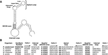
By analysing randomly selected EST clones of the P. westermani adult, we obtained 2 clones (Pw-EST-1, 601 bp-long; Pw-EST-34, 643 bp-long), of which deduced aa sequences shared significant identities with those of GPxs isolated from a variety of organisms, including S. mansoni (AAU34080 and AAC14468), C. sinensis (ABK58679, ABK58680, ABK58681 and ABK58682), Aedes aegypti (AAQ02888), Mus musculus (BAA22780) and human (CAA50793) (E-values <3×10⁻²³). The complete cDNA sequences encompassing each of the clones were determined by consecutive cDNA library screening using the EST clones as probes. The cDNAs, designated PwGPx1 and PwGPx2 (Paragonimus westermani glutathione peroxidase), were comprised of 971 and 918 bp, and contained each single ORF of 189 and 192 codons, from an ATG at nucleotide positions 67–69 and 41–43 to a TAA/TAG at positions 633–635 and 616–618, respectively. In addition to the conventional stop codon, PwGPx2 harboured the second TGA codon within its ORF (nucleotide positions at 233–235), which might decode the 21st aa, Sec (Stadtman, Reference Stadtman1996). In association with the presence of the opal codon, a SECIS motif was detected at nucleotide positions 673–775 within the 3′-untranslated region (UTR) (Fig. 1). The sequence conserved several structural motifs, including 2 helices separated by an internal loop, a SECIS core structure, a quartet located at the base of the second helix and an apical loop; all of which have been well described in a series of GPx genes (Kryukov et al. Reference Kryukov, Castellano, Novoselov, Lobanov, Zehtab, Guigó and Gladyshev2003). In contrast, PwGPx1 contained a standard codon for cysteine (TGC), rather than the Sec codon at the corresponding position and the SECIS was not screened within its nucleotide sequence. Nucleotide sequences of PwGPx1 and PwGPx2 were registered in the GenBank under Accession nos. DQ454159 and DQ454160.
Analysis of selenocysteine insertion sequence (SECIS) motif in PwGPx2 of Paragonimus westermani. (A) The secondary structure SECIS found in the 3′-untranslated region (UTR) of PwGPx2 mRNA. The structure was determined by using the SECISearch program. (B) The primary structures of the SECIS motif. Nucleotide sequences constituting the 3′-UTR SECIS motifs were retrieved from the phospholipid hydroperoxide glutathione peroxidase genes of P. westermani, Schistosoma mansoni, Homo sapiens, Mus musculus and Sus scrofa, and aligned in accordance with the predicted secondary elements. Nucleotides conserved within the invariant SECIS core (Quarter) and apical loop are highlighted by boldface letters.

Determination of primary structures of PwGPx1 and PwGPx2
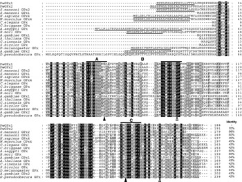
The polypeptides encoded by PwGPx1 (188 aa) and PwGPx2 (191 aa) harboured the predicted molecular masses of 21·7 and 21·6 kDa, respectively, and shared a considerable degree of sequence identity with one another (70%, Fig. 2). The 18 aa residues encompassing the N-terminus of these polypeptides were found to be hydrophobic and predicted as target signals for the translocation of the proteins (underlined sequences in Fig. 2). The Trp residue forming a hydrogen bond with the reactive Sec was replaced by Tyr, another aa with an aromatic side-chain, in the Paragonimus homologues (filled arrowhead in domain C, Fig. 2). Among the 3 aa residues involved in the formation of a salt bridge and hydrogen bond to the glutathione molecule (Epp et al. Reference Epp, Ladenstein and Wendel1983), only the Gln and downstream Arg residues were shown to be tightly conserved among members of PHGPx, including PwGPxs (open arrowheads in Fig. 2). The aa block, which was associated with a tetrameric structure in mammalian GPx proteins other than PHGPxs, was not detected in the aa sequences of PwGPxs (data not shown).
Multiple sequence alignment of PwGPxs with other phospholipid hydroperoxide glutathione peroxidase (PHGPx) members. The deduced amino acid sequences of PwGPx1 and PwGPx2 were aligned with those of Schistosoma mansoni (AAU34080 and AAC14468), Homo sapiens (CAA50793), Mus musculus (BAA22780), Caenorhabditis elegans (CAB03004), Caenorhabditis briggsae (CAE60228), Aedes aegypti (AAQ02888), Bombyx mori (BAE07196), Drosophila melanogaster (AAR96123), Drosophila pseudoobscura (EAL29978), Anopheles gambiae (XP_313166 and XP_313168), Arabidopsis thaliana (CAB78203), Citrus sinensis (CAE46896) and Sorghum bicolor (AAT42166), employing the Clustal X program, then optimized with GeneDoc. The identical amino acids in the alignment are highlighted in black, while similar residues are shown in grey. Three well-conserved domains found in the members of PHGPx are indicated by solid bars with A, B and C. The functional amino acid residues engaged either in the formation of the catalytic centre or in the binding of the glutathione molecule are marked with filled and open arrowheads, respectively. The abbreviation ‘U’ in functional domain A represents selenocysteine. The putative signal peptides are underlined. The identity value of each enzyme against PwGPx1, which was calculated from the alignment after removing positions with a gap as missing data, is provided at the end of the alignment.

Phylogenetic relationships of PwGPxs with their homologues
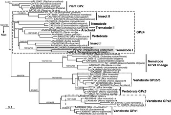
We used the aa sequences of PwGPx1 and PwGPx2 in homology searches with BLAST algorithms and found several hundred entries from the GenBank database. These sequences revealed various degrees of sequence identity ranging 35%–53% (E-values <10−16), of which the majority were PHGPx-like proteins. Information regarding the other GPx families was obtained by a series of subsequent BLAST searches using the aa sequences of human and nematode GPx3. A total of 52 members were finally selected, by considering both the identity values and taxonomical distributions and used in a phylogenetic analysis. In the neighbor-joining tree, the GPxs of higher vertebrates including human and mouse were separately categorized into the 6 GPx families (Fig. 3). With the plant GPxs, proteins isolated from invertebrate animals, much closer relationships with the mammalian PHGPxs were shown, which was consistent with the results of the structural comparison (Fig. 2). However, nematode species were found to express both of the GPx lineages (GPx3 and GPx4). The screenings of the draft genomic sequences of S. mansoni (assembly version 3 at the Sanger Institute) and Anopheles gambiae (NCBI database) showed no evidence reflecting the presence of GPx1/GPx3-like genes in these genomes (data not shown). The tree also revealed that each of the trematode and insect PHGPxs had diverged separately into 2 distinct subclades. In addition to the biased distributions of GPx families, the Sec codon and the associated SECIS motif were recognized largely within the mRNA sequences coding for the trematode and vertebrate GPx members (indicated by † and ‡, respectively, in Fig. 3). Among the arthropod proteins examined, only an arachnidal PHGPx (Boophilus microplus, ABA25916) was found to be Sec-dependent.
The evolutionary relationships of the PwGPx genes with members of the various GPx gene families. The phylogenetic positions of PwGPx1 and PwGPx2 were predicted on the basis of alignment of amino acid sequences. The tree was constructed by a neighbor-joining algorithm using PHYLIP and was rooted with GPxs of Neurospora crassa (CAE76176) and Saccharomyces cerevisiae (NP_009803). Numbers at the major branching nodes indicate their percentages of appearance in 100 bootstrap replicates, and values obtained from other analyses using either maximum parsimony or maximum likelihood algorithms are also presented to ensure the statistical significance of these nodes. The identity of each analysed sequence is distinguished by a protein identification number, followed by the species name from which it was isolated. The presence of selenocysteine (Sec) codon (†) and Sec insertion sequence (‡) within corresponding mRNA sequence is indicated at the end of each sequence identity.

Genomic distribution of PwGPxs and expression of native PwGPx1 and PwGPx2 proteins
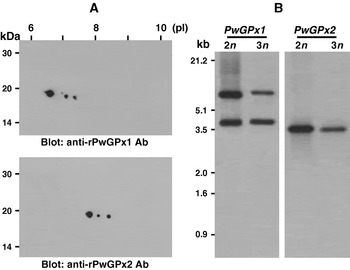
As shown in Fig. 4A, the 3 protein spots, which migrated at approximately 20 kDa, were detected to be strongly reactive against each of the rPwGPx-specific antibodies by the 2-D Western blot analyses of an adult worm extract. The average pI values of the reactive spots were comparable to that of either PwGPx1 (6·04) or PwGPx2 (8·37), which had been predicted theoretically from the deduced aa sequences. In order to determine whether these spots represented paralogues or allelic forms, Southern blot analyses of PwGPx1 and PwGPx2 were carried out with the genomic DNAs from both diploid (2n) and triploid (3n) worms digested with the restriction enzyme, BamH I. The enzyme harboured a single cut in PwGPx1 but no cut in PwGPx2. The PwGPx1 probe revealed 2 signal bands with nearly identical intensity, while that of PwGPx2 detected a single band (Fig. 4B). This result showed that the PwGPx1 and PwGPx2 might exist as a single copy gene in the parasite genome. Therefore, the multiple spots identified by Western blotting seemed to represent the major allelic forms of PwGPx1 and PwGPx2, with considerable frequencies in the P. westermani population. The nucleotide sequences acquired from the cDNA library revealed 3 representative alleles of the PwGPx2 gene (unpublished observation). The pI values of these alleles could be differently calculated as 7·86 (68·3%), 8·31 (12·2%) and 8·37 (14·6%), due to diagnostic base substitutions introduced within their sequences. These values appeared to match well with those of positive spots in the Western blotting. The cDNA sequences corresponding to PwGPx1 also showed a matching pattern similar to that of PwGPx2. Any protein homologous to PwGPx1 or PwGPx2 was not detected in the excretory-secretory products of the parasite, which further suggested that the N-terminal hydrophobic sequences are responsible for the targeting of the proteins into certain organelle(s), rather than into extracellular compartments (data not shown).
Expression and genomic distributions of the PwGPx genes. (A) Two-dimensional (2-D) Western blots of PwGPx1 and PwGPx2. The adult crude extract (50 μg) was isoelectrically focused using IPGphore (pH 3–10), and further resolved by 12% SDS-PAGE. Separated proteins were transferred onto nitrocellulose membranes, and probed with the rPwGPx1- and rPwGPx2-specific mouse antisera (1:3000 dilution), respectively. Molecular mass in kDa and isoelectric points (pI) are shown on the left and top. (B) Southern blot analyses of PwGPx1 and PwGPx2. The genomic DNAs (each of 10 μg) of both diploid (2n) and triploid (3n) Paragonimus westermani were digested with BamH I (one cut in PwGPx1 but no cut in PwGPx2 probes) and probed with digoxigenin-labelled gene-specific DNA fragments. The positions of the DNA size standards (in kb) are also shown.

PwGPx proteins exhibited preferential substrate accessibility toward H2O2 rather than cumene hydroperoxide with an electron donor, GSH
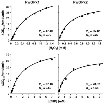
We purified native PwGPx proteins though a series of column chromatographies since the recombinant forms of PwGPxs expressed in the E. coli cells did not show any detectable enzyme activity, probably due to lack of the post-translational phosphorylation process (Arthur, Reference Arthur2000). The purified enzymes efficiently reduced H2O2 and cumene hydroperoxide using GSH as an electron donor. The V max/K m ratios of PwGPx1 and PwGPx2 were determined to be 67·70 and 90·05, respectively, against H2O2, and 14·19 and 17·92, respectively, against cumene hydroperoxide (Fig. 5). These observations collectively suggested that PwGPxs possess preferential substrate accessibility toward H2O2 rather than cumene hydroperoxide, with GSH as an electron donor. The reducing potential of the Sec-dependent PwGPx2 was substantially higher than that of the Sec-independent PwGPx1 at the physiological pH.
Biochemical properties of native PwGPx1 and PwGPx2 proteins. The specificity and catalytic efficiency of the proteins purified by a series of gel chromatographies were examined toward catalytic substrates (H2O2 and cumene hydroperoxide [CHP]). The experiments were repeated 3 times and a representative result presented. The specific activity was calculated as ΔOD340/nmol/min. The steady-state kinetics of the native proteins was analysed by measuring GPx activities using varying concentrations of the catalytic substrates and reduced glutathione. The V max (V m, ΔOD340/nmol/min) and K m (mm) values of each of the proteins are presented.

Oxidative stress-inducible chemicals increased the expression levels of PwGPx1 and PwGPx2 in viable worms
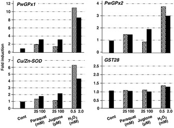
We observed the induction patterns of the PwGPx genes by treating viable adult worms with oxidative stress-inducing chemicals such as paraquat, juglone and H2O2. As shown in Fig. 6, the worms incubated with 0·5 mm H2O2 demonstrated a highly up-regulated expression of PwGPx1 (approximately 11-fold) and PwGPx2 (4-fold). In addition, paraquat and juglone exerted their effects on the induction of these genes, although the extent was less than H2O2; an approximately 2–3 fold increase in PwGPx1 and 1·5–2 fold increment in PwGPx2 were observed. The transcription level of Cu/Zn-SOD was also increased in a manner comparable to that of PwGPx2 upon stimulation, while that of GST28 did not show significant elevation. High concentrations of H2O2 resulted in decreased expressions of these antioxidant genes, which might be attributable to a lethal effect of the chemical (30–40% death after treatment).
Induction patterns of the PwGPx genes under oxidative stresses. Viable Paragonimus westermani adults were incubated for 1 h at 37°C in RPMI 1640 medium supplemented with each of the indicated chemicals. The expression levels of PwGPx1 and PwGPx2 were determined by semi-quantitative RT-PCR. The relative amounts of the PwGPx1 and PwGPx2 transcripts were quantified via determination of the intensities of the electrophoresed amplicon bands. These values were normalized against those of the β-actin gene, which was employed as a quantity control (see Materials and Methods section). The fold inductions were calculated by comparing the intensities between each of the experimental and control groups. The transcription levels of the Cu/Zn-SOD (AY675506) and GST28 (L43919) genes of P. westermani were also employed as comparative target genes. The reproducibility of the responsive inductions was examined by preparing the whole experimental batches 3 times, and a representative data set is shown in this figure.

Expression pattern and localization of PwGPxs were highly associated with the maturation of reproduction-related cells
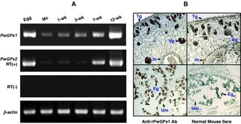
In trematode parasites, expressions of GPxs were increased proportionally according to the maturation and growth of the worm. The GPxs were shown to have specific localities within reproduction-related cells such as vitellocytes (Roche et al. Reference Roche, Liu, LePresle, Capron and Pierce1996; Sayed et al. Reference Sayed, Cook and Williams2006; Cai et al. Reference Cai, Bae, Kim, Sohn, Lee, Jiang, Kim and Kong2008). We observed the expressional regulation of the PwGPx genes in accordance with the developmental stages of P. westermani with the gene-specific primers via a semi-quantitative RT-PCR. The amplification of the target transcripts from total RNAs demonstrated that these genes are transcribed constitutively throughout all of the developmental stages examined, with different induction patterns. The transcription of PwGPx2 was maintained at the basal level from the metacercariae to the 3-week-old juveniles, but the activity increased profoundly in the 7-week-old immature worms, and peaked in the 12-week-old adult worms and eggs. The expression of PwGPx1 gradually increased during those developmental stages and reached its maximal levels at the egg-laying adult worm stage (Fig. 7A).
Spatiotemporal expression of PwGPxs. (A) Expression profiles of PwGPx1 and PwGPx2 during the maturation of Paragonimus westermani. The transcription levels of PwGPx1 and PwGPx2 were examined with total RNAs extracted from the respective developmental stages as indicated on the top, via a semi-quantitative RT-PCR. The amplified products were separated on 2% agarose gels and stained with ethidium bromide. A primer pair for the β-actin gene was used as a quantity control. The absence of any contaminated genomic DNA was confirmed by preparing reactions without reverse transcriptase during the first round of cDNA synthesis (RT-). M, 100-bp DNA size standards; Mc, metacercaria. (B) Histological distribution of PwGPxs in P. westermani adult worm. Adult worm sections (4 μm thickness) were reacted with either the mouse antiserum specific to rPwGPx1 or with normal mouse serum (1:200 dilution). The positive signals are largely restricted within eggs and vitelline glands. Eg, eggs in uterus; In, intestine; Tg, tegument; Um, uterine membrane; Vg, vitelline glands.

Tissue-specific distributions of PwGPx proteins were observed in adult worm sections employing anti-rPwGPx antibodies. As shown in Fig. 7B, immunohistochemical staining with the anti-rPwGPx1 antibody exhibited a strictly restricted localization pattern within the vitelline glands and intrauterine eggs. No positive reaction was detected in the other tissues or organs including ovary, seminal receptacle, testis, intestine and teguments. Antibody specific to the rPwGPx2 protein showed an immunoreactive pattern similar to that of the anti-rPwGPx1 antibody (data not shown).
DISCUSSION
The tissue-invasive helminth parasites may be continuously exposed to dual oxidative stresses, from both ROS generated by endogenous intracellular metabolism and ROS/nitric oxide generated by host inflammatory and immune cells (Selkirk et al. Reference Selkirk, Smith, Thomas and Gounaris1998; Zelck and von Janowsky, Reference Zelck and von Janowsky2004). During their life-span in host tissues, removal of ROS and protection of macromolecules from the oxidative attacks would be inevitable. The antioxidant enzymes might also constitute a major defensive system in these causative parasites (Callahan et al. Reference Callahan, Crouch and James1988). Therefore, the helminths might be taken as a good model for investigation of the functional diversification with antioxidant enzymes. Together with the thioredoxin system, the glutathione-related molecules, such as glutathione, glutathione reductase, glutaredoxin and GPx, have been thought to perform crucial roles in thiol-disulfide redox homeostasis in these parasites, not only by providing electrons to essential enzymes but also by protecting them against oxidative stress (Henkle-Dührsen and Kampkötter, Reference Henkle-Dührsen and Kampkötter2001; Salinas et al. Reference Salinas, Selkirk, Chalar, Maizels and Fernandez2004).
In the present study, we have isolated 2 novel genes that putatively coded for P. westermani GPxs. The deduced aa sequences of these genes revealed the primary structure characteristic to the PHGPx (GPx4) family, including aa conservation, absence of the subunit interaction domain and well-preserved catalytic and glutathione-binding domains. In a phylogenetic analysis, PwGPxs showed tight relationships with the other PHGPx-related members. PwGPx proteins preferred H2O2 as substrate rather than cumene hydroperoxide and an electron donor, GSH. Spatiotemporal expression of these proteins was highly associated with the maturation of reproduction-related cells. The up-regulated expression of PwGPxs induced by exogenous redox-recycling drugs and H2O2 suggested that these enzymes may actively participate in the detoxification of oxidative damage to protect P. westermani in high oxygen-tension status.
The general substrate affinity of GPx for H2O2 has been brought into question, as PHGPx shows a preference for lipid hydroperoxide. Furthermore, certain GPxs of parasitic nematodes have been examined to exhibit no enzymatic activity against H2O2 (Arthur, Reference Arthur2000; Henkle-Dührsen and Kampkötter, Reference Henkle-Dührsen and Kampkötter2001). When we employed H2O2 as a principal substrate, a considerable level of specific GPx activity was detected in the P. westermani adult worm extract (data not shown). In addition, the treatment of viable worms with H2O2 induced significant up-regulation of PwGPx1 and PwGPx2 mRNA and protein expression. These results may suggest that trematodes, unlike the parasitic nematodes, are equipped with the GPx enzyme system, which directly functions as an H2O2 scavenger. Redox-cycling drugs such as juglone and paraquat are capable of crossing cell membranes and can be utilized as intracellular ROS inducers, as they trigger the conversion of oxygen into O2− (O'Brien, Reference O'Brien1991). These chemicals also activated the PwGPx genes, albeit their effect was not as prominent as H2O2. The elevated GPx activity upon treatment of these drugs is likely to result indirectly from the accumulation of H2O2, via the action of SOD, and/or increased lipid peroxidation in response to the generation of ROS.
GPxs have been known to contain Sec at their catalytic site, which is co-translationally inserted in response to the UGA codon, a stop signal in the standard genetic code. The alternative decoding of UGA generally depends on a cis-factor, called SECIS, which is located in the 3′-UTR of these selenoprotein genes (Stadtman, Reference Stadtman1996). In our analysis, however, the selenium-dependent GPxs (sGPxs) showed a biased distributional pattern across diverse eukaryotic domains. The Sec codon and the concurrent SECIS motif were detected exclusively within the mRNA sequences of the trematode and vertebrate GPxs. Among the trematode enzymes obtainable, only PwGPx1 was determined to be a selenium-independent GPx (siGPx). The GPx genes retrieved from the other taxa did not contain the Sec-related sequence factors, with an exception of one isolated from an arachnidal tick. Regarding catalytic activity, the lower redox potential of selenol in Sec (pKa=5·2) than thiol in cysteine (pKa⩾8·0) is favourable for the conversion of the reduced enzyme into the oxidized form, and thus sGPx seems to be more catalytically active than siGPx, under physiological conditions (Stadtman, Reference Stadtman1996). The siGPx has been postulated either to comprise a second-line defence or to cooperate with sGPx in an as yet unknown novel manner, to cope with cellular oxidative stresses (Utomo et al. Reference Utomo, Jiang, Furuta, Yun, Levin, Wang, Desai, Green, Chen and Lee2004). It is also possible that siGPx performs a role as an antioxidant enzyme in special microenvironments, considering the effect of the surrounding pH on the ionizing potential of the normal thiol in cysteine. The siGPxs of filarial nematodes, of which functions are mainly confined at the cuticular matrix, are likely to be an example of the second suggestion (Cookson et al. Reference Cookson, Blaxter and Selkirk1992). Our results demonstrated that the induction level of Sec-independent PwGPx1 was much higher than that of Sec-dependent PwGPx2, either during development of P. westermani or against exogenous oxidative stresses. We are currently designing a study to observe whether the difference in the transcriptional activity is associated with the differential catalytic potentials and/or whether the two enzymes exert their effects within each of specific microniches in vitellocytes.
Among the 6 families of GPx isoenzymes, the PHGPxs have been exclusively isolated in trematodes and arthropods, whereas the GPx3 homologues have been identified as a major GPx family responsible for anti-oxidative defence in the parasitic nematodes. In contrast to the inconsistently conserved structures of the PHGPx genes across taxa, nematode and vertebrate genes encoding the GPx3-like proteins possessed an intron shared by all of the genes examined (unpublished data). The differentially preserved genomic structures across eukaryotic domains reflected that the GPx genes have multiple origins in the evolutionary episode of diverse eukaryotic domains; several paralogues, which are multiplied during early stage of eukaryotic evolution, may have evolved variably and one or a few paralogue lineages are likely to have been maintained in each of the taxonomical clades. The 2 subclade genes found in trematodes seem to have been duplicated after the divergence of Trematoda from the other animal domains. Investigations on these biased evolutionary pathways of the GPx genes along with the lower animal taxa, especially in association with their specialized functions, will provide us more detailed knowledge regarding the biological implications of this antioxidant enzyme system.
This work was supported by the Anti-Communicable Diseases Control Program of the National Institute of Health (NIH 348-6111-215), National Research and Development Program, Ministry of Health and Welfare, Korea and also by a research grant from the Samsung Biomedical Research Institute, #SBRI B-A2-006. Seon-Hee Kim was financially supported by the Brain Korea 21.